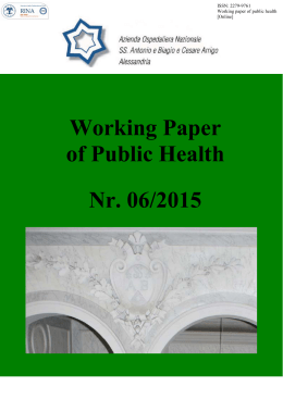
STUDIO “LUME” LONG SURVIVORS IN PLEURAL MESOTHELIOMA Study protocol 1 STEERING COMMITTEE This study was born from a multidisciplinary collaborative groups of epidemiologists, oncologists, surgeons and pathologists of the Fondazione IRCCS Istituto Nazionale dei Tumori (INT) and of the Istituto Superiore di Sanità (ISS). The INT host the Rete Tumori Rari (www.retetumorirari.it) and the oldest Italian cancer registry (Varese CR). A Steering Committee (SC), which consist of all the experts of the INT and of the ISS involved in the study, is envisioned to supervise the study. The SC will be the decision making body; it will be responsible for all strategic planning, ensuring that the timetable is maintained, that the milestones are met and that corrective actions will be taken if necessary. It will receive update on progresses and it will decide on any actions which will be taken cooperatively. Gemma Gatta (Evaluative Epidemiology unit, INT) is the principal investigator, the other experts of the INT and of the ISS involved in the SC are listed below. Annalisa Trama (Evaluative Epidemiology unit, INT) Laura Botta (Evaluative Epidemiology unit, INT) Marina Chiara Garassino (Medical Oncology Division, INT) Eva Regina Haspinger (Medical Oncology Division, INT) Filippo De Braud (Medical Oncology Department, INT) Giuseppe Pelosi (Pathology Unit, INT) Silvana Canevari (Genomic Department) Loris Cecchini (Genomic Department) Flavio Crippa (Nuclear Medicini Unit, INT) Alfonso Vittorio Marchianò (Radiology Unit, INT) Ugo Pastorino (Thoracic Surgery Unit, INT) Rossella Bertulli/Paolo Casali (Cancer Medicine Unit for Adult Sarcoma, Rete Tumori Rari, INT) Riccardo Capocaccia/Roberta De Angelis (Cancer Epidemiology Unit, ISS). 2 BACKGROUND Mesothelioma is a rare cancer. In Italy, the most recent data are published in the Fourth Report of the national registry of mesothelioma (ReNaN) which report on cases diagnosed in the period 1993-2008 [1]. According to these data, 93% of cases of mesothelioma occured in the pleura, the median age at diagnosis was 69 years with no appreciable differences by gender (70 years in women and 69 in men). Up to 45 years, mesothelioma was very rare (only 2.3% of the total registered cases) and the percentage of cases with an age at diagnosis less than 55 years was equal to 9.4% of the total. The majority of cases (71.6% of the 15,845) were male. The standardized rate (per 100,000 residents) for malignant pleural mesothelioma in 2008 was 3.55 in men and 1.35 in women. In practice, there have been 1,422 cases among the different Italian regions in 2008 [1]. There is a huge variability in incidence rates across Italy with a ratio between lowest (Ragusa, Salerno) and highest rates (Genova, Friuli) of about 10-20 [2]. It is well known that this cancer is due to (mainly occupational) asbestos exposure, and surveillance of its incidence falls under a specific law. Survival for mesothelioma is very poor with 50% of mesothelioma patients which die within 280 days from diagnosis [3]. Time trends relative survival did not show improvements over the last decades [4]. However, the project rare cancers in Italy (RITA) observed 193 cases of mesothelioma long survivors (alive after > 3 years from diagnosis) in its pool of 19 Italian CRs in the period 1995-2000 corresponding to the 11% of all malignant mesothelioma registered by the network of general population-based cancer registry (CR). The same proportion of long term survivors (12%) were found in the wider database of the project Surveillance of Rare Cancers in Europe (RARECARE) which contains data from 89 population-based CRs in 21 European countries (www.rarecare.eu). The project RARECARE undertook a quality study asking CRs to verify the diagnosis and the follow-up of mesothelioma long-term survival. The results confirmed that out of the 678 cases reported in the 34 CRs participating to the European study, 578 (85%) were confirmed mesothelioma long-term survivors [5]. In the pool of Italian CRs that participated to the revision, 90% of cases were confirmed mesothelioma long-term survivors. Thus, mesothelioma long-term survivors do exist and the majority are malignant pleural mesothelioma (MPM). For MPM, only few treatment seems to have a certain activity, however the optimal strategy is far to be standardised. In fact, the management of this disease needs a multidisciplinary team which comprises at least dedicated surgeons, oncologists and radiotherapist. At present, the role of surgery is highly debated. On the basis of some encouraging results from large clinical series, extra pleural pneumonectomy (EPP) in which the lung and ipsilateral parietal pleura, pericardium, and hemidiaphragm are resected, became in some centers within multimodal treatment regimen, the standard of care in the management of patients with resectable MPM. However, the recent MARS study suggested that radical surgery in the form of EPP within trimodal therapy offers no benefit and possibly harm patients. According to this study, the hazard ratio [HR] for overall survival between the EPP and no EPP groups was 2·75 (1·21–6·26; p=0·016) after adjustment for sex, histological subtype, stage, and age at randomisation. Median survival was 14·4 months (5·3–18·7) for the EPP group and 19·5 months (13·4 to time not yet reached) for the no EPP group. In addition, there were ten serious adverse events reported in the EPP group and two in the no EPP group [6]. The evidence supporting debulking surgery is limited too and recommendations are to consider pleurectomy/decortication in patients to obtain symptom control [7]. With regard to chemotherapic drugs, the last decade was marked by the introduction of pemetrexed and raltitrexed however no major improvements were obtained. First-line combination chemotherapy including cisplatin and pemetrexed or raltitrexed demonstrated greater activity than cisplatin alone in phase III trials, with higher response rates and improved survival. However, in the BTS study, there was no survival advantage of chemotherapy over best supportive care alone. In the light of limited evidence of efficacy of chemotherapy, the 3 available guidelines on mesothelioma management recommend to discuss with the patients and their relatives on a case-by-case basis, the decision to administer chemotherapy and also second line treatments are poorly active [7;8]. Because of limited data on the best combination treatment, it is recommended that patients who are considered candidates for a multimodal approach should be included in a prospective trial at a specialised centre. In reality, the management of MPM patients diverges quite substantially among hospitals in Italy (treatments are hospital/institution dependent). In this context, it seems important to describe how pattern of care differs among hospital and to what extend such differences have an impact on the prognosis of MPM. MPM diagnosis and staging are also challenging. Actually MPM staging is based on non invasive (Computed Tomography (CT), Positron Emission Tomography (PET), Magnetic Resonance Imaging (MRI)) and invasive (video-assisted thoracoscopy, mediastinoscopy) techniques. However, CT, MRI and PET are reliable for determining extra thoracic extension of disease but lack sensitivity and specificity in predicting pathologic T and N status. Staging describes the anatomical extent of a tumour. There are at least five staging systems available in pleural mesothelioma, the latest one actually in use is the 7 th edition of the TNMbased UICC classification. The main drawback of the classifications is the inaccuracy in describing T- and N-extent by current imaging techniques. Moreover, even when patients are classified based on pathologic analysis, current staging systems do not effectively stratify prognosis. Because of this, an international panel of experts strongly advocated the development of a new robust and uniform clinical staging system [7]. Three dimensional display of axial images may more accurately portray overall disease extent, given the complex morphologic features of pleural mesothelioma. Thus, recent evidence proposed that CT-derived tumour volume can be used to stratify survival of patients with epithelial mesothelioma after extrapleural pneumonectomy and should be included in prognostic evaluation of patients for whom resection is being considered [9]. However, further research are needed to confirm this hypothesis. To conclude, MPM patients have a poor outcome and an optimal treatment is not clearly defined, staging procedures are inaccurate and prognostic factors of clinical importance in the management of MPM are scarce. MPM is a rare cancers and low incidence is a major obstacle to conducting clinical trials to develop effective treatments. However, MPM long term survivors do exist thus, to further study and to improve the quality of care for these cancers is a public health priority. One way of doing this would be to concentrate treatment in specialised centres and establish collaborative groups among such centres to thereby achieve necessary organisational structure, critical mass, patients and tissue samples for carrying out clinical trials, developing alternative study designs and methodological approaches to clinical experimentation and improving accuracy of staging procedures for MPM. AIMS Against this background, our study aims to promote the collaboration of research and clinical groups to study clinical practices and, more important to identify prognostic factors and other factors contributing to long term survival. In details, the study will: - describe patterns of care for MPM in selected Italian populations covered by populationbased cancer registries (CRs) analyse MPM survival per different patterns of care analyse the association between outcome and hospital case volume propose new criteria for staging patients on the basis of CT and PET to be prospectively tested. The new staging system should have a prognostic role of clinical importance to guide the management of MPM 4 - study clinical and biological characteristics of MPM long term survivors (alive after > 3 years from diagnosis) to understand if and how they differ from those with a worse prognosis collaborate with the National Network of Rare Cancers (Rete Tumori Rari) to establish a virtual network of centres/research groups to collect and share clinical information, diagnostic imaging and tissue samples of MPM. STUDY DESIGN The description of the work will be provided considering 3 major activities: 1) population-based study to describe the management of MPM in Italy and to analyse MPM survival by different and new prognostic factors 2) pilot study on biological characteristics of MPM long-term survivors 3) development of a repository of CT and PET images to generate new hypotheses to develop new staging system including also volume assessment and SUV value. These activities are not independent and will be carried out in parallel with an high integration. 1) Population-based study This study is based on the data provided by CRs. To the study will collaborate the general cancer registry adhering to the Association of Italian Cancer Registries (AIRTum) (http://www.registritumori.it) and the network of mesothelioma dedicated cancer registry included in the Italian network of the National Registry of Mesothelioma (ReNaM) [http://www.ispesl.it/renam/Index.asp]. The ReNaM is organised in a regional network. In each region there is an operative centre (COR) with the task of identifying all cases of mesothelioma in its territory and identifying the mode of exposure to asbestos. Completeness of cases is routinely assessed, national diagnostic protocols are used to standardize the criteria for the diagnosis of mesothelioma in each COR, the exposure to asbestos history is carried out with a standard questionnaire by trained staff. The general cancer registration in Italy started at the end of the seventies. Today, in the AIRTum there are 32 general CRs covering 40% of the Italian population (more than 20 millions of citizens). CRs adhere to and store their data in a common data base. To describe the pattern of care, clinical information on staging procedures, treatment, and recurrence will be collected for all MPM cases diagnosed in the period 2003-2008 in a subgroup of CRs. CRs do not routinely collect clinical information, thus CRs will be requested to check again the clinical files to collect these information. These type of studies (high resolution studies) are new for rare cancers such as mesothelioma. All AIRTum CRs are well experienced with high resolution study and have easily access to the clinical dossiers. This is not the case for all COR which were established with the objective of identifying all cases of mesothelioma and studying the association of the cases with the asbestos exposure. Thus, the latter will be essential for collecting information on exposure and other information collected only by COR such as smoking history. In collaboration with the divisions of thoracic surgery and medical oncology of the INT, a protocol for the data collection will be developed. CRs will collect information included in the protocol (please refer to Annex 1) and will verify the availability of histological specimen of MPM long term survivors. - Criteria of inclusion in the high resolution study The accurate diagnosis of mesothelioma can be difficult because mesothelioma is a very heterogeneous cancer which creates various misleading histopathological pitfalls. MPM may resemble benign pleural lesions, metastatic lesions, and benign inflammatory or reactive lesions of the pleura that may occur in patients at approximately the same age as in MPM (pleural effusion during cardiac failure, collagen disease, pneumonia, etc.). 5 In our study, we will include ONLY MPM with a histological or cytological diagnosis. Major markers that will be considered are [7]: markers used to separate epithelioid mesothelioma from adenocarcinoma o Markers with positive diagnostic value for mesothelioma (anticalretinin and antiWilms tumour antigen-1 or the membrane marker anti-epithelial membrane antigen (EMA); for epithelioid mesothelioma, anti-cytokeratin(CK)5/6, antiD2-40 (podoplanin) or anti-mesothelin). o Markers with negative diagnostic value (anti-Ber-EP4, a membrane marker; antithyroid transcription factor-1, a nuclear marker; or monoclonal anticarcinoembryonic antigen, anti-B72-3, anti-MOC-31, antioestrogen/ progesterone, anti-EMA, cytoplasmic staining). markers used to separate sarcomatoid mesothelioma from squamous and transitional cell carcinoma (broad-spectrum anti-cytokeratin antibodies and markers with negative predictive value (such as anti-CD34 and anti-B-cell lymphoma 2 marker, anti-desmin, anti-S100) All tumour subtype will be included: mesothelioma, NOS; fibrous, mesothelioma; epithelioid mesothelioma and mesothelioma, biphasic and sarcomatoid (ICD-O3 M9050-9053). A mesothelioma diagnosed after another primary cancer is to be included only if the first tumour occurred in a different organ. A panel of experts will review the histological specimens of MPM long term survivors to verify whether are real MPM. The experts will include INT pathologists as well as external pathologists with experience in mesothelioma registration in general cancer registries or in mesothelioma dedicated registries. CRs will be requested to verify the follow-up of MPM long term survivors. CR will be requested to verify the availability of the histological specimens of MPM long term survivors in the hospitals. The INT experts in collaboration with CR and the Rete Tumori Rari will contact the different hospitals requesting the histological specimens for the central revision. Handling of histological specimens Once identified, the physician in charge of the contributing hospital will be asked to contact the INT pathologist in order to organize the specimens submission. In particular, the samples (paraffin block and stained slide) should be sealed within a proper container to avoid damages. Collection is scheduled through TNT courier in a final single way. TNT courier will pick up the material directly at the centre and will deliver it to the Pathology Department of Fondazione IRCSS Istituto Nazionale dei Tumori Via Venezian, 1 20133 Milano (Italy) Histological specimens Samples should consist of tissue fragments from the primary tumour, or from any metastatic site of disease, obtained by incisional or excisional biopsies, or by major surgical procedures. A copy of the original pathology reports should be included. The paraffin block should contain enough tumour tissue as to allow additional cutting of at least 20 new and representative 4 micron-thick paraffin sections; in particular, the neoplastic area should measure at least 2mm2 on the tissue section. Paraffin sections can be sent as a substitute of the paraffin block provided that they fulfil the above pre-requisites. - Criteria of exclusion from the high resolution study 6 Cases with ONLY a clinical diagnosis, cases notified by their death certificate only (DCO) and cases discovered at autopsy will not be included in the study. - Number of cases The high resolution study will describe pattern of care and survival for all MPM patients and for long term vs not long term MPM survivors. To perform stratified analyses for MPM long term survival, we need at least 300 cases of long term survivors [10]. To have 300 cases we have to identify 350 MPM long term survivors. This because, MPM long term survivors might not be real long term survivors. Assuming, on the basis of the RARECARE project experience, that 85% of MPM long term survivors are real long term survival we will have to identify 350 cases of MPM long term survivors to have 300 real MPM long term survivors. Finally, assuming that long term survivors are 11% of all cases of mesothelioma (as in the RITA experience) we will have to include in our high resolution study 3200 MPM. - Study period The study will include MPM diagnosed in Italy in the period 2003-2008. All the included cases should have been followed until the end of 2011. - CRs included in the study All AIRTum and ReNaM CRs will be invited to adhere to the study. CRs will be selected on the basis of the following criteria: - Availability of data for the period considered in the study Possibility to collect all the clinical information included in the protocol of the high resolution study Representativeness of different geographical areas The protocol for the data collection will be shared and discussed with CRs and a meeting will be organised to train CR staff to properly read/search the information requested in the clinical files. After this training, CRs will be requested to collect information for 20 cases diagnosed in the period 2003-2008. This small exercise will allow to identify CRs with the best information available and therefore to select those that will contribute to the study. - Data to be collected (please refer to Annex 1) - Data collection, storage and analysis The evaluative epidemiology unit of the INT which is the coordinator of this study will develop an electronic case report form (eCRF) in collaboration with the INT trial office. Designated investigator staff will access the eCRF with a username and a password and will enter the data required by the protocol into the eCRF. The coordinator will be responsible for assuring that the data entered into the eCRF is complete, accurate, and that entry and updates are performed in a timely manner. The methodology of collecting data will be done by software Heavybase. The eCRF system is based on a peer to peer system. This system allows an installation of softwear in local (on the utent’s client) enabling utents to work offline, without an internet connection. The system will be automatically update in presence of an internet connection, synchronizing datas of all centers, leaving visible for each center records (patient) relevant for the center. 7 The coordinator will merge, clean and store CR data in a unique database. Quality checks used in previous high resolution studies [11-13] will be performed to identify possible incongruences. In case of problems the record will be sent back to the CRs for correction. 2) Pilot study on biological characteristics of MPM long-term survivors A pilot study will be undertaken using the cases of MPM available in the INT database and collected by the CRs interested in contributing to this pilot study. The INT has a prospective database since 2002, which includes patients who underwent radical surgery before or after chemotherapy and patients who were treated with medical therapy only. For all patients, information are available on: TNM stage, sex, age, exposure to asbestos, smoking history, date of diagnosis, date of first administration of chemotherapy and time elapsed to surgery. Patients who had chemotherapy were divided in pre- and post-pemetrexed. The same information will be available for CRs cases. Handling of histological specimens arriving from contributing hospitals outside the INT Hospitals with specimens available will be identified by CRs (as previously explained during the high resolution study). CRs in collaboration with the coordinator will assess which of these hospitals are interested in providing their histological specimens also for this pilot study. Those interested will be requested to provide histological specimen of MPM long term survivors and not long term survivors. The hospital interested, will identify a physician in charge of this study which will be asked to contact the INT pathologist in order to organize the specimens submission. In particular, the samples (paraffin block and stained slide) should be sealed within a proper container to avoid damages. Collection is scheduled through TNT courier in a final single way. TNT courier will pick up the material directly at the centre and will deliver it to the Pathology Department of Fondazione IRCSS Istituto Nazionale dei Tumori Via Venezian, 1 20133 Milano (Italy). Histological specimens Samples should consist of tissue fragments from the primary tumour, or from any metastatic site of disease, obtained by incisional or excisional biopsies, or by major surgical procedures. A copy of the original pathology reports should be included. The paraffin block should contain enough tumour tissue as to allow additional cutting of at least 20 new and representative 4 micron-thick paraffin sections; in particular, the neoplastic area should measure at least 2mm2 on the tissue section. Paraffin sections can be sent as a substitute of the paraffin block provided that they fulfil the above pre-requisites. Genomic analyses performed on tissues Determination of mutational status From the INT database, forty-five samples are envisioned for the pilot study: 15 long term survivors of MPM and 30 not long term survivors of MPM matched for confounding factors (age, sex, MPM variant, stage). The study is designed as a two-steps study relying upon: a) Exome deep sequencing of 15 long-term survivors, which will be retrospectively analyzed in formalin-fixed, paraffin-embedded tissues to identify recurrent genetic alterations, and 30 non long-term survivors serving as tumor control groups (training set) Exome sequencing will be done on the SOLiD sequencer (Life Technologies) and copied off instrument for processing using Applied Biosystems BioScope v1.2.1 mapping and analysis software suite. b) b) The obtained molecular profiles will concur to implement a tumor gene panel based on the Ion AmpliSeq technology running upon a IonTorrent personal genome machine (PGM) for 8 studying further independent series (validation set). Molecular profiles will also be correlated with clinical information and survival in the retrospective series and with the response rate and time to progression in prospectively collected patients benefiting from therapies (clinical set). Mutated tumors and all cases showing mutation rate lower than 5-10% will also be studied by Direct sequencing according to the Sanger’s method to further confirm the obtained results. Multiplex PCR amplification, and deep and Sanger sequencing: using the Ion AmpliSeqTM technology (Life Technologies) upon IonTorrent PGM and 3500Dx Genetic Analyzer (both from Life Technologies) for confirming candidate genes with prognostic or predictive relevance, in duplicates, in the validation or clinical set 3. Develop a repository of CT and PET images to generate new hypotheses for improving the staging system CRs will identify hospital of diagnosis and treatment of the registered MPM cases and will verify the availability of the CT images at the hospital. If the CT images are available, the hospital will be contacted and invited to join the Rete Tumori Rari and to share the DICOM-CT and PET images of their MPM cases included in the study. The CT and PET images collected will be reviewed centrally by a panel of experts including radiologists of the INT and other experts extra INT. The panel will define an accurate estimate of the tumour volume and the SUV value. This information will allow to assess whether the CT-derived tumour volume can be used as a prognostic factor of clinical importance. Collection is scheduled through TNT courier in a final single way. TNT courier will pick up the material directly at the centre and will deliver it to the Radiology Department of Fondazione IRCSS Istituto Nazionale dei Tumori Via Venezian, 1 20133 Milano (Italy). In order to support the clinical and molecular analyses we will generate a trial specific database and eCRF. This will be curated by an independent Database Engineer plus our statistical team within a multi-purpose secure web based database. The data base allows for all patient data and files to be securely uploaded (CT/PET, scans, PDFs, notes and all biomarker analyses). Data will be centrally locked but each centre will have the ability to generate and maintain their own private data base, releasing only study relevant information. All patient data will be anonymously labelled in system once entered into the database. Anonymity will be maintained by the Data-Base engineer and only the Lead Statistician will have access to all data sets. In order to provide a technical support to store the histological specimens of the pilot study on the biological characteristics of MPM long term survivors and the CT images, we will generate a trial specific database and eCRF. This will be curated by an independent Database Engineer plus our statistical team within a multi-purpose secure web based database. The data base allows for all patient data and files to be securely uploaded (CT/PET, scans, PDFs, notes and all biomarker analyses). Data will be centrally locked but each centre will have the ability to generate and maintain their own private data base, releasing only study relevant information. All patient data will be anonymously labelled in system once entered into the database. Anonymity will be maintained by the Data-Base engineer and only the Lead Statistician will have access to all data sets. 9 STUDY OVERSIGHT A Monitoring Committee will be established to discuss and properly interpret the results of the study. This Committee will include: epidemiologists, statisticians, pathologists, medical oncologists, registrars, molecular biologist and patient representative) Descriptive analysis will be performed for the most relevant clinical information collected (stage at diagnosis, diagnostic procedure and type of treatment) and will be provided for the pool of CRs participating to the high resolution study and per each CR. Descriptive analyses will be provided overall and separating MPM long term survivors from those who died within 3 years from diagnosis. Relative survival will be estimated using the method by Ederer [14] and will be analysed by stage, treatment and other relevant prognostic factors. It will be estimated for all MPM cases and separating long term survivors from those who died within 3 years from diagnosis. Relative survival will be given also by CR, hospital volume (low, medium, high hospital volume), age, sex, period of diagnosis, ICD-O morphology. Multivariate analysis will be performed in order to provide relative excess risk of dying taken into account region (CRs), age, sex, stage at diagnosis or tumour volume, hospital volume, histopathological subtype and treatment. To solve the problem of the stage migration effect also the determinant of stage, that is the examination performed to define stage at diagnosis, will be considered as further covariate in the multivariate model. - Authorship and rules for accessing the database (please refer to Annex 2) 10 TIMETABLE OF THE STUDY Activites Tasks Population based study Identification of CR contributing to the population-based study and training Data collection by CR for the population based study Histological specimens collection Central revision of histological specimen of MPM long term survivors Pilot study on biological Repository of charactheristis of MPM long-term CT images survivors population based study data check and analyses Selection of the 45 cases with clinical information from the INT database Pre-Clinical Discovery (training set) Pre-Clinical (Validation set) Clinical Validation Statistical analysis Radiological Collection Central revision of CT and PET images Dissemination/publication Months 0 1 2 3 4 5 6 7 8 9 10 11 12 13 14 15 16 17 18 19 20 21 22 23 24 25 26 27 28 29 30 31 32 33 34 35 36 ETHICS AND GOOD CLINICAL PRACTICE The responsible Investigator will ensure that this study is conducted in compliance with the protocol, following the instructions and procedures described in it, adhering to the principles of Good Clinical Practice and with current local legislation and in accordance with: ICH Harmonized Tripartite Guidelines for Good Clinical Practice 1996 Directive 2001/20/EEC of the European Parliament and of the Council Declaration of Helsinki concerning medical research in humans (Helsinki 1964, amended Tokyo 1975, Venice 1983, Hong Kong 1989, Somerset West 1996 and Edinburgh) Patient protection The names of patients will not be recorded. A sequential identification number will be automatically attributed to each patient registered in the trial. This number will identify the patient and must be included on all CRFs. In order to avoid identification errors, patient initials (maximum of 4 letters) and date of birth will also be reported on the CRFs. Investigators will guarantee that all persons involved in this study will respect the confidentiality of any information concerning the trial subject. All parties involved in this clinical trial will maintain the strict confidentiality to assure that neither the person nor the family privacy of the patient participating in the trial is violated; appropriate measures shall be taken to avoid the access of non-authorized persons to the trial data. The processing of the personal data of patients taking part in the trial, and in particular regarding data concerning consent, shall comply with local law on the privacy and with the European Directive on the Privacy of data (95/46/EC). INFORMED CONSENT The investigator must explain to each patient (or legally authorised representative) the nature of the study, its purpose, the procedures involved, the expected duration, the potential risks and benefits involved and any discomfort it may entail. Each patient must be informed that participation in the study is voluntary and that she/he may withdraw from the study at any time and that withdrawal of consent will not affect her/his subsequent medical treatment or relationship with treating physician. The informed consent will be given by means of standard written statement, using non-technical language. The patient should read and consider the statement before signing and dating it, and should be given a copy of the signed document. If the subject cannot read or sign the document, oral presentation may be made or signature given by the subject’s legally appointed representative, if witnessed by a person not involved in the study, mentioning that the patient could not read or sign documents. No patient can enter the study before her informed consent has been obtained. The informed consent is part of the protocol and must be submitted by the investigator to the local ECs. Please refer to Annex 3 12 STUDY FINANCING The study is sponsored by AIRC (Associazione Italiana per la Ricerca sul Cancro) which plays the role of notfor-profit Sponsor. No funds will be provided to Ethical Committees in accordance to the D.Lgs 24-06-2003 and DM 17-12-2004 Results derived from the study are property of the Fondazione IRCSS Istituto Nazionale dei Tumori, Milano which shares it and decide on publication with the Steering Committee. 13 REFERENCES 1. Marinaccio A, Binazzi A, Branchi C, at al. Quarto rapporto: Il registro Nazionale mesotelioma. INAIL, Settore Ricerca Dipartimento Medicina del Lavoro Laboratorio di Epidemiologia e Statistica Sanitaria Occupazionale. Edizione, 2012. 2. AIRT working group. I Tumori in Italia, Rapporto 2006. I dati di incidenza e mortalità dei Registri tumori per il periodo 1998-2002. Epidemiologia e prevenzione, supplemento 2, 2006 3. Montanaro F, Rosato R, Gangemi M, Roberti S, Ricceri F, Merler E, Gennaro V, Romanelli A, Chellini E, Pascucci C, Musti M, Nicita C, Barbieri PG, Marinaccio A, Magnani C, Mirabelli D. Survival of pleural malignant mesotelioma in Italy: a population-based study. International Journal of Cancer 2009 Jan 1;124(1):201-7. 4. AIRTUM WG. Italian Cancer Figures, report 2011. Survival of cancer patients in Italy. Epidemiologia e prevenzione 2011;3:105. 5. Martinez C, Gatta G, Trama A, et al. Report with quality considerations on the available data on rare cancers. Available on line at the rarecare web-site www.rarecare.eu (accessed May 18th 2013) 6. Treasure T, Lang-Lazdunski L, Waller D et al. Extra-pleural pneumonectomy versus no extra-pleural pneumonectomy for patients with malignant pleural mesothelioma: clinical outcomes of the Mesothelioma and radical Surgery (MARS) randomized feasibility study. Lancet Oncol 2011; 12: 763-72 7. Scherpereel, P. Astoul, P. Baas et al. Guidelines of the European Respiratory Society and the European Society ofnThoracic Surgeons for the management of malignant pleural mesothelioma. Eur Respir J 2010; 35: 479–495. 8. Zucali PA, Simonelli M, Michetti G, et al. Second-line chemotherapy in malignant pleural mesothelioma: Results of a retrospective multicenter survey. Lung Cancer 2012;75(3):360-7. 9. Gill RR, Richards WG, Yeap BY et al. Epithelial Malignant Pleural Mesothelioma After Extrapleural Pneumonectomy: Stratification of Survival With CT-Derived Tumor Volume. AJR 2012; 198:359–363. 10. Montanaro F, Rosato R, Gangemi M et al. Survival of pleural malignant mesothelioma in Italy: A population-based study. Int. J. Cancer 2009;124: 201–207 11. Gatta G, Capocaccia R, Sant M, et al. Understanding variations in survival for colorectal cancer in Europe: a EUROCARE High-Resolution Study. GUT, 47(4) 533-538, 2000. 12. Sant M et al Stage at diagnosis is a key explanation of differences in breast cancer survival across Europe. Int J Cancer 2003; 106, 416- 422. 13. Gatta G, Zigon G, Buemi et al. Prostate cancer treatment in Europe at the end of 1990s. Acta Oncol. 2009 Feb 24:1-7. 14. Hakulinen T, Seppä K, Lambert PC. Choosing the relative survival method for cancer survival estimation. Eur J Cancer 2011;47(14):2202-10. 14 ANNEX 1: PROTOCOL FOR THE DATA COLLECTION OF THE HIGH RESOLUTION STUDY IDPZ Identificativo del paziente nel registro IDRT Identificativo del registro (che verrà fornito dal centro di coordinamento) IDTUMORE Identificativo del tumore, se diverso o in aggiunta all’identificativo del paziente SESSO 1=maschio; 2=femmina DATA NASCITA gg/mm/anno COMUNE RESIDENZA codice ISTAT a 6 cifre ALTRI TUMORI Altri tumori maligni (precedenti o contemporanei) 1=si; 2=no; 8=informazione non riportata in cartella 9=non è stato possibile reperire l’informazione da parte del registro SEDE TUMORI PRECEDENTI Se il paziente ha una storia di tumori precedenti o contemporanei, riportare in chiaro la sede tumorale del primo tumore diagnosticato al paziente SEDE TUMORI PRECEDENTI ICD-O3 Se il paziente ha una storia di tumori precedenti o contemporanei, riportare il Codice ICD-O3 per la sede tumorale del primo tumore diagnosticato al paziente (riportare fino alla 3a cifra. Esempio C61.9). 1=non fumatore 2=fumatore < 20 sigarette/die FUMO DI SIGARETTA 20 sigarette/die 3=ex fumatore 8=informazione non riportata in cartella 9=non è stato possibile reperire l’informazione da parte del registro 1=no 2= si (possibile o accertata) ESPOSIZIONE ASBESTO 3=sconosciuto TITOLO DI STUDIO 1=licenza elementare, nessun titolo 15 2=licenza media 3=diploma 2-3 anni (qualifica professionale) 4=diploma 4-5 anni (maturità) 5=laurea e post-laurea 8=informazione non riportata in cartella 9=non è stato possibile reperire l’informazione da parte del registro DATA INCIDENZA Come riportata dal registro tumori: gg/mm/anno ALTRE MALATTIE POLMONARI CONCOMITANTI 1= nessuna 2= asbestosi 3= broncopolmonite cronica ostruttiva 4= altro (specificare in chiaro) 8=informazione non riportata in cartella 9=non è stato possibile reperire l’informazione da parte del registro DIAGNOSI (accertamenti vicini alla data di incidenza) DIAGNOSTICA PER IMMAGINI RADIOGRAFIA DEL TORACE 1=non eseguita 2=negativa 3=dubbia 4=positiva 5=tecnicamente non valutabile 8=informazione non riportata in cartella 9=non è stato possibile reperire l’informazione da parte del registro TAC TORACE 1=non eseguita 2=negativa 3=dubbia 4=positiva 5=tecnicamente non valutabile 16 8=informazione non riportata in cartella 9=non è stato possibile reperire l’informazione da parte del registro RISONANZA MAGNETICA DEL TORACE 1=non eseguita 2=negativa 3=dubbia 4=positiva 5=tecnicamente non valutabile 8=informazione non riportata in cartella 9=non è stato possibile reperire l’informazione da parte del registro PET SCAN 1=non eseguito 2=negativo 3=dubbio 4=positivo 5=tecnicamente non valutabile 8=informazione non riportata in cartella 9=non è stato possibile reperire l’informazione da parte del registro MODALITA’ DI CONFERMA ISTOLOGICA (ve ne possono essere più di una, inserire informazione per tutte quelle effettuate) conferma istologica non eseguita toracentesi (indicare in cc la quantità di liquido tolto) Data ago aspirato Data esito (riportare in chiaro la sede della biopsia in particolare pleura mediastinica, diaframmatica, parietale, viscerale, pericardio) ago biopsia Data esito (riportare in chiaro la sede della biopsia in particolare pleura mediastinica, diaframmatica, parietale, viscerale, pericardio) pleuroscopia/toracoscopia 17 Data esito (riportare in chiaro le sedi dove si sono osservati noduli sospetti in particolare pleura mediastinica, diaframmatica, parietale, viscerale, pericardio) Numero di prelievi bioptici toracotomia Data esito (riportare in chiaro le sedi dove si sono osservati noduli sospetti in particolare pleura mediastinica, diaframmatica, parietale, viscerale,pericardio, pericardio) Numero di prelievi bioptici mediastinoscopia Data esito (riportare in chiaro la sede dei linfonodi sospetti e se evidenza di infiltrazione della pleura mediastinica) Numero di prelievi bioptici broncoscopia Data esito (riportare in chiaro se infiltrazione dei bronchi, se evidenza di infiltrazione della pleura mediastinica) videotoracoscopia (VATS) Data esito (riportare in chiaro le sedi dove si sono osservati noduli sospetti in particolare pleura mediastinica, diaframmatica, parietale, viscerale, pericardio) Numero di prelievi bioptici biopsia non specificata Data esito (riportare in chiaro la sede della biopsia in particolare pleura mediastinica, diaframmatica, parietale, viscerale, pericardio) informazione non riportata in cartella 18 non è stato possibile reperire l’informazione da parte del registro TIPO di DIAGNOSI Istologica (data ospedale esito: certo, possibile, incerto, suggestivo, compatibile, verosimile… parole da cercare nel referto ) Citologica (specificare se toracentesi o ago aspirato) (Istologica (data ospedale esito: certo, possibile, incerto, suggestivo, compatibile, verosimile ) Non possibile distinguere se citologico o istologico DIAGNOSI CITOLOGICA riportare in chiaro la morfologia e la certezza della diagnosi (Riportare anche le formule dubbie, come: possibile, suggestivo, compatibile, verosimile. DATA DIAGNOSI CITOLOGICA gg/mm/anno ESAMI IMMUNOISTOCHIMICI WT1 Podoplanina 1=non eseguito; 2=negativo; dubbio; 3=positivo; 4=tecnicamente non valutabile CALRETININA 1=non eseguito; 2=negativo; dubbio; 3=positivo; 4=tecnicamente non valutabile CLAUDINA-4 1=non eseguito; 2=negativo; dubbio; 3=positivo; 4=tecnicamente non valutabile BeREP4 1=non eseguito; 2=negativo; dubbio; 3=positivo; 4=tecnicamente non valutabile TTF-1 1=non eseguito; 2=negativo; dubbio; 3=positivo; 4=tecnicamente non valutabile CEA 1=non eseguito; 2=negativo; dubbio; 3=positivo; 4=tecnicamente non valutabile VIMENTINA 1=non eseguito; 2=negativo; dubbio; 3=positivo; 4=tecnicamente non valutabile 19 CITOCHERATINA 5/6 1=non eseguito; 2=negativo; dubbio; 3=positivo; 4=tecnicamente non valutabile PAS 1=non eseguito; 2=negativo; dubbio; 3=positivo; 4=tecnicamente non valutabile EMA 1=non eseguito; 2=negativo; dubbio; 3=positivo; 4=tecnicamente non valutabile 5= descrivere pattern se presente B72-3 1=non eseguito; 2=negativo; dubbio; 3=positivo; 4=tecnicamente non valutabile LEU-M1 1=non eseguito; 2=negativo; dubbio; 3=positivo; 4=tecnicamente non valutabile MICRO ELETTR. 1=non eseguito; 2=negativo; dubbio; 3=positivo; 4=tecnicamente non valutabile FISH p16 1=non eseguito; 2=negativo; dubbio; 3=positivo; 4=tecnicamente non valutabile D40 1=non eseguito; 2=negativo; dubbio; 3=positivo; 4=tecnicamente non valutabile PAS-DIASTASI 1=non eseguito; 2=negativo; dubbio; 3=positivo; 4=tecnicamente non valutabile ALTRO (specificare) OSPEDALE DIAGNOSI Indicare per esteso la denominazione dell’ospedale e del reparto in cui il caso era ricoverato al momento dell’esame (istologico o citologico) più probante per la diagnosi. Per i casi sospetti, ma di cui non si ha documentazione istologica o citologica, indicare ospedale e reparto dove è stata posta la 20 diagnosi clinica PRECEDENTI RICOVERI (ENTRO 1 ANNO DALLA DIAGNOSI) Riportare eventuali ricoveri per accertamenti/patologie collegate alla diagnosi di mesotelioma e/o a problemi pleurici/polmonari. Sulla base delle SDO disponibili al registro MORFOLOGIA TUMORE La morfologia è quella alla diagnosi Debbono essere riportate anche le formule dubbie, come: possibile, incerto tra, molto verosimilmente, probabile ecc. (in chiaro) ICD-O3M Codice ICD-O3 per morfologia tumorale (riportare fino alla 5a cifra senza inserire “/”). La morfologia è quella alla diagnosi. Stadio Tumore localizzato Mesotelioma confinato alla pleura Pleura omolaterale e/o viscerale Noduli al di sotto della pleura viscerale Localizzato, NAS Tumore esteso a livello regionale con interessamento diretto di organi e strutture adiacenti Esteso agli organi/strutture adiacenti: Parete toracica Tessuti molli Diaframma Fascia endotoracica Miocardio Polmone, NAS Organi/tessuti mediastinici Pericardio Costole Noduli di mesotelioma che hanno invaso la pleura viscerale fino al polmone Tumore esteso a livello regionale con invasione dei solo linfonodi omolaterali 21 Linfonodi regionali: Aortici, NAS Peri/para aortici, NOS: Aorta ascendente Linfonodi nella finestra aorta-polmonare Linfonodi trachea-bronchiali inferiori o carenali Linfonodi bronco-polmonari o ilari Linfonodi polmonari o intrapolmonari, NAS Vasi linfatici interlobulari e lobulari Mediastino, NAS Peri/parabronchiali Linfonodi peri/para esofagei Linfonodi peri/para tracheali Linfonodi regionali, NAS Estensione regionale per invasione diretta di organi e strutture adiacenti E dei linfonodi omolaterali Tumore esteso a livello regionale, NAS Metastasi a distanza Linfonodi a distanza Controlaterale/bilaterale ilare Controlaterale/bilaterale mediastinico Linfonodo cervicale profondo o scaleno Linfonodi opraclavicolari Altri linfonodi a distanza Organi Tessuti molli del collo Polmone controlaterale Pleura controlaterale 22 Organi intra-addominali Peritoneo Versamento pleurico Estensione e metastasi sconosciuta TNM patologico Da completare con le informazioni post intervento chirurgico. Per chi ha effettuato intervento chirurgico va compilato sia il TNM clinico che il patologico. pT TX tumore primitivo non definibile T0 tumore primitivo non evidenziabile T1 Tumore che invade la pleura omolaterale parietale, con o senza focale coinvolgimento della pleura viscerale T1a tumore che invade la pleura omolaterale parietale (mediastinica, diaframmatica). Nessun coinvolgimento della pleura viscerale T1b tumore che invade la pleura omolaterale parietale (mediastinica, diaframmatica) con focale coinvolgimento della pleura T2 Tumore che invade qualsiasi foglietto pleurico omolaterale, con almeno una delle seguenti caratteristiche (tumore della pleura viscerale confluente, coinvolgimento diaframma, coinvolgimento parenchima polmonare) T31 Tumore che invade qualsiasi foglietto pleurico omolaterale, con almeno una delle seguenti caratteristiche (coinvolgimento della fascia endotoracica, coinvolgimento del tessuto adiposo mediastinico, focolaio unico che invade i tessuti molli della parete toracica) T42 Tumore che invade qualsiasi foglietto pleurico omolaterale, con almeno una delle seguenti caratteristiche (coinvolgimento diffuso o multifocale dei tessuti molli della parete toracica, interessamento costale, invasione del peritoneo attraverso il diaframma, interessamento di qualsiasi organo mediastinico, estensione diretta alla pleura contro laterale, coinvolgimento midollo spinale, foglietto viscerale del pericardio, versamento pericardico con cellule tumorali, interessamento miocardio, coinvolgimento del plesso brachiale) pN NX linfonodi regionali non valutabili N0 linfonodi regionali liberi da metastasi N1 Metastasi nei linfonodi omolaterali peribronchiali e/o omolaterali ilari, 1 2 T3 descrive una neoplasia localmente avanzata ma potenzialmente operabile T4 descrive una neoplasia localmente avanzata e tecnicamente non operabile 23 comprendendo anche i casi di invasione diretta N2 Metastasi nei linfonodi sottocarenali e/o mammari interni omolaterali o mediastinici omolaterali N3 Metastasi nei linfonodi controlaterali mediastinici, mammari interni o ilari e/o omo o controlaterali sovraclaveari o scalenici pM MX Metastasi a distanza non accertabili M0 Metastasi a distanza assenti M1 Metastasi a distanza presenti TERAPIA INTERVENTO CHIRURGICO Verificare nel referto e nella descrizione dell’intervento se ha raggiunto la radicalità /se l’esportazione è stata completa. 1=intervento chirurgico non eseguito 2= pleurectomia (rimozione di tutta la pleura parietale, diaframmatica e mediastinica) radicalità: si, no, informazione non disponibile in cartella, non reperita dal registro 3= pleurectomia-decorticazione (P/D) associa la pleurectomia alla decorticazione viscerale e comporta la resezione della superficie pleurica viscerale con la conservazione del parenchima polmonare radicalità: si, no, informazione non disponibile in cartella, non reperita dal registro 4= pleuropneumonectomia extrapleurica (EPP) include la rimozione “enblock” di pleura, pericardio, diaframma e di tutto il polmone coinvolto dalla malattia radicalità: si, no, informazione non disponibile in cartella, non reperita dal registro 5= talcaggio 8=Informazioni non disponibili in cartella 9=non è stato possibile reperire l’informazione da parte del registro DATA INT CHIRURGICO Data dell’intervento chirurgico: gg/mm/anno 8=informazione non riportata in cartella 9=non è stato possibile reperire l’informazione da parte del registro 24 OSPEDALE INTERVENTO CHIRURGICO Ospedale in cui è stato effettuato l’intervento chirurgico (riportare in chiaro) 8=informazione non riportata in cartella 9=non è stato possibile reperire l’informazione da parte del registro INTERVENTO CHIRURGICO NON ESEGUITO (MOTIVO) Se l’intervento chirurgico non è stato eseguito, specificarne le cause (anche più di una): CHEMIOTERAPIA (CT) - comorbidità - stadio avanzato - altro o motivi non specificati - informazione non riportata in cartella - non è stato possibile reperire l’informazione da parte del registro Prima linea PRIMA LINEA 1=chemioterapia non eseguita 2=monochemioterapia (indicare farmaco: cisplatino, doxorubicina (epirubicina) , methotrexate, raltitrexed (tomudex), pemetrexed (alimta), carboplatino, vinorelbina (navelbina), adriblastina (adriamicina), taxolo (paclitaxel), taxotere (docetaxel), gemcitabina (gemzar) farmaco non disponibile) 3= combinazione di (pemetrexed/cisplatino, pemetrexed/carboplatino, cisplatino/adriamicina, combinazione non disponibile) raltitrexed/cisplatino, carboplatino/taxolo, 4= Farmaco sperimentale (specificare quale) 8=informazione non riportata in cartella 9=non è stato possibile reperire l’informazione da parte del registro DATA INIZIO CT PRIMA LINEA gg/mm/anno OSPEDALE Ospedale in cui è stata effettuata la chemioterapia (riportare in chiaro) CHEMIOTERAPIA PRIMA 8=informazione non riportata in cartella LINEA 9=non è stato possibile reperire l’informazione da parte del registro CHEMIOTERAPIA (CT) 1= non eseguita SECONDA LINEA 2=monochemioterapia (indicare farmaco: cisplatino, doxorubicina (epirubicina) , methotrexate, raltitrexed (tomudex), pemetrexed (alimta), carboplatino, vinorelbina (navelbina), adriblastina (adriamicina), taxolo (paclitaxel), taxotere (docetaxel), gemcitabina (gemzar) farmaco non disponibile) 3= Farmaco sperimentale (specificare quale) 25 8=informazione non riportata in cartella 9=non è stato possibile reperire l’informazione da parte del registro DATA INIZIO CT SECONDA LINEA gg/mm/anno OSPEDALE CT SECONDA LINEA SE DIFFERENTE DA QUELLO DELLA PRIMA LINEA RADIOTERAPIA 1=non eseguita 2=curativa 3=non curativa Dose (Gy) Sede 1=pleura totale 2=pleura parziale 3=tragitti dei drenaggi 8=informazione non riportata in cartella 9=non è stato possibile reperire l’informazione da parte del registro Tipo di radioterapia 1=RT convenzionale 2=Tomoterapia 3=Cyberknife 4=True beam 5=Radioterapia 3d 8=informazione non riportata in cartella 9=non è stato possibile reperire l’informazione da parte del registro DATA RADIOTERAPIA gg/mm/anno OSPEDALE RADIOTERAPIA APPROCCIO MULTIMODALE 1=non eseguito 2= chemioterapia neoadiuvante, seguito da EPP e RT postoperatoria 26 3= chemioterapia neoadiuvante, seguito da EPP 4= chemioterapia nei adiuvante, seguito da P/D 5= chemioterapia nei adiuvante, seguito da P/D e RT 6=altro (riportare in chiaro) 8=informazione non riportata in cartella 9=non è stato possibile reperire l’informazione da parte del registro DATA INIZIO APPROC MULTIMODALE Riportare la data del primo intervento descritto tra la sequenza di quelli elencati sopra: gg/mm/anno OSPEDALE APPROCCIO MULTIMODALE Ospedale in cui sono stati effettati i diversi interventi descritti 8=informazione non riportata in cartella 9=non è stato possibile reperire l’informazione da parte del registro INCLUSIONE IN CLINICAL 1=Sì TRIALS 2=No 8=informazione non riportata in cartella 9=non è stato possibile reperire l’informazione da parte del registro CASO DISCUSSO IN GRUPPO MULTIDISCIPLINARE 1=Sì 2=No 8=informazione non riportata in cartella 9=non è stato possibile reperire l’informazione da parte del registro FOLLOW-UP CLINICO E STATO IN VITA PRIMA PROGRESSIONE DELLA MALATTIA inserire l’evento più vicino alla diagnosi 0= nessuna progressione 1= progressione (pleura, polmone, linfonodi mediastinici, pericardio, diaframma, fegato) 8=informazione non riportata in cartella 9=non è stato possibile reperire l’informazione da parte del registro DATA PRIMA PROGRESSIONE gg/mm/anno STATO Stato in vita all’ultima data del controllo del follow-up: 27 1=vivo; 2=deceduto; 3=perso di vista DATA FOLLOW-UP O DECESSO La data dell’ultimo controllo del follow-up è la data alla quale il registro ha eseguito l’ultimo controllo In caso di decesso la data di morte va riportata come data di follow-up. data: gg/mm/anno CAUSA MORTE Se il paziente è deceduto, riportare il Codice ICD-9 per la causa di decesso ICD-9 28 ANNEX 2: RULES FOR ACCESSING THE DATABASE AND AUTHORSHIP Accessing the database of the high resolution study LUME is a research collaboration between a number of Italian population-based cancer registries, the INT and the ISS. LUME has been planned and will be supervised by a Steering Committee composed by all the experts of the INT and of the ISS. LUME will have a Working Group composed of representatives of the cancer registries which will participate to the project and of a few other experts contributing to the study (Monitoring Committee and panel of experts for the centralised revision of MPM long term survivors). The Evaluative Epidemiology Unit of the INT is the coordinating centre, and provides the secretariat for the Steering Committee, the panel of experts and the Working Group. The LUME database will be developed by the coordinating centre and will be stored at the INT however: - data remain the property of the contributing registries, whose consent is required before they can be used for purposes other than those originally envisaged in LUME all members of the Working Group and of the Steering Committee can access the database to perform analyses (envisioned by the LUME protocol or new and ad hoc) all members that provide data must be informed of any analysis, not envisioned by LUME, being carried out, and each registry has the right to oppose the inclusion of its data in analyses with which it does not agree. The Working Group agrees that any future developments of LUME will be in line with the spirit of collaboration. The Working Group also agrees that collaboration of LUME with other projects/biobank/clinical registry on mesothelioma is to be encouraged. Authorship 1. All publications based on pooled data must mention the LUME Working Group among the authors (or as the author), a suitable authorship formula being: >Authors A, B, C, ... and the LUME Working Group=, with all members listed in a footnote or appendix to the article. 2. The Steering Committe will propose a publication plan to the Working Group. 3. However, analyses and publications can be proposed and carried out by any member of the Working Group or by other researchers not belonging to the Working Group, and will be supervised by the Steering Committee. 4. In general, the researchers who performed the analyses and/or wrote the paper will be first authors in the publication. 5. The first author must provide a justification to the Steering Committee for the names of all authors appearing separately in the authorship list. The list of individual authors should be kept short. 6. The functions of the Steering Committee are to plan and supervise analyses and publications, to ensure adherence to this publication policy. Availability of LUME publications Publications from the LUME project will be sent to the Coordinating centres which will circulate them to the Steering Committee and the Working Group members. 29 ANNEX 3: INFORMED CONSENT Numero dello studio clinico presso la Fondaz. IRCCS Istituto Nazionale dei Tumori di Milano: Titolo dello studio osservazionale biologico: STUDIO “LUME” LUNGO SOPRAVVIVENTI NEL MESOTELIOMA Versione 1 del 23 maggio 2013 ________________________________________________________________________________ Modulo n. 1 INFORMAZIONE PER IL PAZIENTE Questo modulo, che Le sarà consegnato con anticipo rispetto alla Sua decisione finale, riporta le informazioni essenziali sullo studio biologico a cui Le viene chiesto di partecipare. E’ importante che Lei legga queste informazioni e che le discuta con il Medico prima di firmare il consenso alla partecipazione allo studio (Modulo n. 2: “Consenso Informato alla Partecipazione allo Studio”). Partecipano allo studio solo i Pazienti che accettano. Il Paziente può ritirare il suo consenso in ogni momento. Gentile Signora/e, La ricerca scientifica negli ultimi anni ha acquisito nuove informazioni sui geni e sulla loro capacità di influire sulle condizioni di salute e sull’esito delle terapie con farmaci. I geni sono costituiti da DNA, si aggregano nei cromosomi e sono contenuti nel nucleo di tutte le nostre cellule (Figura 1). 30 Figura 1. DNA, geni e cromosomi Le alterazioni dei geni nel tempo, in seguito a cause che non sono ancora bene note, possono avere dato nel tempo esito alla formazione del suo tumore. L’ analisi pertanto dei geni nelle cellule del tumore può fornire informazioni indispensabili per capire come questi processi si sono creati e come il tumore ha potuto nascere, crescere ed evolversi. La malattia. In questo studio saranno inclusi pazienti affetti da mesotelioma pleurico seguiti presso l’ Istituzione dove lei è seguito. Il mesotelioma pleurico è una malattia che origina dalla pleura e la sua insorgenza sembra essere correlata in numerosi casi a un’ esposizione all’ amianto con una latenza circa 30-40 anni. In alcuni pazienti invece, nonostante le accurate indagini, non è possibile trovare una sicura correlazione con questa esposizione. Sappiamo che alcuni pazienti hanno una prognosi particolarmente buona e noi ricercatori crediamo che alla base di questo comportamento della malattia ci possa essere qualche fattore genetico che ne riduca l’ aggressività. L’obiettivo di questo studio. L’ obiettivo dello studio è innanzitutto fare una fotografia della realtà italiana per capire se malattie come la Sua vengano trattate in modo omogeneo. Come Lei saprà, la Sua malattia, per la possibile correlazione con l’ amianto, deve essere sottoposta a registrazione obbligatoria. E’ quindi possibile che l’identificazione del Suo caso sia avvenuta attraverso i Registri; Raccoglieremo pertanto informazioni in forma anonima sul’ estensione della sua malattia tramite TAC e PET, sui trattamenti chirurgici e medici a cui è stato sottoposto e sui loro risultati. Il secondo obiettivo che vorremmo raggiungere è raccogliere quando disponibile un pezzo del tessuto che le hanno asportato e che è servito ai medici per fare la Sua diagnosi. Su tale pezzo cercheremo di ottenere le massime informazioni possibili attraverso una tecnica che si chiama sequenziamento genico per cercare di identificare quei geni che possano caratterizzare un andamento diverso della malattia e una risposta diversa alle terapie a cui è stato sottoposto. Questo studio è di tipo osservazionale, prospettico e non interventistico. Lo studio sarà condotto solo dopo che Lei avrà letto attentamente e capito il Modulo di Consenso Informato e previa approvazione del Comitato Etico. 31 Il disegno di questo studio biologico e raccolta dati genetici. Ogni paziente che accetta di partecipare deve dare la propria adesione scritta allo studio dopo aver letto in ogni sua parte il Modulo di Consenso Informato, chiedendo tutte le spiegazioni che riterrà opportune, al medico dello studio, al fine di capire chiaramente ogni parola o informazione. Apponendo la sua firma al presente Consenso Informato, Lei acconsente che i ricercatori della Fondazione IRCCS Istituto Nazionale Tumori di Milano utilizzino le sue informazioni cliniche e il materiale ottenuto dal Suo campione per le ricerche indicate. Le finalità dell’analisi genetica alla quale le chiediamo di sottoporsi è quella di potere acquisire informazioni utili per chiarire i meccanismi della formazione del mesotelioma e degli effetti terapeutici per fornire nel futuro ulteriori informazioni per una scelta razionale delle terapie. A tal fine, verrà utilizzato materiale proveniente dalle biopsie diagnostiche o dai pezzi chirurgici di pazienti affetti come Lei da mesotelioma pleurico. Tale materiale è stato prelevato durante le normali indagini diagnostiche e di stadiazione della malattia, cui Lei è stato sottoposto presso il Suo Istituto, e sarà utilizzato per l’analisi dello stato mutazionale dei suoi geni. Tutte queste indagini saranno effettuate presso la Fondazione IRCCS Istituto Nazionale dei Tumori di Milano. Lei non avrà benefici diretti nel sottoporsi a questa procedura. I possibili benefici di questo studio si riferiscono alle conoscenze scientifiche che esso fornirà, che potranno concorrere al miglioramento e all’ottimizzazione delle cure per il mesotelioma pleurico. I suoi campioni biologici saranno conservati presso l’ Anatomia patologice della Fondazione IRCCS Istituto Nazionale Tumori di Milano Il materiale biologico conservato (nel rispetto della legge italiana sulla Privacy) verrà utilizzato esclusivamente per fini di ricerca scientifica e mai a fini di lucro e/o commerciali e previa autorizzazione del Comitato Etico della Fondazione IRCCS Istituto Nazionale dei Tumori di Milano Verrà elaborata una lista centralizzata dei pazienti ed una scheda di registrazione presso l’ Epidemiologia dell’ Istituto Nazionale di Tumori di Milano.. Il materiale prelevato verrà utilizzato esclusivamente per condurre tutte le ricerche previste nella sperimentazione che Le stiamo proponendo. Nell’ambito di questo studio il Suo materiale genomico potrà essere estratto dai campioni prelevati per la determinazione dello stato mutazionale dei suoi coinvolti nelle via di trasmissione dei processi di cancerogenesi, ma in nessun caso verrà analizzato il suo intero patrimonio genetico. Se Lei desidera saperne di più su questo Progetto di Ricerca non esiti a chiedere ulteriori informazioni al personale del Nostro Centro, che allo stesso tempo provvederà ad informarLa in modo tempestivo qualora divengano disponibili informazioni che possano influenzare la Sua volontà a continuare la partecipazione allo studio. La copertura assicurativa. Data la natura osservazionale dello studio, ovvero trattandosi di atti medici, la polizza assicurativa stipulata dalla Fondazione garantisce apposita copertura, ai sensi della normativa vigente. 32 Garanzie a tutela del Paziente partecipante allo studio clinico. L’effettuazione di questo studio è stata approvata dal Comitato Etico Indipendente presso la Fondazione IRCCS Istituto Nazionale Tumori di Milano. I campioni saranno processati, conservati ed analizzati sotto la responsabilità di: Dr Gemma Gatta e dott.ssa Annalisa Trama, presso la Fondazione IRCCS Istituto Nazionale Tumori di Milano Epidemiologia Dr.ssa Marina Chiara Garassino, Dirigente Medico presso la Fondazione IRCCS Istituto Nazionale Tumori di Milano e del Professor Giuseppe Pelosi, Direttore del Dipartimento di Anatomia Patologica della Fondazione IRCCS Istituto Nazionale Tumori di Milano. La sua partecipazione allo studio è totalmente libera. Questo significa che Lei può liberamente decidere di non partecipare allo studio, dunque di non firmare questo consenso, senza pregiudicare in nulla l’assistenza che riceverà in seguito presso questa istituzione. Una copia di questa Scheda Informativa e una copia dell’eventuale Consenso restano in possesso del Paziente che accetti di partecipare allo studio. Informativa e manifestazione del consenso al trattamento dei dati personali (ai sensi della deliberazione . 52 del 24/7/2008 Linee guida per i trattamenti di dati personali nell’ambito delle sperimentazioni cliniche di medicinali) Titolari del trattamento e relative finalità La Fondazione IRCCS Istituto Nazionale dei Tumori tratterà i Suoi dati personali, in particolare quelli sulla salute, soltanto nella misura in cui sono indispensabili alla realizzazione dello studio. L’elenco aggiornato dei responsabili del trattamento dei dati dei pazienti partecipanti allo studio e di tutti i soggetti che li trattano – inclusi i laboratori d’analisi – è sempre per Lei disponibile: potrà esserLe fornito, previa richiesta, dallo stesso medico che La sta seguendo. Natura dei dati Il medico che La seguirà nello studio La identificherà con un codice: i dati che La riguardano raccolti nel corso dello studio, ad eccezione del Suo nominativo, saranno, registrati, elaborati e conservati unitamente a tale codice, alla Sua data di nascita, al sesso, al Suo peso e alla Sua statura etc. Soltanto il medico e i soggetti autorizzati potranno collegare questo codice al Suo nominativo. Modalità del trattamento I dati, trattati mediante strumenti anche elettronici, saranno diffusi solo in forma rigorosamente anonima, ad esempio attraverso pubblicazioni scientifiche, statistiche e convegni scientifici. La Sua partecipazione allo studio implica che, in conformità alla normativa sulle sperimentazioni cliniche dei medicinali, il personale coinvolto nello studio, il Comitato Etico e le Autorità Sanitarie Italiane e straniere potranno conoscere i dati che La riguardano, contenuti anche nella Sua documentazione clinica originale, con modalità tali da garantire la riservatezza della Sua identità. Esercizio dei diritti Potrà esercitare i diritti di cui all'art. 7 del Codice (es. accedere ai Suoi dati personali, integrarli, aggiornarli, rettificarli, opporsi al loro trattamento per motivi legittimi, ecc.) rivolgendosi direttamente al centro di sperimentazione (Dr.ssa Marina Chiara Garassino 02/23902656 esterno). Potrà interrompere in ogni momento e senza fornire alcuna giustificazione la Sua partecipazione allo studio: in tal caso, i campioni biologici a Lei correlati verranno distrutti. Non saranno inoltre raccolti ulteriori dati che La riguardano, ferma restando l'utilizzazione di quelli eventualmente già raccolti per determinare, senza alterarli, i risultati della ricerca. Fornendo il presente consenso, Lei autorizza a trattare campioni biologici e informazioni sanitarie che La riguardano per gli scopi dello studio da coloro che vi sono coinvolti (personale sanitario, data manager, statistici, personale ispettivo di organismi regolatori e quanti abilitati dal protocollo di studio e/o dalle 33 normative vigenti). In questo caso, qualunque informazione che La riguardi verrà, nell’eventualità, pubblicata solo in forma anonima, evitando accuratamente e rigorosamente qualunque dettaglio che possa in qualche modo consentire a terzi di risalire alla sua identità. Inoltre, i dati che la riguardano verranno conservati secondo le misure di protezione previste dal Codice. 34 Numero dello studio clinico presso la Fondaz. IRCCS Istituto Nazionale dei Tumori di Milano: Titolo dello studio osservazionale biologico: STUDIO “LUME” LUNGO SOPRAVVIVENTI NEL MESOTELIOMA Versione 1 del 23 maggio 2013 ______________________________________________________________________________ Modulo n. 2 CONSENSO INFORMATO ALLA PARTECIPAZIONE ALLO STUDIO BIOLOGICO AL TRATTAMENTO DATI PERSONALI Questo modulo deve essere firmato da Lei solo nel caso decida di partecipare allo studio clinico. E’ importante che Lei abbia discusso approfonditamente con il Medico prima di firmare questo consenso, anche sulla base della scheda riassuntiva a cui esso si riferisce (Modulo n. 1: “Informazione per il Paziente”). Partecipano allo studio solo i Pazienti che accettano. Il Paziente può ritirare il suo consenso in ogni momento. Dichiaro di essere stato/a esaurientemente informato/a a proposito di questo studio, di aver ricevuto una copia dell’informativa e di averne discusso adeguatamente con i Medici che ne sono responsabili presso questa istituzione. Esprimo il mio consenso libero ed informato: a prendere parte allo studio clinico in oggetto al trattamento di dati che mi riguardano, in formato cartaceo ed elettronico, nel rispetto delle normative vigenti ai sensi fra l’altro del Decreto Lgs. 196/2003 e successive modifiche ad integrazione del consenso da me già sottoscritto al trattamento dei dati sensibili presso l’Istituto Nazionale Tumori di Milano alla raccolta, conservazione e utilizzo dei campioni biologici per eseguire ricerche alle condizioni precedentemente dettagliate Ogni determinazione che non sia specificata nell’allegata scheda informativa non potrà essere effettuata se non dopo nuova approvazione da parte del Comitato Etico Indipendente, che deciderà se sia necessario il rilascio di un nuovo consenso da parte mia; I risultati delle determinazioni effettuate saranno pubblicati solo in forma anonima e aggregata con i risultati delle determinazioni riguardanti altri pazienti. Ho diritto a richiedere la distruzione dei campioni, se non già utilizzati, e limitatamente al periodo in cui questo è tecnicamente possibile, in particolare fino a che non siano ancora stati resi completamente anonimi. 35 Non è prevista una informazione a me od ai miei medici circa i risultati delle determinazioni effettuate sui campioni biologici, dato il loro carattere sperimentale, ma potrò richiedere (fin da ora) di essere informato circa i risultati di questo studio biologico, così come dello studio clinico principale. ………………………………….……… Firma del Ricercatore Nominativo ………………………………….……… Data: …../…../………. ………………………………….……… Firma del Paziente Nominativo ………………………………….……… Data: …../…../………. Firma del Rappresentante legale o del Tutore (se appropriato) Nominativo ………………………………….……… ………………………………….……… Data: …../…../………. Firma di un Testimone (se appropriato) Nominativo ………………………………….……… ………………………………….……… Data: …../…../………. 36
Scaricare