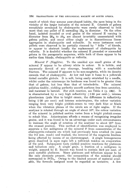
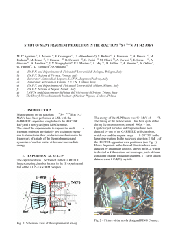
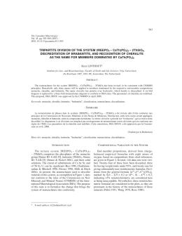
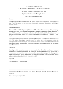
Eur. J. Mineral.
2007, 19, 229–245
Chopinite, [(Mg,Fe)3䊐](PO4)2, a new mineral isostructural with sarcopside,
from a fluorapatite segregation in granulite-facies paragneiss,
Larsemann Hills, Prydz Bay, East Antarctica
EDWARD S. GREW1,*, THOMAS ARMBRUSTER2, OLAF MEDENBACH3, MARTIN G. YATES1
and CHRISTOPHER J. CARSON4
1Department
of Earth Sciences, University of Maine, 5790 Bryand Research Center, Orono, Maine 04469-5790, USA
*Corresponding author, e-mail: [email protected]
2Laboratorium für chemische und mineralogische Kristallographie, Universität Bern, Freiestrasse 3,
3012 Bern, Switzerland
3Institut für Geowissenschaften/Mineralogie, Ruhr-Universität Bochum, 44780 Bochum, Germany
4Research School of Earth Sciences, Australian National University, Canberra, ACT 0200, Australia
Abstract: Chopinite, the Mg-dominant analogue of sarcopside, is a new mineral corresponding to synthetic Mg3(PO4)2-II, a highpressure polymorph of the meteoritic mineral farringtonite. A representative electron-microprobe analysis is SiO2 0.32, P2O5 47.32,
Al2O3 0.05, MgO 30.35, MnO 0.15, FeO 20.99, CaO 0.35, F 0.02, Cl 0.01, Sum 99.54 wt %, which gives
Ca0.02Mg2.20Fe0.86Mn0.01Si0.02P1.95O8. Single-crystal X-ray diffraction gives monoclinic symmetry, P21/c, a = 5.9305(7) Å, b =
4.7583(6) Å, c = 10.2566(10) Å, q = 90.663(9)°, V 289.41(6) Å3, calculated density 3.34 g/cm3, Z = 2. Chopinite is of the olivine
structure type, but with ordered vacancies and strongly distorted octahedra due to the valence 5+ for P, which results in marked
ordering of Mg at M2, whereas Fe2+ concentrates at M1, most likely because of its axial symmetry. The strongest lines in the powder
pattern [d in Å, (Icalc), (hkl)] are 5.92 (42) (100), 3.84(100) (102), 3.48(52) (111, 012, 111), 2.51(72) (113, 113), 2.44 (73) (211, 211).
Chopinite is colorless and transparent, biaxial (–), [ 1.595(2), q 1.648(2), * 1.656(2) (589 nm). 2Vx (meas.) = 40(2)°, 2Vx (calc.) =
41°; X // b, Z ^ a ~55°. Chopinite is found as four inclusions isolated in a fluorapatite segregation in a quartz mass in a paragneiss from
Brattnevet, Larsemann Hills, East Antarctica. Grains are mostly anhedral and range from 0.1 × 0.3 mm to 0.2 × 0.6 mm in size.
Minerals present in the chopinite-bearing specimen include wagnerite-Ma5bc, xenotime-(Y), stornesite-(Y), P-bearing K-feldspar
and plagioclase, Ti-rich biotite, sillimanite, orthopyroxene, sapphirine, hercynite, and corundum. It is inferred to have formed as a
result of high melt P concentrations by reaction of biotite with an anatectic melt in which P/Ca ratio exceeded that buffered by apatite
saturation due to the very slow diffusion of P relative to Ca in anatectic melt.
Key-words: phosphate, new mineral, Antarctica, Larsemann Hills, electron microprobe, crystal structure, granulite facies, anatexis.
Introduction
Sarcopside, (Fe, Mn, Mg)3(PO4)2, is an uncommon mineral
in granite pegmatites and in IIIAB meteorites; there are also possible occurrences in metamorphic environments and
pallasites. Sarcopside is invariably dominated by Fe, with
the maximum XMg = Mg/(Mg+Fe) = 0.37 and maximum
XMn = Mn/(Mn+Fe) = 0.36. With rare exceptions terrestrial
sarcopside forms intergrowths with graftonite or triphylite.
The Mg-analogue of sarcopside was first synthesized as a
metastable phase by a solid-state exchange process at
660°C and 1 bar by Berthet et al. (1972), and subsequently
at 600 °C and 30 kbar, by Annersten & Nord (1980). Brunet & Vielzeuf (1996) and Brunet et al. (1998) showed that
the Mg-analogue of sarcopside is a high-pressure polymorph of the meteoritic mineral farringtonite and deterDOI: 10.1127/0935-1221/2007/0019-1712
mined the equilibrium reaction to lie at 6.32–8.30 kbar for
566–824 °C.
Here we report Mg-dominant analogue of sarcopside as
the new mineral chopinite from a terrestrial metamorphic
environment: a fluorapatite segregation in granulite-facies
(800–860 °C, 6–7 kbar) biotite-quartz-plagioclase paragneiss at 69° 24.437’ S, 76° 15.057’ E on Brattnevet Peninsula, Larsemann Hills, Prydz Bay, East Antarctica. The mineral and name were approved by the Commission on New
Minerals and Mineral Names, International Mineralogical
Association (2006-004). The name is for Christian Chopin
(born 1955) of the Ecole Normale Supérieure, Paris, France,
for his major contributions to the mineralogy of phosphates.
Holotype material (sample no. 121401E and thin section
121401E4) is deposited in the Musée de Minéralogie, Ecole
des Mines de Paris as catalogue number M 73096.
0935-1221/07/0019-1712 $ 7.65
ˇ 2007 E. Schweizerbart’sche Verlagsbuchhandlung, D-70176 Stuttgart
230
E.S. Grew, T. Armbruster, O. Medenbach, M.G. Yates, C.J. Carson
Analytical methods
The optical properties of chopinite grain #3 were measured
at the Ruhr-Universität Bochum by routine immersion procedure using a microrefractometer spindle-stage (Medenbach, 1985).
X-ray powder-diffraction data were obtained from chopinite grain #3 with a 57.3 mm diameter Gandolfi camera and
CuK [ radiation at the Ruhr-Universität Bochum (Table 1).
Cell dimensions of chopinite grain #3 (Table 2) were refined from reflections at scattering angles 13 < ’ < 20° obtained with graphite-monochromated MoK [ radiation on an
ENRAF NONIUS CAD4 diffractometer equipped with a
point detector at the Universität Bern. CAD4 instrumentation gives higher accuracy albeit similar precision as the
CCD instrument.
Single-crystal X-ray intensity data were collected on
chopinite grain #3 with a 3-circle SMART BRUKER CCD
1K, graphite-monochromated MoK [ radiation at the Universität Bern. Like sarcopside, chopinite is pseudo-orthorhombic, but truly monoclinic. We chose the standard set-
ting P21/c. Twinning (imitating orthorhombic symmetry) is
evident from optical inspection and structure refinement
leaving the choice between (100) and (001) as twin planes.
Due to q = 90.66° (close to 90°) the twin law was not obvious from the single-crystal diffraction pattern. The halfwidths of hk0 and 0kl reflections were checked and the hk0
reflections were systematically found to be more asymmetric and broadened compared to 0kl reflections, which suggests that the twin plane is {100}, i.e., identical to the twin
plane {001} reported by Hurlbut (1965) for sarcopside in
the P21/a setting. In order to deal with the extensive polysynthetic twinning, a large window for intensity integration
for each reflection was chosen. It was then possible to collect the intensity of all twin domains. The structure was
solved by direct methods and refined using the SHELXTL
Version 6.12 program package (Sheldrick, 1997). All sites
were refined with neutral atom scattering-factors (Mg, Fe, P,
and O) and anisotropic displacement parameters. In addition, variation of Mg and Fe on the octahedral sites M1 and
M2 was allowed assuming complete occupancy. The octahedral site M1’ at 0,0,1/2, which is occupied in forsterite
Table 1. X-ray powder-diffraction data for chopinite (grain 3).
Iest
dmeas (Å)
Icalc
dcalc (Å)
hkl
st
w
st
vw
st
5.92
4.31
3.84
3.65
3.48
42
29
100
5.9306
4.3162
3.8571
100
011
102
4
3.4979
111
11
3.4879
012
37
3.4817
111
w
2.97
25
2.9653
200
m
2.77
46
2.7764
013
st
2.51
59
2.5236
113
13
2.5055
113
st
2.44
40
2.4497
211
33
2.4385
211
m
2.26
11
2.2680
212
17
2.2572
014
7
2.2505
212
w
2.15
7
2.1604
121
13
2.1566
121
w
1.79
8
1.8006
311
12
1.7898
115
m
1.74
17
1.7490
222
8
1.7440
024
16
1.7409
222
vw
1.65
15
1.6696
124
13
1.6476
106
w
1.47
10
1.4884
206
14
1.4827
400
8
1.4736
206
w
1.40
6
1.4003
017
7
1.3997
133
vw
1.35
5
1.3511
232
5
1.3474
232
Note: Intensities are visual estimates. Five strongest lines are given
in bold. Calculated data from LAZY PULVERIX (Yvon et al., 1977)
based on single-crystal refinement: a = 5.9305(7) Å, b = 4.7583(6)
Å, c = 10.2566(10) Å, q = 90.663(9)°
Table 2. Parameters for X-ray data collection and crystal-structure
refinement of chopinite.
Diffractometer
X-ray radiation
X-ray power
Temperature
Crystal size (mm3)
Detector to sample distance
Rotation axis
Rotation width
Total number of frames
Frame size
Time per frame
Space group
Cell dimensions
Collection mode
Reflections collected
Maximum 2 ’
Index range
Unique reflections
Reflections > 2 c (I)
Rint
Rc
Number of least squares parameters
GooF
R1 , I > 2 c (I)
R1 , all data
wR2 (on F2)
+ 2 e (e/Å3)
– 2 e (e/Å3)
Siemens Smart CCD
MoK [ (0.71073 Å)
50 kV, 40 mA
293 K
0.1 × 0.1 × 0.03
5.4 cm
K
0.3°
1362
512 × 512 pixels
120 sec
P21/c (Nr. 14)
a = 5.9305(7) (Å)
b = 4.7583(6) (Å)
c = 10.2566(10) (Å)
q = 90.663(9)°
V = 289.41(6) Å3
Z=2
automated hemisphere
1543
55.43
–7 e h e 5
–6 e k e 4
–11 e l e 12
617
567
0.028
0.027
64
1.034
0.024
0.027
0.058
0.4
0.4
231
Chopinite from the granulite-facies, Prydz Bay, Antarctica
and fayalite, was found to be vacant, or very nearly so. The
twin contribution was refined to 17.1(2) %. Anisotropic and
isotropic displacement parameters, atomic coordinates and
occupancies are given in Tables 3–5.
Chopinite and associated minerals in sections of sample
121401E were analyzed with a Cameca SX-100 electron
microprobe at the University of Maine and with a Cameca
SX-50 at the Centre de Microanalyse Camparis, Paris. Analytical conditions for analysis of phosphates at the University of Maine were 15 kV accelerating voltage, 10 nA beam
current and 20 µm spot diameter, and data were processed
using the X-Phi correction of Merlet (1994). The standards
used for chopinite and wagnerite were fluorapatite (FK [ ),
tugtupite (NaK [ ), synthetic Mg3(PO4)2 (MgK [ ), albite
(AlK [ ), albite (SiK [ ), synthetic Mg3(PO4)2 (PK [ ), tugtupite (ClK [ ), fluorapatite (CaK [ ), rutile (TiK [ ), rhodonite
(MnK [ ), almandine (FeK [ ); additionally for apatite, fluorapatite (PK [ ), barite (SK [ ), celestine (SrL [ ), synthetic YAl garnet (YL [ ), synthetic rare-earth element phosphates
(REE L [ ), and U metal (UM q ). Three grains of chopinite
were analyzed at twenty spots where each constituent was
counted for 5 seconds, but 4 to 9 analyses were rejected because of impurities and alteration. Fluorine in biotite was
analyzed using the TAP crystal and a polylithionite standard. Zr content of rutile was counted for a total of 1600 seconds using a zirconia standard (SPI-47) and 4 spectrometers
simultaneously to improve the statistics.
Analytical conditions for analysis of silicates and oxides
at the Centre de Microanalyse Camparis were 15 kV accelerating voltage, 10 nA beam current and 5 µm spot diameter, and data were processed using PAP corrections. The
standards and counting times used for chopinite, wagnerite
and biotite were Durango fluorapatite or topaz (15 s, FK [ ),
albite (10 s, NaK [ ), synthetic Mg3(PO4)2, (10 s, MgK [ ), orthoclase (15 s, AlK [ ), diopside (15 s, SiK [ ), synthetic
Mg3(PO4)2 (10 s, PK [ ), scapolite (15 s, ClK [ ), orthoclase
(10 s, KK [ ), diopside (10 s, CaK [ ), MnTiO3 (10 s, TiK [ ),
MnTiO3 (10 s, MnK [ ), hematite (10 s, FeK [ ), sphalerite
(15 s, ZnK [ ), and barite (10 s, BaL [ ).
Identification of minerals not analyzed with WDS was
confirmed by taking an element scan using energy-dispersive spectroscopy at the University of Maine.
Crystal structure
Chopinite and sarcopside are of the olivine structure type,
but with ordered vacancies and strongly distorted octahedra due to the valence 5+ for P (Fig. 1). In olivine each oxygen atom is coordinated to 3 M and 1 Si yielding the “ideal” bond strength of 2 for oxygen (3 x 2/6 + 4/4). In sarcopside O1, O2, and O3 (our numbering) are bonded 2× to M
and 1× to P yielding 2 × 2/6 + 5/4 = 1.917 indicating slight
underbonding. However, O4 bonds 3 × to M and 1 × to P
yielding 3 × 2/6 + 5/4 = 2.25 indicating significant overbonding. For this reason all M-O4 and P-O4 bonds are
rather long (Table 5), which results in strong distortions
and a marked ordering of Mg at the M2 site (89 % Mg).
Fe2+ is ordered at M1 (52 % Fe), most likely because of axial symmetry of the M1 octahedron, that is, the presence of
three 180° O-M1-O angles (Table 5). This symmetry
seems to be more appropriate for arrangement of d-electron orbitals than the M2 octahedron, which lacks this axial
symmetry (with 152°, 153°, and 169°). The M1 octahedron
Table 3. Anisotropic displacement parameters Uij with standard deviations in parentheses
for chopinite.
Site
U11
U22
U33
U12
U13
U23
Fe1
Mg2
P
O1
O2
O3
O4
0.0126(4)
0.0093(5)
0.0112(3)
0.013(1)
0.014(1)
0.011(1)
0.013(1)
0.0065(4)
0.0088(5)
0.0078(3)
0.009(1)
0.0115(9)
0.011(1)
0.0096(9)
0.0103(4)
0.0086(4)
0.0099(3)
0.012(1)
0.0101(9)
0.015(1)
0.014(1)
–0.0006(3)
–0.0001(3)
0.0000(3)
0.0009(8)
–0.0010(8)
–0.0008(8)
0.0000(8)
0.0024(3)
0.0009(4)
0.0005(3)
0.0014(8)
–0.0003(9)
0.0033(7)
0.0003(8)
0.0001(3)
0.0001(3)
–0.0002(3)
–0.0007(7)
–0.0002(7)
–0.0019(8)
–0.0014(7)
Table 4. Atomic coordinates and isotropic displacement parameters, with standard deviations in parentheses, for chopinite.
site
x/a
y/b
z/c
Beq (Å2)
Occupancy
Fe1
1/2
0
1/2
0.77(1)
0.518(6)
Mg1
1/2
0
1/2
0.77(1)
0.482
Fe2
0.2377(2)
0.0118(1)
0.77910(8)
0.70(1)
0.107(4)
Mg2
0.2377(2)
0.0118(1)
0.77910(8)
0.70(1)
0.893
P
0.2562(1)
–0.4293(2)
0.59884(6)
0.76(1)
1
O1
0.2756(3)
0.2519(4)
0.6058(2)
0.89(3)
1
O2
0.2552(3)
–0.3189(4)
0.4588(2)
0.92(3)
1
O3
0.0612(3)
–0.3153(4)
0.6789(2)
0.97(3)
1
O4
0.5334(3)
0.2781(4)
0.3373(2)
0.95(3)
1
Anisotropically refined atoms are given in the form of the isotropic equivalent displacement parameter defined as Beq = (8/3) ‘ 2 7 i( 7 j( Uij ai* aj* ai.aj))
232
E.S. Grew, T. Armbruster, O. Medenbach, M.G. Yates, C.J. Carson
Table 5. Bond lengths (Å) and angles (°).
Length
Angles
Fe1
O1_$1
2.102(2)
O1
2.102(2)
180
O2_$1
2.139(2)
94.25(7)
85.75(7)
O2
2.139(2)
85.75(7)
94.25(7) 180.00(7)
O4
2.140(2)
83.41(7)
96.59(7)
69.30(7) 110.70(7)
O4_$1
2.141(2)
96.59(7)
83.40(7) 110.70(7) 69.30(7)
180
Mean
2.127
O1_$1
O1
O2_$1
O2
O4
Vacancy
O2
2.187(2)
O2_$5
2.187(2) 180.00(7)
O1_$5
2.289(2)
92.10(6) 87.90(6)
O1
2.289(2)
87.90(6) 92.10(6)
180
O3_$5
2.394(2) 112.67(7) 67.33(7)
82.17(6) 97.83(6)
O3
2.394(2)
67.33(7) 112.67(7)
97.83(6) 82.17(6)
180
Mean
2.290
O2
O2_$5
O1_$5
O1
O3_$5
Mg2
O3_$4
2.006(2)
O2_$3
2.061(2)
91.50(9)
O4_$5
2.100(2) 118.74(9) 85.66(8)
O1
2.127(2)
93.52(8) 169.11(9)
83.45(8)
O3
2.133(2)
88.45(6) 97.21(8) 152.66(8) 92.59(8)
O4_$1
2.282(2) 153.24(9) 100.06(8)
86.38(5) 79.52(7)
66.32(7)
Mean
2.118
O3_$4
O2_$3
O4_$5
O1
O3
P
O1_$2
1.523(2)
O3
1.526(2) 112.74(11)
O2
1.529(2) 112.70(11) 112.85(12)
O4_$1
1.576(2) 112.11(11) 102.43(10) 103.15(11)
Mean
1.539
O1_$2
O3
O2
Symmetry codes. $1: –x+1,–y,–z+1. $2: x, y–1, z. $3: x, –y–1/2, z+1/2. $4: –x, y+1/2,
–z+3/2. $5: x, –y+1/2, z+1/2.
is slightly larger (<M1-O> = 2.217 Å) than M2 (<M2-O> =
2.118 Å). In addition, M1 is more regular: the difference
between the largest and shortest M-O bond is 0.038 Å for
M1 but 0.274 Å for M2. Lastly, M1 is adjacent to the vacant octahedral site, thus the M1 octahedron is freer to
adopt a favorable coordination for Fe2+.
The largest octahedral site (<M-O> = 2.290 Å) in the
structure is M1’, which is vacant in sarcopside and chopinite. Local occupation of M1’ would increase the bond
strength of O1, O2, and O3 by additional 2/6, which is energetically not favorable. Nevertheless, the highest positive
peak in the final difference-Fourier map of 0.4 e/Å3 was
found at M1’. This residual density was not considered in
the refinement because the highest negative peak close to
the tetrahedral P sites was also –0.4 e/Å3. One may speculate
that the minor substitution of P5+ by Si4+ determined by electron microbe-analyses (Table 6) is charge balanced by very
minor occupation of M1’ by additional Mg or Fe.
There is a minor difference between XMg obtained from
the structure refinement (XMg = 0.756) vs. the corresponding
value (XMg = 0.720) determined by electron microprobe
analyses (Table 6). There are several possible explanations
for this discrepancy. (1) X-ray single-crystal diffraction
yields the 3-dimensional average bulk composition of the
entire crystal, whereas the electron-microprobe beam does
not penetrate very far below the surface of the section, i.e.,
the different results could be due to compositional zoning.
(2) Because Fe is the atom with the strongest scattering
power in the structure, there is also a minor correlation in the
structure refinement between Fe occupancy on one hand,
scale factor and displacement parameters on the other. (3)
The very minor (0.4 e/Å3) residual density at M1’ was not
considered. Furthermore, the twinning model with {100} as
twin plane maps M1 on M1’ (Fig. 1). Thus there is some
correlation between M1’ occupancy and the twinning contribution. Another possible explanation is the ionization level of oxygen. However, Armbruster et al. (1990) tested the
effect of choosing neutral vs. ionic scattering factors on displacement parameters (Beq), which are even more sensitive
than occupancies, and found no significant variation in Beq
of T sites in models with Si4+ and Al+3 compared to those
with neutral Si and Al in albite.
The synthetic analogue Mg3(PO4)2 of chopinite was first
synthesized by Berthet et al. (1972) as a metastable product
of the following solid state reaction at 660°C: 2×LiMg
(PO4) (olivine structure-type) + MgSO4 → Mg3(PO4)2 (sarcopside structure-type) + Li2SO4. The above authors determined from X-ray powder data the correct symmetry (P21/b;
Chopinite from the granulite-facies, Prydz Bay, Antarctica
233
fraction and found Fe enriched at M2 and Ni dominating at
M1. This Fe order scheme is the opposite of that found in
chopinite. However, in Fe2Ni(PO4)2 two transition metal
ions are competing with each other for the most favorable
coordination. In this case Ni occupies the less distorted site.
Previous crystallographic studies of solid solutions of
Me3(PO4)2 with Me = Co2+, Mg, Zn, Mn2+, Fe2+ in sarcopside-like Ni3(PO4)2 yielded the preference Ni > Co >Mg, Zn >
Mn >> Fe for M1 over M2 (Nord, 1984). A neutron powder
structure-refinement of synthetic sarcopside Fe3(PO4)2 at
59 K (Warner et al., 1992) yielded <M1-O> = 2.147 Å
and <M2-O> = 2.159 Å, whereas a single-crystal X-ray
structure refinement (Moore, 1972) of natural (Fe0.78
Mn0.21Mg0.01)3(PO4)2 sarcopside gave <M1-O> = 2.130 Å
and <M2-O> = 2.164 Å. Due to the similarity of Mn and Fe
scattering factors for X-rays Moore (1972) did not determine whether Fe and Mn order in sarcopside, but instead assumed equal distribution of both elements at M1 and M2.
However, comparison of <M1-O> and <M2-O> bond
lengths with those of Warner et al. (1992) suggests that the
larger Mn2+ will order at M2.
Physical and optical properties of chopinite
Fig. 1. Projection of the chopinite structure along b. M2 octahedra
(Mg enriched) form unbroken edge-sharing chains parallel to a,
whereas M1 octahedra (Fe enriched) alternate with vacant polyhedra
(dark with line and cross pattern). PO4 tetrahedra are marked with
crosses.
their setting: a = 5.912(2), b = 10.21(3), c = 4.73(2), Å,
* = 90°60’) and derived in analogy to olivine a correct structure model with ordered octahedral vacancies. At 820°C this
metastable chopinite analogue transformed to Mg3(PO4)2
having the farringtonite structure. Farringtonite is not based
on a hexagonal closest-package of oxygen (as the olivine
and sarcopside structure-type) and has for this reason also a
larger unit-cell volume, 316.6 Å3 (Nord & Kierkegaard,
1968) vs. 285.8 Å3. In farringtonite Mg occurs in fivefold
and octahedral coordination.
Recently Henry et al. (2003) refined the sarcopside-like
structure of synthetic Fe2Ni(PO4)2 by neutron powder dif-
Most physical properties cannot be determined because of
the small grain size and the very limited amount of available
material. Two good cleavages are evident in photomicrographs and back scattered electron images (see below);
these could {001} and {100}, the two good cleavages reported for sarcopside (Hurlbut, 1965). Density calculated
with empirical formula is 3.34 g/cm3.
Chopinite is colorless and transparent, biaxial (–), [
1.595(2), q 1.648(2), * 1.656(2) (589 nm). 2Vx (meas.) =
40(2)°, 2Vx (calc.) = 41°. Dispersion was not visible, but the
interference figure was poor due to inclusions of brownish
material. Assuming that the twin plane is {100}, orientation
of the principal vibration directions derived from stereographic projection is X // b, Z /\ a ~55°. Larsen & Berman
(1934) reported Z // b, X /\ c = 45° for sarcopside, but they
gave the cleavages as perfect in {010} and {100}. Hurlbut
(1965) was unable to confirm the Larsen & Berman orientation and reported an apparent optical orientation for sarcopside characteristic of a triclinic crystal with Y closest to b.
Hurlbut (1965) failed to resolve the obvious (to him) inconsistency with Larsen & Berman (1934), and we have no explanation why the orientation for chopinite differs from both
of those reported for sarcopside.
Chemical composition and compatibility index
of chopinite
The three analyzed chopinite grains are fairly homogeneous
and virtually identical in composition (Table 6). Chopinite is
nearly pure ferromagnesian phosphate containing but minor
Ca, Mn and Si. We did not analyze Li, which was reported in
sarcopside (Hurlbut, 1965) and is an essential constituent of
the structurally related triphylite-lithiophyllite series. Fluorine contents obtained at University of Maine are negligible,
234
E.S. Grew, T. Armbruster, O. Medenbach, M.G. Yates, C.J. Carson
Table 6. Analyses of chopinite in section 121401E4-1.
Grain
Probe
No. spots
1
UM
12
wt %
0.13(4)
SiO2
P2 O 5
48.00(50)
b.d.
TiO2
Al2O3
0.02(3)
MgO
30.85(25)
MnO
0.11(4)
FeO
20.75(32)
ZnO
n.a
0.01(4)
Na2O
n.a
K 2O
CaO
0.11(10)
BaO
n.a
F
0.04(6)
Cl
b.d.
Sum
99.99
Formulae per 8 Oxygen
Si
0.006
P
1.965
Al
0.001
Mg
2.224
Mn
0.005
Fe
0.839
Zn
–
Na
0.001
Ca
0.006
Sum
5.046
0.726
XMg
1
Cam
7
0.10(3)
47.80(49)
0.01(2)
0.02(4)
30.29(42)
0.14(6)
20.34(42)
0.03(6)
b.d.
0.01(1)
0.02(2)
0.11(18)
0.19(30)
b.d.
98.88
0.005
1.977
0.001
2.206
0.006
0.831
0.001
0.000
0.001
5.029
0.726
2
UM
16
0.23(6)
47.84(53)
b.d.
0.01(2)
30.87(31)
0.14(4)
21.11(38)
n.a
0.01(4)
n.a
0.10(10)
n.a
0.01(11)
b.d.
100.30
0.011
1.956
0.001
2.223
0.006
0.853
–
0.001
0.005
5.055
0.723
2
Cam
5
3*
UM
11
0.16(4)
47.54(17)
0.01(2)
0.01(2)
29.92(50)
0.11(4)
20.53(45)
0.05(6)
0.01(2)
b.d.
0.03(2)
0.02(3)
0.18(33)
0.01(1)
98.41
0.32(3)
47.32(44)
b.d.
0.05(4)
30.35(41)
0.15(3)
20.99(38)
n.a
b.d.
n.a
0.35(17)
n.a
0.02(4)
0.01(2)
99.54
0.008
1.976
0.001
2.190
0.005
0.843
0.002
0.001
0.001
5.028
0.722
0.016
1.952
0.003
2.204
0.006
0.855
–
0.000
0.018
5.055
0.720
3*
SREF
49.64
31.96
18.40
100.00
2.0000
2.2675
0.7325
5.000
0.756
Notes: Probe: UM – University of Maine, Cam – Camparis, SREF – single-crystal structure refinement. n.a.
– not analyzed; b.d. – below detection. All Fe as FeO. Numbers in parentheses are 1 c standard deviations of
last 1 or 2 digits in the averages. Totals does not include F, Cl. Formulae do not include Ba, F or Cl. XMg =
atomic Mg/(Mg+Fe). * Grain used for optical and crystallographic studies.
whereas those obtained at the Centre de Microanalyse Camparis vary with the standard used, ranging from below detection to 0.10 wt % with the topaz standard and from 0.62 to 0.76
wt % with the fluorapatite standard. Averages of individual
grains in Table 6 include measurements with both F standards, resulting in high standard deviations for F. Analyses at
the two laboratories gave virtually identical formulae, except
the Centre de Microanalyse Camparis analyses gave a closer
approach to ideal stoichiometry in terms of (P+Si):(Mg+
Fe+Mn+Ca) ratio and lower Ca contents. Both sets of analyses gave a significantly lower Mg:(Fe+Mn) ratio than the
crystal structure refinement (see above).
The Gladstone – Dale relation (Mandarino, 1981) gives a
compatibility index 1 – (KP/KC) = 0.001 (superior) for grain
no. 3.
Relationship of chopinite to other (Mg, Fe,
Mn)3(PO4)2 phases
Only two terrestrial minerals are composed almost exclusively of (Mg, Fe, Mn)3(PO4)2: the isostructural phases chopinite and sarcopside (Fig. 2). Graftonite (Fe > Mn) and
beusite (Mn > Fe) are (Fe, Mn, Ca, Mg)3(PO4)2 phosphates
containing significant Ca. Graftonite has a higher Mn/Fe ratio and lower Mg/Fe ratio than associated sarcopside. The
Brattnevet chopinite is far more magnesian than any known
sarcopside or graftonite, and unlike most terrestrial sarcopside, it is not intergrown with either graftonite or triphylite.
Experiments suggest that there is no break in solid solution
between synthetic end-member chopinite and sarcopside
(e.g. Annersten & Nord, 1980; Charalampides et al., 1988).
An Mg-dominant analogue of graftonite has not been reported in nature or experiment.
Meteoritic Mg-Fe-Mn phosphates include not only graftonite and sarcopside, but also farringtonite (Fig. 2). Synthetics with the farringtonite structure extend to 60 % Feend-member (Annersten et al., 1980). Graftonite and sarcopside in IVA and IIIAB irons are Fe-Mn solid solutions
containing negligible Mg; graftonite is richer in Mn than associated sarcopside; identification by X-ray diffraction was
possible in a few cases (Bild, 1974; Olsen et al., 1999). In
contrast, phosphates in the Graves Nunatak (GRA) 95209
lodranite are Mn-poor (Mg, Fe)3(PO4)2 phases that range
from Mg-rich to Fe-rich, referred to as “farringtonite” and
“Mg-graftonite” or “graftonite/sarcopside”, respectively
235
Chopinite from the granulite-facies, Prydz Bay, Antarctica
(Floss, 1999; McCoy et al., 2006). A Fe-Mg phosphate in
the Brahin pallasite also has the stoichiometry of sarcopside
(Buseck & Holdsworth, 1977). Although information other
than chemical was not obtained to identify these ferromagnesian phosphates, there is reason to suspect that two or
three phases are present in GRA 95209 (see below).
1.0
Mg3(PO4)2
Occurrence and associated minerals
Chopinite occurs in one (Fig. 3) of many fluorapatite segregations in a quartz mass roughly 10 cm thick and 3 m long in
biotite-quartz-plagioclase paragneiss, which also contains
segregations of prismatine and cordierite at this locality on
Meteoritic phosphates
Farringtonite
GRA 95209
Brahin
0.8
chopinite
0.6
0.4
terrestrial
sarcopside
sarcopside
in IIIAB irons
terrestrial
graftonite
0.2
0.0
0.0
0.2
0.4
0.6
0.8
1.0
Fegraftonite in IIIAB irons Fe3(PO4)2
Fig. 2. Plot of natural farringtonite, graftonite, sarcopside, and chopinite compositions in terms of the divalent cations excluding Ca. The
fields for terrestrial graftonite (dashed line) and sarcopside (solid line) are largely from intergrowths (Černý et al., 1998; Corbella i Cordomı́
& Melgarejo i Draper, 1990; Fontan & Fransolet, 1986; Fransolet, 1977; Fransolet et al., 1986; Hurlbut, 1965; Huvelin et al. (1971); Lindberg
(1950); Livingstone, 1980; Malló et al., 1995; Palache et al. (1951); Povondra et al., 1987; Roda et al., 2004; Smeds et al.,1998; Stalder &
Rozendaal, 2002; Zhang, 1995). Sources of other data: chopinite (this paper); farringtonite (Fuchs et al., 1973; Bild, 1974; Buseck & Holdsworth, 1977); phosphates in meteorite GRA 95209 (Floss, 1999; McCoy et al., 2006); graftonite and sarcopside in IVA and IIIAB iron meteorites (Bild, 1974; Olsen et al., 1999); phosphate (sarcopside?) in the Brahin pallasite (Buseck & Holdsworth, 1977).
Mn3(PO4)2
Section 121401E4-1
Ap
Qz
4
3
2
1
Ap
Bt 1
Qz
Bt 2
Fig. 3. Photomicrograph of fluorapatite segregation (Ap) containing four grains of chopinite (numbered, Fig. 4) in section 121401E4. Matrix
is quartz (Qz); Bt 1 and 2 – biotite grains closest to chopinite (Table 9); only a tip of the second grain is visible in this photograph. Plane polarized light.
236
E.S. Grew, T. Armbruster, O. Medenbach, M.G. Yates, C.J. Carson
Qz
200 Pm
Qz
Ap
Cpn
Ap
(a)
(b)
200 Pm
Qz
Cpn
Ap
Ap
200 Pm
Qz
Xnt
Qz
100 Pm
Cpn
Ap
(d)
(c)
Ap
(e)
Cpn
(f )
Fig. 4. Chopinite (Cpn) enclosed in fluorapatite (Ap) in section 121401E4. (a) Photomicrograph of grain 1. Plane polarized light. (b) Back
scattered electron image of this grain. (c) Photomicrograph of grain 3. Plane polarized light. (d) Back scattered electron image of this grain.
(e) Back scattered electron image of grain 2. (f) Back scattered electron image of grain 4. Qz – quartz, Xnt – xenotime-(Y). Darker areas in
the photomicrographs are secondary minerals.
Brattnevet Peninsula. The segregation is 8 mm long, variable in thickness and highly irregular in outline (only a portion is shown in Fig. 3). Overall, 31 sections were cut from
22 different slices of sample 121401E, but only one section
contained chopinite, four grains in all (section 121401E4).
These grains are anhedral, or show some roughly planar surfaces, and range from 0.1 × 0.3 mm to 0.2 × 0.6 mm (Fig. 4).
The mineral is partially altered and its original form is obscured; the present grains could have been aggregates of two
or three single crystals. Cores of unaltered material range
from less than 0.1 × 0.1 mm to 0.2 × 0.4 mm and constitute
single crystals, albeit one crystal appears to have been bro-
ken. Alteration of chopinite resulted in a mixture of phosphates, including secondary fluorapatite, a phase similar to
the isokite-like mineral reported by Grew et al. (2007), and
xenotime-(Y), the last in specks (<10 µm) along grain
boundaries (Fig. 4e). Other secondary phases appear green
or brown in thin section, but could not be identified. No unaltered chopinite is in contact with quartz, but altered chopinite contacts quartz at one spot (Fig. 4f).
Wagnerite-Ma5bc, biotite, albite, pyrite, and monazite(Ce) are also present in the chopinite-bearing section, but
none contacts chopinite. Overall, a diverse suite of minerals
was found in specimen 121401E, but many very sparingly
Chopinite from the granulite-facies, Prydz Bay, Antarctica
0.04
Y + REE per 12.5 O
Fluorapatite in 121401E
0.03
0.02
75
=
E)
Na
0.
E
0.01
0.00
0.00
,R
(Y
0.01
0.02
4 Chp
4 Wag
1 Sto, Wag
Ideal
0.03
0.04
0.05
0.06
Na per 12.5 O
Fig. 5. Plot of fluorapatite Na, Y and total rare earth element (REE)
contents in specimen 121401E, including data from Grew et al.
(2006). Legend gives section number and identity of nearby ferromagnesian phosphate mineral (abbreviations in Table 7). Line is for
reference only.
Table 7. Minerals and their occurrence in specimen 121401E.
Mineral
Ap seg*
Qz matrix
Phosphates
Fluorapatite (Ap)
XX
XX
Wagnerite (Wag)
X
–
Stornesite-(Y) (Sto)
T
–
Chopinite (Chp)
R
–
Monazite-(Ce) (Mnz)
T
–
Xenotime-(Y) (Xnt)
T
–
“Souzalite-like”
2nd
–
Silicates
Quartz (Qz)
X
XX
Plagioclase (Pl)
X
X
K-feldspar (Kfs)
x
–
Sillimanite (Sil)
T
T
Orthopyroxene (Opx)
–
R
Sapphirine (Spr)
–
R
Biotite (Bt)
X
X
Chlorite
2nd
2nd
Sericite
2nd
2nd
Zircon (Zrn)
–
R
Oxides
Hercynite (Hc)
x
T
Corundum (Crn)
–
R
Rutile (Rt)
–
R
Magnetite (Mgt)
x
T
Ilmenite (Ilm)**
x
T
Miscellaneous
Pyrite
T
–
Celestine
R
–
Fe carbonate
2nd
–
Note: XX – very abundant; X – abundant throughout; x – present
throughout; T – in trace amounts; R – rare, i.e., <5 grains total in 1 or
2 sections; 2nd – a secondary phase, generally sparse. *In the fluorapatite segregations, with or without quartz, in contrast to minerals not
found within the segregations. **Includes exsolved hematite and
secondary “pseudorutile”.
237
(Table 7). The phosphates occur exclusively as inclusions in
apatite, although contacts with quartz are common; in a few
cases, wagnerite is found between apatite and silicates.
The coarse-grained fluorapatite hosting chopinite and
wagnerite in section 121401E4 is relatively rich in Mg, Cl,
Fe and Y (Table 8), features characteristic of fluorapatite in
stornesite-(Y)-bearing sections of 121401E and of specimens from the type locality (Grew et al., 2006). Yttrium and
total rare earth elements (REE) increase regularly with Na in
sample 121401E, but not at the ratio predicted by the substitution (Y, REE) + Na = 2Ca (Fig. 5). Halogen contents of apatite vary little from spot to spot (Fig. 6a).
Next to fluorapatite, the most abundant phosphate is wagnerite. Two inclusions of partially altered pale-yellow wagnerite, up to 1.5 mm long, in a second, larger apatite segregation in the chopinite-bearing thin section 121401E4 are
5b polytype and contain little Ti (Table 8). Two other grains
in this section average 0.33 wt % TiO2 and XMg = 0.92. In
other sections of 121401E wagnerite is commonly developed around K-feldspar or biotite and its break down products.
Brown flakes of Ti-rich biotite (Table 9) are commonly
enclosed in fluorapatite or in quartz near fluorapatite (e.g.
Fig. 3). Cl/(Cl+F) ratio (Fig. 6b) and Ti content vary
inversely with XMg. Distributions of Cl, F and OH between
biotite and apatite can be used as geothermometers (Zhu &
Sverjensky, 1992), which we applied to the data obtained
on the U Maine electron microprobe assuming a constant
apatite XCl throughout sample 121401E (Fig. 6a) and P = 6
kbar. Chlorine distribution [ln KD = 4.35 – 4.95, where
KD = (Cl/OH)Ap/(Cl/OH) Bt] gives less than 500 °C, whereas F distribution [ln KD = 3.32 – 4.02] gives 698–788 °C
(cf. 800–860 °C for the metamorphic peak, Grew et al.,
2006).
Perthitic K-feldspar occurs as selvages formed from
breakdown of biotite (Grew et al., 2006) and as coarser
grains enclosing oriented vermicules and rounded blebs of
quartz (Fig. 7). P2O5 contents of K-feldspar and plagioclase
(An9-18) enclosed in fluorapatite or contiguous to fluorapatite range from 0.09 to 0.51 wt % (Grew et al., 2006; this paper, Table 10). Albite (An0.3) is a minor component of the
quartz matrix in the chopinite-bearing section.
Orthopyroxene is largely altered to phyllosilicates; it is
separated from apatite and from small grains of wagnerite
enclosed in apatite by a quartz-K-feldspar-oligoclase corona (Fig. 8a; Table 11). Sapphirine is found in direct contact
with quartz (Fig. 8b) or enclosed in sillimanite, which isolates it from fluorapatite and quartz. In most cases, sillimanite appears to be a later-formed mineral, being found with
the low-Ti skeletal biotite and as an overgrowth on corundum (Fig. 8c,d; Table 11); a prism is also enclosed in fluorapatite.
Hercynite (Table 12), magnetite (Table 12) and ilmenite
are found both as vermicules and tiny grains (several tens
micrometers) in K-feldspar resulting from breakdown of biotite enclosed in fluorapatite, and as very rare independent
coarser grains several hundreds micrometers across in
quartz, either enclosed in the fluorapatite segregations or in
the quartz matrix. In contrast, corundum (Table 11) has only
been found in quartz, from which it is separated by a thin
238
E.S. Grew, T. Armbruster, O. Medenbach, M.G. Yates, C.J. Carson
Table 8. Selected analyses of fluorapatite (Ap) and wagnerite-Ma5bc (Wag) in the chopinite-bearing section 121401E4-1.
Mineral
Grain
No. spots
wt %
SiO2
P2 O 5
SO3
TiO2
Al2O3
MgO
MnO
FeO
Na2O
CaO
SrO
Y2O3
La2O3
Ce2O3
Nd2O3
Yb2O3
UO2
F
Cl
H2O calc
O=F, Cl
Sum
Formulae
O (anhyd.)
Si
P
S
Ti
Al
Mg
Mn
Fe
Na
Ca
Sr
Y
La
Ce
Nd
Yb
U
Sum
F
Cl
Hcalc
Sum
XMg
Ap
10
10
Ap
20
17
Ap
50
18
Wag*
4
13
Wag**
5
20
b.d.
42.02
b.d.
b.d.
0.03
0.69
0.22
2.44
0.31
50.43
0.03
0.56
0.10
0.24
0.06
0.01
0.08
2.37
2.09
0.10
–1.47
100.32
b.d.
41.98
b.d.
b.d.
b.d.
0.86
0.27
2.74
0.29
50.27
0.02
0.47
0.07
0.21
0.06
0.06
0.04
2.28
2.10
0.14
–1.43
100.43
b.d.
41.44
0.01
b.d.
b.d.
0.61
0.29
3.10
0.31
49.88
b.d.
0.55
0.10
0.29
b.d.
0.06
b.d.
2.36
2.07
0.09
–1.47
99.71
0.02
41.90
n.a.
0.08
b.d.
45.42
0.12
6.69
b.d.
0.13
n.a.
n.a.
n.a.
n.a.
n.a.
n.a.
n.a.
10.65
b.d.
0.32
–4.48
100.87
0.03
42.10
n.a.
0.09
0.01
45.28
0.14
6.68
b.d.
0.11
n.a.
n.a.
n.a.
n.a.
n.a.
n.a.
n.a.
10.75
b.d.
0.29
–4.53
100.95
12.5
0
3.019
0
0.000
0.003
0.087
0.016
0.173
0.051
4.585
0.001
0.025
0.003
0.008
0.002
0
0.002
7.975
0.637
0.300
0.063
1.000
0.335
12.5
0
3.014
0
0
0
0.109
0.020
0.194
0.047
4.567
0.001
0.021
0.002
0.007
0.002
0.001
0.001
7.986
0.613
0.302
0.086
1.000
0.360
12.5
0
3.005
0.001
0.000
0.000
0.078
0.021
0.222
0.052
4.578
0.000
0.025
0.003
0.009
0.000
0.001
0.000
7.997
0.640
0.300
0.059
1.000
0.259
4.5
0.001
0.983
–
0.002
0
1.876
0.003
0.155
–
0.004
–
–
–
–
–
–
–
3.023
0.933
0
0.067
1
0.924
4.5
0.001
0.986
–
0.002
0.000
1.868
0.003
0.155
–
0.003
–
–
–
–
–
–
–
3.018
0.940
0
0.060
1
0.924
Notes: n.a. – not analyzed; b.d. – below detection. All Fe as FeO. H2O calculated from
stoichiometry: F + Cl + H = 1. XMg = atomic Mg/(Mg+Fe).
Analyses done at the University of Maine.
0
Near to chopinite grains 1 and 2, wagnerite grain 5, respectively
*a = 9.670(3), b = 31.74(1), c = 11.924(4) Å, q = 108.25(2)°, V = 3481(2) Å3
**a = 9.681(3), b = 31.685(8), c = 11.919(3) Å, q = 108.14(2)°, V = 3474(2) Å3
239
Chopinite from the granulite-facies, Prydz Bay, Antarctica
selvage of sillimanite (e.g. Fig. 8c,d, Table 11). A rutile
grain (Table 12) occurs with zircon along the boundary of
quartz with a sillimanite aggregate enclosing corundum.
Temperatures calculated from the 386 ppm Zr measured in
rutile are 751 °C (calibration of Zack et al., 2004) and
663 °C (calibration of Watson et al., 2006).
Origin of chopinite
Chopinite is distinct from sarcopside not only in the dominance of Mg over Fe, but also in being found as discrete
grains enclosed in fluorapatite in a metamorphic assemblage. There are few terrestrial occurrences of sarcopside
that is not intergrown with graftonite or triphylite (e.g.
Table 9. Selected analyses of biotite in sample 121401E.
Sect / gr
Probe
F std
No. spots
4-2 / 1
UM
Pol
10
wt %
37.08
SiO2
0.04
P2 O 5
5.07
TiO2
15.18
Al2O3
0.15
Cr2O3
9.63
K 2O
0.10
Na2O
CaO
b.d.
BaO
0.13
MgO
14.10
MnO
0.02
FeO
14.63
ZnO
n.a.
F
1.32
Cl
0.41
3.32
H2O calc
O=F, Cl
–0.65
Total
100.52
Formula per 22 O anhydrous
Si
5.495
P
0.005
IVAl
2.500
Sum IV
8.000
Ti
0.565
VIAl
0.152
Cr
0.018
Mg
3.116
Mn
0.002
Fe
1.813
Zn
–
Sum VI
5.666
K
1.820
Na
0.028
Ca
0.000
Ba
0.007
Sum XII
1.856
Total
15.522
F
0.619
Cl
0.103
3.278
Hcalc
Sum
4.000
0.632
XMg
4-3 / 1
UM
Pol
10
19 / 2
UM
Pol
7
4-1 / 1o
Cam
Ap
2
4-1 /2o
Cam
Ap
2*
36.64
0.07
6.19
15.09
0.09
9.58
0.08
0.02
0.11
12.03
0.04
16.47
n.a.
1.10
0.52
3.36
–0.58
100.83
36.92
0.03
5.58
14.83
0.10
9.58
0.17
b.d.
0.07
13.09
b.d.
16.12
n.a.
1.46
0.60
3.19
–0.75
101.01
37.03
0.03
5.37
16.07
n.a.
9.81
0.10
0.01
b.d.
14.08
0.01
12.78
b.d.
0.92
0.44
3.51
–0.48
99.67
37.22
0.04
5.43
15.59
n.a.
9.67
0.12
0.01
b.d.
14.71
0.03
13.09
0.09
0.82
0.37
3.59
–0.43
100.34
5.464
0.009
2.527
8.000
0.694
0.125
0.011
2.675
0.005
2.054
–
5.564
1.823
0.023
0.004
0.007
1.857
15.420
0.521
0.132
3.347
4.000
0.566
5.490
0.004
2.506
8.000
0.625
0.093
0.012
2.902
0.000
2.005
–
5.637
1.818
0.049
0.000
0.004
1.870
15.507
0.685
0.152
3.163
4.000
0.591
5.480
0.004
2.516
8.000
0.598
0.286
–
3.107
0.001
1.582
0.000
5.573
1.852
0.029
0.001
0.000
1.882
15.455
0.429
0.109
3.461
4.000
0.663
5.477
0.005
2.518
8.000
0.601
0.186
–
3.227
0.003
1.610
0.010
5.637
1.815
0.033
0.001
0.000
1.849
15.486
0.382
0.091
3.527
4.000
0.667
Notes: Sect/gr – section/grain. Probe: UM – University of Maine, Cam – Camparis. n.a. – not
analyzed; b.d. – below detection. F standards: Pol – polylithionite, Ap – fluorapatite. All Fe as
FeO. H2O calculated from stoichiometry: F + Cl + H = 4. XMg = atomic Mg/(Mg+Fe). oGrains
1 and 2 are the closest biotite grains to chopinite (Fig. 3). *Only one spot for F.
X Cl = Cl/(Cl+F)
240
E.S. Grew, T. Armbruster, O. Medenbach, M.G. Yates, C.J. Carson
0.35
(a)
4C
4W
1
Ave
Fluorapatite
0.33
0.31
0.25
0.30
0.35
0.40
X Mg = Mg/(Mg+Fe)
0.28
4 -- 2
4 -- 3
19
3 -- 1
3 -- 4
3 -- 6
All
Linear (All)
Biotite
X Cl = Cl/(Cl+F)
0.24
0.20
0.16
X Cl = -1.1933X Mg + 0.8753
2
R = 0.9407
0.12
(b)
0.08
0.50
0.55
0.60
0.65
X Mg = Mg/(Mg+Fe)
Fig. 6. Plots of Cl/(Cl+F) vs. Mg/(Mg+Fe) for primary fluorapatite and
biotite in sample 121401E, including data from Grew et al. (2006). (a)
Fluorapatite. Numbers in legend refer to section; C indicates spots near
chopinite, W indicates spots near wagnerite. (b) Numbers in legend refer to three sections of sample 121401E (sections 4 – 2 and 4 – 3 are
from the same slice as the chopinite-bearing section, but do not contain
chopinite).
Table 10. Analyses of feldspars in sample 121401E.
Sect / gr
Mineral
No. spots
19 / 1*
Pl
11
19 / 1a*
Kfs
3
10 / 2**
Pl
10
10 / 6
Kfs
10
Wt %
SiO2
63.92
64.74
65.82
64.73
Al2O3
22.77
18.69
21.71
18.46
P2O5
0.31
0.33
0.34
0.19
Fe2O3
0.32
0.23
0.01
0.19
MnO
0.01
0.02
b.d.
b.d.
MgO
b.d.
0.01
b.d.
0.03
Na2O
9.75
1.64
10.24
1.00
K2O
0.08
14.21
0.54
14.99
CaO
3.38
0.11
1.98
b.d.
BaO
b.d.
b.d.
0.02
0.07
Sum
100.54
99.96
100.66
99.65
Formulae per 8 O
Si
2.806
2.972
2.875
2.988
Al
1.178
1.011
1.118
1.004
P
0.012
0.013
0.013
0.008
Fe
0.010
0.008
0.000
0.007
Mn
0.000
0.001
0.000
0.000
Mg
0.000
0.000
0.000
0.002
Na
0.830
0.146
0.867
0.089
K
0.004
0.832
0.030
0.883
Ca
0.159
0.005
0.092
0.000
Ba
0.000
0.000
0.000
0.001
Sum
5.000
4.988
4.996
4.981
Notes: Sect / gr – section / grain. n.a. – not analyzed. All Fe as Fe2O3.
Analyses done at the University of Maine. *In corona between orthopyroxene and fluorapatite (Fig. 8a). **Adjacent to K-feldsparquartz intergrowth (Fig. 7b).
Zhang, 1995), and only one other metamorphic occurrence
if “mineral A” reported from Lake Quoich, Scotland (Livingstone, 1980) is sarcopside, as suggested by its composition (Černý et al., 1998). Associations of fluorapatite and
sarcopside are not common, and in some cases fluorapatite is
later (e.g. Fransolet et al., 1986). The closest analogues to the
Brattnevet assemblage of chopinite, wagnerite and stornesite-(Y) are assemblages of the Fe-dominant analogues, respectively, sarcopside, zwieselite, and johnsomervilleite,
as well as the Mn-dominant phosphate graftonite, in granites
and granitic pegmatites (e.g. London et al., 1999; Roda et al.,
2004). This analogy supports the suggestion that the origin
of chopinite and associated ferromagnesian phosphates is
closely tied to granitic melts resulting from anatexis (Grew
et al. 2006), a scenario we would like to further develop here
based on new information obtained on the Brattnevet sample
and rethinking after constructive reviews.
Table 11. Selected analyses of sapphirine, corundum, sillimanite and
orthopyroxene in sample 121401E.
Sect / gr
4-2 / 1
4-2 / 3
4-2 / 3
19 / 2
Mineral
Spr
Crn*
Sil*
Opx
No. spot
10
10
10
9
wt %
11.65
n.a.
36.47
48.91
SiO2
b.d.
n.a.
0.03
n.a.
P2 O 5
0.04
0.02
0.03
0.20
TiO2
61.56
98.92
61.98
5.67
Al2O3
0.03
b.d.
0.01
n.a.
V2O3
0.02
b.d.
0.01
0.07
Cr2O3
3.32
1.07
1.00
n.a.
Fe2O3
FeO
9.99
n.a.
n.a.
23.61
MnO
0.06
b.d.
0.00
0.19
MgO
13.17
b.d.
0.03
20.94
CaO
b.d.
n.a.
0.02
0.06
0.01
n.a.
b.d.
0.02
Na2O
b.d.
n.a.
b.d.
0.02
K 2O
Sum
99.85
100.02
99.58
99.68
Formulae
O / cations
20 / 14
3
5
6
Si
1.420
–
0.992
1.841
P
0.000
–
0.001
–
Ti
0.003
0.000
0.001
0.006
Al
8.846
1.986
1.987
0.251
V
0.003
0.000
0.000
–
Cr
0.002
0.000
0.000
0.002
0.304
0.014
0.020
–
Fe3+
1.019
–
–
0.743
Fe2+
Mn
0.006
0.000
0.000
0.006
Mg
2.394
0.000
0.001
1.175
Ca
0.000
–
0.000
0.002
Na
0.002
–
0.000
0.001
K
0.000
–
0.000
0.001
Sum
14.000
2.000
3.003
4.028
0.702
–
–
0.613
XMg
Notes: Sect / gr – section / grain. n.a. – not analyzed; b.d. – below detection; dash – not calculated. All Fe as FeO or Fe2O3, except sapphirine, for which FeO and Fe2O3 were calculated by stoichiometry.
XMg = atomic Mg/(Mg+Fe2+). *Fig. 8c,d. Analyses done at the University of Maine.
241
Chopinite from the granulite-facies, Prydz Bay, Antarctica
Ap
W
(b)
(a)
Ap
Pl
Qtz
Q
250Pm
200Pm
Fig. 7. Back scattered electron images of perthite-quartz intergrowths in section 121401E10. (a) K-feldspar host (light) with quartz blebs (Q,
mostly vertical) and plagioclase lamellae (diagonal) surrounded by partially altered wagnerite (W). (b) Mesoperthite (light and dark) with
quartz globules (Qtz) and plagioclase (Pl, Table 10). Ap – fluorapatite.
Qz
Fsp
+Q
Opx
Bt
Spr
Ap
Bt
Hc
Hc
W
100 Pm
(a) (b)
0.2 mm
Qz
S
0.2 mm
200 Pm
S
Qtz
Qz
S
Qtz
(c)
(d)
Crn
S
Fig. 8. (a) Back scattered electron image in section 121401E19 of orthopyroxene (Opx) partially altered to phyllosilicates, including biotite,
and separated from fluorapatite (Ap) and wagnerite (W) by a corona consisting of K-feldspar (lighter), quartz and oligoclase (both darker).
(b) Photomicrograph of sapphirine in direct contact with quartz in section 121401E8. Hercynite (Hc) in grain next to sapphirine is largely altered to a transparent material (diaspore?). Plane light. (c) Photomicrograph of corundum in quartz in section 121401E4 – 2 (2nd cut of slice
yielding chopinite). Plane light (d) Back scattered electron image of (c) showing corundum (Crn) mantled by sillimanite (S) in quartz (Qtz).
Analyses are given in Table 11. Sillimanite is partially altered to phyllosilicate, resulting in speckled material.
The scenario proposed by Grew et al. (2006) is based on
the premise that the fluorapatite segregations and plagioclase-bearing quartz masses could be restitic bodies remaining after melt had been extracted from P-bearing biotite-plagioclase paragneiss. Additional perspective on this scenario
is provided by orthopyroxene, sapphirine and corundum, all
three of which were discovered in the Brattnevet specimen
after the Grew et al. (2006) manuscript was in press. Although the first two minerals have stabilities with quartz,
none has been confirmed for corundum. One possible explanation that Harlov & Milke (2002) forwarded for the juxta-
position of corundum and quartz is that the two phases
formed in separate locations and were forced together by deformation; subsequent reaction between the two was sufficiently slowed by low water activities or difficulty of nucleation that only a narrow rim of sillimanite developed, armoring the corundum.
Failure to attain equilibrium could also explain the appearance of ferromagnesian phosphates in anatectic melts in
which the phosphorus content was supposedly buffered by
apatite. Grew et al. (2006) proposed that melts permeating
the restitic apatite reacted with it and incorporated P. The
242
E.S. Grew, T. Armbruster, O. Medenbach, M.G. Yates, C.J. Carson
F
Table 12. Analyses of oxides in sample 121401E.
Sect / gr
Mineral
No. spots
19 / 1
Mgt
10
4-2 / 2
Rt
5**
10 / 4
Hc*
10
wt %
0.09
93.49
b.d.
TiO2
n.a.
0.05
b.d.
ZrO2
b.d.
1.32
b.d.
SnO2
0.17
0.06
57.31
Al2O3
0.58
0.80
0.29
V2O3
0.67
b.d.
0.03
Cr2O3
67.79
n.a.
4.53
Fe2O3
MgO
0.01
0.01
5.16
MnO
0.01
b.d.
0.01
FeO
31.38
1.55
32.48
NiO
n.a.
n.a.
0.14
ZnO
0.01
b.d.
0.85
0.02
2.34
b.d.
Nb2O5
b.d.
0.11
b.d.
Ta2O5
b.d.
0.22
b.d.
WO3
Total
100.73
99.95
100.79
Formulae
O / cations
4/3
2
4/3
Ti
0.003
0.957
0.000
Zr
–
0.000
0.000
Sn
0.000
0.007
0.000
Al
0.008
0.001
1.897
V
0.018
0.009
0.006
Cr
0.020
0.000
0.001
1.948
–
0.096
Fe3+
Mg
0.000
0.000
0.216
Mn
0.000
0.000
0.000
1.002
0.018
0.763
Fe2+
Ni
–
–
0.003
Zn
0.000
0.000
0.018
Nb
0.000
0.014
0.000
Ta
0.000
0.000
0.000
W
0.000
0.001
0.000
Total
3.000
1.007
3.000
0.000
–
0.221
XMg
Notes: Sect / gr – section / grain. n.a. – not analyzed; b.d. – below detection; dash – not calculated. All Fe is given either as FeO (rutile) or
apportioned between FeO and Fe2O3 by assuming stoichiometry
(Mgt, Hc). XMg = atomic Mg/(Mg+Fe2+). Analyses done at the University of Maine. *Adjacent to quartz. **Except ZrO2 (see text)
distinctive quartz-microperthite intergrowths (Fig. 7) are
inferred to be remains of this melt. Under equilibrium conditions, P and Ca content of the melt would be buffered by
apatite and melt P/Ca ratio would never be great enough for
ferromagnesian (or manganese) phosphates to be stabilized
in addition to apatite according to the model developed by
London et al. (1999). However, Wolf & London (1994) reported experiments in which the low diffusivity of P in granitic melts resulted in buildup of P adjacent apatite to several times the saturation, whereas Ca diffused away more
quickly. These experiments suggest the possibility of high P
concentrations in small melt bodies surrounded by apatite.
If Ca diffused away more rapidly than P, then P/Ca ratio
might have been sufficiently high for ferromagnesian phos-
F
(a)
0.3
Experimental
0.2
0.1
"Src"
Bt
0.0
Crd 0.0
0.1
0.2
0.3
0.4
0.5
0.6
0.7
0.8
0.9
(Mg,Fe)O
1.0
PO2.5
F
F
(b)
0.3
Wag
0.2
Brattnevet
Bt
0.1
Chp
0.0
Opx
0.0
0.1
(Mg,Fe)O
0.2
0.3
0.4
0.5
0.6
0.7
0.8
0.9
1.0
PO2.5
Fig. 9. Schematic relationships of phases in the (Mg,Fe)O-F-PO2.5 system coexisting with granitic melt for (a) experiments at 2 kbar and
650–750 °C (London et al. 1999) and (b) Brattenevet at 6–7 kbar and
800–860 °C (Fig. 10). Silicate phase at (Mg,Fe)O corner depends on
availability of SiO2 and Al2O3 in the melt. The component KAlSi3O8 is
presumed to be in excess, either in the melt or in K-feldspar, so that biotite compositions can be projected into this system. The maximum
and minimum F contents of biotite are plotted in both diagrams.
phates to appear. Thus, the experimental results of London
et al. (1999) on Ca-absent compositions at 650–750 °C,
2 kbar could be applied to Brattnevet [where XF is F/(F + OH
+ Cl)]:
Bt (XMg = 0.47–0.68, XF = 0.06–0.12) + Al2SiO5 (melt) +
SiO2 (melt) + P2O5 (in melt) = Kfs + cordierite (XMg = 0.72–
0.82) + (Mg,Fe)3(PO4)2 (XMg = 0.76–0.85) + H2O
(1)
The corresponding reaction for sample 121401E from Brattnevet is:
Bt (XMg = 0.52–0.67; XF = 0.12–0.22) + P2O5 (in melt) = Kfs
+ Hc + Mgt + Ilm ± Spr ± Crn ± Opx (XMg = 0.59–0.61) +
Wag (XMg = 0.92–0.93) ± Sto (XMg = 0.68–0.71) ± Chp (XMg
= 0.72–0.73) + H2O
(2)
XMg of initial biotite compositions in the experiments and
nature overlap, but the natural biotite is much richer in Ti
(5.0–6.6 vs. 0.9–1.0 wt % TiO2) and in halogens. The association of highly magnesian wagnerite with Fe-dominant oxides might balance the less magnesian synthetic cordierite +
(Mg,Fe)3(PO4)2 assemblage in terms of XMg, and Ti would
account for ilmenite in the natural product assemblage.
However, the natural product assemblage differs from the
experimental because the most abundant phosphate is wagnerite rather than (Mg,Fe)3(PO4)2, and cordierite is absent
from the Brattnevet sample (only a trace of what could be altered cordierite was found in the Stornes specimens, Grew
et al., 2006).
Figure 9 compares the Brattnevet assemblage to the experimental assemblage reported by London et al. (1999).
The appearance of wagnerite in the natural assemblage is
243
Chopinite from the granulite-facies, Prydz Bay, Antarctica
Bt + Chp + 2Qtz + 1.4F = 2Opx + 2Wag + KAlSi3O8 +
0.8H2O
(3)
However, there is no evidence from fluorapatite and biotite
halogen contents within the Brattnevet specimen (Fig. 6) for
variation in F contents from one part of the specimen to another, and thus this reaction would not explain the appearance of chopinite in the Brattnevet specimen. Fluorapatite
associated with chopinite has a higher XMg than other fluorapatite in the Brattnevet specimen (Fig. 6a), and nearby biotite is more magnesian than other biotite in the sample (e.g.
Table 9), suggesting that increasing Mg/Fe ratio could favor
Bt + Chp. However, a local increase in P could be more important than either F or Mg/Fe, i.e., a shift rightward of the
bulk composition in the triangles in Fig. 9. If the presence of
wagnerite is due to a non-equilibrium excess of P in the anatectic melt over that resulting from fluorapatite saturation,
an even greater deviation from equilibrium would be needed
to attain the higher P contents necessary for chopinite formation. Stornesite-(Y) presumably also results from a localized excess of P. It is not as rare as chopinite, implying that
another condition necessary for chopinite formation is localized depletion of Ca and Na.
Chopinite-farringtonite relations
Brunet & Vielzeuf (1996) and Brunet et al. (1998) showed
that their Mg3(PO4)2-II (chopinite) is a high-pressure polymorph of the meteoritic mineral farringtonite and predicted
that Fe2+ would stabilize chopinite-sarcopside solid solutions to lower pressures. Calculation of isopleths (Fig. 10)
requires data on Mg-Fe distribution between farringtonite
and chopinite-sarcopside as well as the volume of reaction
determined by Brunet & Vielzeuf (1996) and Brunet et al.
(1998). Grew et al. (2006) calculated this distribution coefficient, KD = (Mg/Fe)Far/(Mg/Fe)Src = 2.30, from the distribution coefficients between each of these two minerals and
johnsomervilleite or chladniite calculated from compositions in terrestrial and meteoritic minerals (Fig. 11a). We assumed that the most magnesian compositions that McCoy et
al. (2006) called farringtonite are indeed farringtonite. More
ferroan compositions show different Fe-Mg distributions,
i.e., the “Mg-graftonite” of Floss (1999) behaves like sarcopside-chopinite.
The isopleth for 70 % chopinite solid solution passes
through the box indicating peak conditions in the Larsemann Hills, but the isopleth for 40 % chopinite is 2 kbar
higher than the synthesis of this composition by Charalampides et al. (1988) at 0.8 kbar, 500 °C (Fig. 10). This discrepancy implies the calculated KD is too low, that is, the
isopleths are too closely spaced; variation of KD with composition could also play a role.
London et al. (1999) referred to their (Mg0.8Fe0.2)3(PO4)2
phase as “sarcopside” but cited no evidence other than com-
Ky
9
8
l
Si
Larsemann
7
Pressure (kbar)
most likely due to greater availability of F, which is evidenced in the higher F contents in the Brattnevet biotite and
is consistent with a reaction relating Chp + Bt and Wag +
Opx assemblages based on simplified compositions from
the Brattnevet specimen:
6
5
4
I
Chp 1
Far 1
III
0
Chp 0.7
4
.8
0
r
a
F
Sil
An
Hills
II
d
0
Chp 0.4
1
.6
0
r
a
F
3
2
1
0
500
Chp 0.10
Far 0.20
Chp 0.4
synthetic (Mg 0.8 Fe0.2 ) 3 (PO4 )2
600
700
800
900
Temperature (ºC)
Fig. 10. Schematic pressure-temperature diagram showing isopleths
calculated from the reaction and volume change in the Mg3(PO4)2
system (Chp 1-Far 1, Brunet & Vielzeuf 1996; Brunet et al. 1998) for
chopinite-sarcopside solid solution associated with farringtonite solid solution and assuming both solid solutions are ideal and a Kd =
(Mg/Fe)Far/(Mg/Fe)Chp = 2.30 (Fig. 11a). Charalampides et al.
(1988) synthesized sarcopside containing 40 % chopinite at 0.8 kbar,
500 °C (arrow Chp 0.4). Numbered gray-filled boxes indicate peak
(I) and post peak (II, III) metamorphic conditions for the Larsemann
Hills based on P-T estimates reported for the Larsemann Hills and
nearby exposures (Thost et al., 1994; Fitzsimons, 1996; Carson et
al., 1997). Unnumbered gray-filled box indicates synthesis conditions for (Mg0.8Fe0.2)3(PO4)2 (average composition) by London et al.
(1999). Al2SiO5 relations are from Pattison (1992). And – andalusite, Chp – chopinite, Far – farringtonite, Ky – kyanite, Sil – sillimanite.
position to back this identification. Even allowing for discrepancy noted above, Fig. 10 shows that it is highly unlikely that their phase is chopinite; more likely, it is farringtonite. This identification is consistent with Mg-Fe distribution between biotite and their synthetic phosphate
(Mg,Fe)3(PO4)2: KD = (Mg/Fe)P/(Mg/Fe)Chp = 2.28
(Fig. 11b), virtually identical for KD for farringtonite-sarcopside (Fig. 11a).
Acknowledgments: We wish to thank the leader, Bob Jones,
and other members of the 2003–2004 Australian National
Antarctic Research Expedition for logistics support during the
summer field season. Assisted by Michel Fialin, Christian
Chopin graciously carried out analyses of his namesake, biotite, wagnerite, apatite and albite in section 121401E4 at the
Centre de Microanalyse Camparis, for which we are deeply
grateful. We thank Fabrice Brunet for fruitful discussions, David London for comments, and Fabrice Brunet and an anonymous referee for thoughtful reviews of the manuscript. CJC’s
and ESG’s fieldwork in the Larsemann Hills was supported by
Antarctic Science Advisory Committee Project no. 2350.
ESG’s and MGY’s research was supported by U.S. National
Science Foundation grants OPP-0228842 and MRI-0116235
to the University of Maine.
244
E.S. Grew, T. Armbruster, O. Medenbach, M.G. Yates, C.J. Carson
7
6
Mg/Fe in (Mg, Fe)3(PO4)2
K D = 2.30
Sarcopside
Farringtonite
"Mg-graftonite"
graftonite/sarcopside
Linear (Sarcopside)
Linear (Farringtonite)
5
4
(Mg/Fe)Far = 1.354(Mg/Fe)Fil
2
R = -1
3
2
(Mg/Fe)Src = 0.588(Mg/Fe)Fil
1
2
R = 0.93
(a)
0
0
1
2
3
4
5
Mg/Fe in fillowite group
7
Brattnevet
Experiment
Linear (Brattnevet)
Linear (Experiment)
Mg/Fe in (Mg,Fe)3(PO4)2
6
K D = 2.28
5
(Mg/Fe)P =
3.0158(Mg/Fe)Bt
4
R = -0.2
2
3
2
(Mg/Fe)Chp = 1.3231(Mg/Fe)Bt
1
2
R = not calc.
0
0.0
0.5
1.0
1.5
2.0
(b)
2.5
Mg/Fe in biotite
Fig. 11. Distribution of Fe and Mg between (Mg, Fe)3(PO4)2 phases,
fillowite group minerals and biotite. (a) KD = (Mg/Fe)Far / (Mg/
Fe)Scp. Sources of data: Livingstone (1980), Černý et al. (1998), and
Corbella i Cordomı́ & Melgarejo i Draper (1990) for sarcopside and
johnsomervilleite; Floss (1999) and McCoy et al. (2006) for farringtonite, “Mg-graftonite”, graftonite/sarcopside and chladniite in lodranite GRA 95209. (b) KD = (Mg/Fe)P / (Mg/Fe)Chp, where P is the
(Mg, Fe)3(PO4)2 phosphate reported by London et al. (1999) as sarcopside. Sources of data: Camparis analyses of chopinite (average
from Table 6), Camparis analyses of biotite in grains 1 and 2 (Table
9), and analyses of synthetic (Mg, Fe)3(PO4)2 and biotite from London et al. (1999).
References
Annersten, H. & Nord, A.G. (1980): A high pressure phase of magnesium orthophosphate. Acta Chem. Scand., A34, 389–390.
Annersten, H., Ericsson, T., Nord, A.G. (1980): The cation ordering
in iron-containing zinc and magnesium orthophosphates determined from Mössbauer spectroscopy. J. Phys. Chem. Solids, 41,
1235–1240.
Armbruster, T., Bürgi, H.B., Kunz, M., Gnos, E., Brönnimann, S.,
Lienert, C. (1990): Variation of displacement parameters in structure refinements of low albite. Am. Mineral., 75, 135–140.
Berthet, G., Joubert, J.C., Bertaut, E.F. (1972): Vacancies ordering in
new metastable orthophosphates [Co3䊐]P2O8 and [Mg3䊐]P2O8
with olivin-related structure. Z. Kristallogr., 136, 98–105.
Bild, R.W. (1974): New occurrences of phosphates in iron meteorites. Contrib. Mineral. Petrol, 45, 91–98.
Brunet, F. & Vielzeuf, D. (1996): The farringtonite / Mg3(PO4)2-II
transformation: A new curve for pressure calibration in pistoncylinder apparatus. Eur. J. Mineral., 8, 349–354.
Brunet, F., Chopin, C., Seifert, F. (1998): Phase relations in the MgO–
P2O5–H2O system and the stability of phosphoellenbergerite: petrological implications. Contrib. Mineral. Petrol., 131, 54–70.
Buseck, P.R. & Holdsworth, E. (1977): Phosphate minerals in pallasite meteorites. Mineral. Mag., 41, 91–102.
Carson, C. J., Powell, R., Wilson, C.J.L., Dirks, P.H.G.M. (1997):
Partial melting during tectonic exhumation of a granulite terrane:
an example from the Larsemann Hills, East Antarctica. J. Metamorphic Geol., 15, 105–126.
Černý, P., Selway, J.P., Ercit, T.S., Breaks, F.W., Anderson, A.J., Anderson, S.D. (1998): Graftonite – beusite in granitic pegmatites of
the Superior Province: A study in contrasts. Can. Mineral., 36,
367–376.
Charalampides, G., Ericsson, T., Nord, A.G., Khangi, F. (1988):
Studies of hydrothermally prepared (Fe, M)3(PO4)2-sarcopsides.
N. Jahrb. Mineral. Mh., 1988, 324–336.
Corbella i Cordomı́, M. & Melgarejo i Draper, J.-C. (1990): Caracterı́sticas y distribución de los fosfatos de las pegmatitas granı́ticas de la penı́nsula del Cap de Creus (Pirineo oriental catalán).
Bol. Soc. Españ. Mineral., 13, 169–182.
Fitzsimons, I.C.W. (1996): Metapelitic migmatites from Brattstrand
Bluffs, East Antarctica–Metamorphism, melting and exhumation
of the mid crust. J. Petrol., 37, 395–414.
Floss, C. (1999): Fe,Mg,Mn-bearing phosphates in the GRA 95209
meteorite: Occurrences and mineral chemistry. Am. Mineral., 84,
1354–1359.
Fontan, F. & Fransolet, A.-M. (1986): Les phosphates de Fe et Mn
des pegmatites de Valmy, Massif des Albères (Pyrénées Orientales), France. Bol. Soc. Españ. Mineral., 9, 391–396.
Fransolet, A.-M. (1977): Intercroissances et inclusions dans les associations graftonite-sarcopside-triphylite. Bull. Soc. Franç.
Minéral. Cristallogr., 100, 198–207.
Fransolet, A.-M., Keller, P., Fontan, F. (1986): The phosphate mineral associations of the Tsaobismund Pegmatite, Namibia. Contrib.
Mineral. Petrol., 92, 502–717.
Fuchs, L.H., Olsen, E., Gebert, E. (1973): New X-Ray and compositional data for farringtonite, Mg3(PO4)2. Am. Mineral., 58, 949–
951.
Grew, E.S., Armbruster, T., Medenbach, O., Yates, M.G., Carson,
C.J. (2006): Stornesite-(Y), (Y, Ca)䊐2Na6(Ca,Na)8(Mg,Fe)43
(PO4)36, the first terrestrial Mg-dominant member of the fillowite
group, from granulite-facies paragneiss in the Larsemann Hills,
Prydz Bay, East Antarctica. Am. Mineral., 91, 1412–1424
Grew, E.S., Armbruster, T., Medenbach, O., Yates, M.G., Carson,
C.J. (2007): Tassieite, (Na,䊐)Ca2(Mg,Fe2+,Fe3+)2(Fe3+,Mg)2
(Fe2+,Mg)2(PO4)6.2H2O, a new hydrothermal wicksite-group
mineral in fluorapatite nodules from granulite-facies paragneiss
in the Larsemann Hills, Prydz Bay, East Antarctica. Can. Mineral., 45, in press
Harlov, D.E. & Milke, R. (2002): Stability of corundum + quartz relative to kyanite and sillimanite at high temperature and pressure.
Am. Mineral., 87, 424–432.
Henry, P.F., Weller, M.T., Wilson, C.C. (2003): Determination of the
cation distribution in Fe2Ni(PO4)2 using isotopic substitution and
powder neutron diffraction. J. Appl. Crystallogr., 36, 1361–1367.
Hurlbut, C.S. (1965): Detailed description of sarcopside from East
Alstead, New Hampshire. Am. Mineral., 50, 1698–1707.
Huvelin, P., Orliac, M., Permingeat, F. (1971): Graftonite et sarcop-
Chopinite from the granulite-facies, Prydz Bay, Antarctica
side de Sidi-bou-Othmane (Jebilet, Maroc). Notes Serv. Géol.
Maroc, 31, 277–284.
Larsen, E.S. & Berman, H. (1934): The microscopic determination
of the nonopaque minerals. U.S. Geol. Surv. Bull. 848, 266 p.
Lindberg, M.L. (1950): Arrojadite, hühnerkobelite, and graftonite.
Am. Mineral., 35, 59–76.
Livingstone, A. (1980): Johnsomervilleite, a new transition-metal
phosphate mineral from the Loch Quoich area, Scotland. Mineral. Mag., 43, 833–836.
London, D., Wolf, M.B., Morgan, G.B., VI, Garrido, M.G. (1999):
Experimental silicate – phosphate equilibria in peraluminous granitic magmas, with a case study of the Alburquerque Batholith at
Tres Arroyos, Badajoz, Spain. J. Petrol., 40, 215–240.
Malló, A., Fontan, F., Melgarejo, J.-C., Mata, J.M. (1995): The Albera zoned pegmatite field, Eastern Pyrenees, France. Mineral.
Petrol., 55, 103–116.
Mandarino, J.A. (1981): The Gladstone – Dale relationship: Part IV.
The compatibility concept and its application. Can. Mineral., 19,
441–450.
McCoy, T.J., Carson, W.D., Nittler, L.R. Stroud, R.M. Bogard, D.D.,
Garrison, D.H. (2006): Graves Nunataks 95209: A snapshot of
metal segregation and core formation. Geochim. Cosmochim. Acta, 70, 516–531.
Medenbach, O. (1985): A new microrefractometer spindle-stage and
its application. Fortschr. Mineral. 63, 111–133.
Merlet, C. (1994): An accurate computer correction program for
quantitative electron-probe microanalysis. Mikrochimica Acta,
114, 363–376.
Moore, P.B. (1972): Sarcopside: its atomic arrangement. Am. Mineral., 57, 24–35.
Nord, A.G. (1984): Crystallographic studies of olivine-related sarcopside-type solid solutions. Z. Kristallogr., 166, 159–176.
Nord, A.G., & Kierkegaard, P. (1968): The crystal structure of
Mg3(PO4)2. Acta Chem. Scand., 22,1466–1474.
Olsen, E.J., Kracher, A., Davis, A.M., Steele, I. M., Hutcheon, I.D.,
Bunch, T.E. (1999): The phosphates of IIIAB iron meteorites.
Meteoritics & Planetary Science, 34, 285–300.
Palache, C., Berman, H., Frondel, C. (1951): The System of Mineralogy of James Dwight Dana and Edward Salisbury Dana, Yale
University. 7th ed, v. II. Halides, Nitrates, Borates, Carbonates,
Sulfates, Phosphates, Arsenates, Tungstates, Molybdates, etc.
Wiley, New York, 1124 p.
Pattison, D.R.M. (1992): Stability of andalusite and sillimanite and
the Al2SiO5 triple point: Constraints from the Ballachulish aureole, Scotland. J. Geol., 100, 423–446.
245
Povondra, P., Pivec, E., Čech, F., Lang, M., Novák, F., Prachař, I.,
Ulrych, J. (1987): Přibyslavice peraluminous granite. Acta Univ.
Carol., Geol., 3, 183–283.
Roda, E., Pesquera, A., Fontan, F., Keller, P. (2004): Phosphate mineral associations in the Cañada pegmatite (Salamanca, Spain):
Paragenetic relationships, chemical compositions, and implications for pegmatite evolution. Am. Mineral., 89, 110–125.
Sheldrick, G.M. (1997) SHELXS97 and SHELXL97. University of
Göttingen, Germany..
Smeds, S.-A., Uher, P., Černý, P., Wise, M.A., Gustafsson, L., Penner, P. (1998): Graftonite – beusite in Sweden: Primary phases,
products of exsolution, and distribution in zoned populations of
granitic pegmatites. Can. Mineral., 36, 377–394.
Stalder, M. & Rozendaal, A. (2002): Graftonite in phosphatic iron
formations associated with the mid-Proterozoic Gamsberg Zn-Pb
deposit, Namaqua Province, South Africa. Mineral. Mag., 66,
915–927.
Thost, D. E., Hensen, B. J., Motoyoshi, Y. (1994): The geology of a
rapidly uplifted medium and low pressure granulite facies terrane
of Pan-African age: the Bolingen Islands, Prydz Bay, Eastern Antarctica. Petrology, 2, 293–316.
Warner, J.K., Cheetham, A.K., Nord, A.G., von Dreele, R.B., Yethiraj, M. (1992): Magnetic structure of iron (II) phosphate, sarcopside, Fe3(PO4)2. J. Materials Chem., 2 (2), 191–196.
Watson, E.B., Wark, D.A., Thomas, J.B. (2006): Crystallization
thermometers for zircon and rutile. Contrib. Mineral. Petrol.,
151, 413–433.
Wolf, M.B. & London, D. (1994): Apatite dissolution into peraluminous haplogranitic melts: An experimental study of solubilities
and mechanisms. Geochim. Cosmochim. Acta, 58, 4175–4145.
Yvon, K., Jeitschko, W., Parthe, E. (1977): LAZY PULVERIX, a
computer-program, for calculating X-ray and neutron-diffraction
powder patterns. J. Appl. Crystallogr., 10, 73–74.
Zack, T., Moraes, R., Kronz, A. (2004): Temperature dependence of
Zr in rutile: empirical calibration of a rutile thermometer. Contrib. Mineral. Petrol., 148, 471–488.
Zhang, R. (1995): Mg-sarcopside, a new variety of sarcopside
group. Kuangwu Yanshi = J. Mineral. Petrol., 15, 6–10 (in Chinese with English abstract).
Zhu, C. & Sverjensky, D.A. (1992): F-Cl-OH partitioning between
biotite and apatite. Geochim. Cosmochim. Acta, 56, 3435–3467.
Received 22 July 2006
Modified version received 21 November 2006
Accepted 18 December 2006
246
E.S. Grew, T. Armbruster, O. Medenbach, M.G. Yates, C.J. Carson
Scaricare