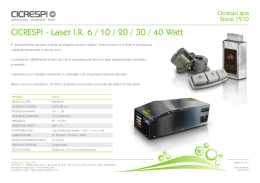
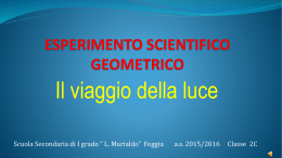
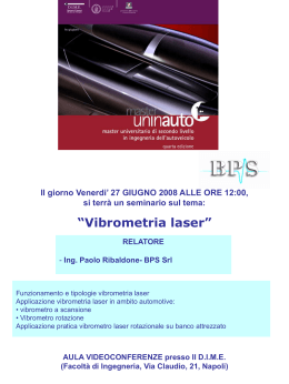
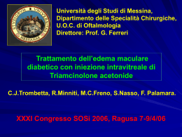
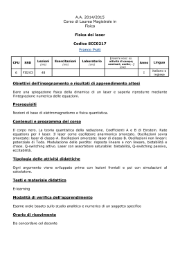
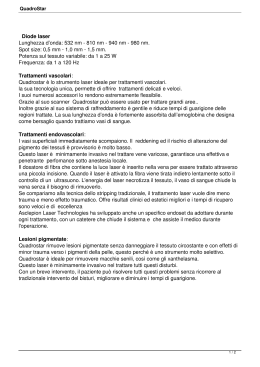
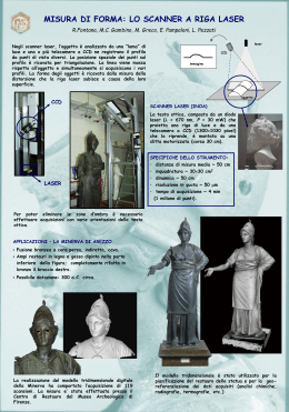
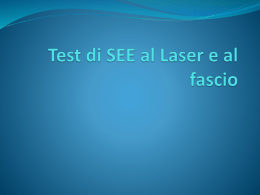
caso clinico
Trattamento dell’edema maculare diabetico
con bevacizumab intravitreale
e fotocoagulazione laser FAG-guidata
Treatment of diabetic macular edema with intravitreal
bevacizumab and fluorescein angiography guided laser
photocoagulation
M. Leozappa, T. Micelli Ferrari
Riassunto
Scopo: valutare l’efficacia della terapia combinata con bevacizumab intravitreale (IVB) e fotocoagulazione laser a guida fluorangiografica nel trattamento
dell’edema maculare diabetico (EMD).
Metodi: 44 pazienti con EMD; 24 (gruppo A) trattati con IVB (1,25 mg) seguita
da fotocoagulazione laser FAG-guidata; 20 pazienti (gruppo B) trattati con sola
IVB. Il follow-up è stato di 10 ± 4 mesi.
Risultati: un mese dopo il trattamento l’AV era significativamente migliorata e
lo spessore medio foveale era diminuito (p < 0,001) in entrambi i gruppi.
All’ultimo controllo, nel gruppo A, l’AV era migliorata significativamente
(p = 0,01) e il valore medio dello spessore della retina era significativamente
diminuito (p < 0,01) rispetto al pre-operatorio; nel gruppo B, il miglioramento
morfologico e funzionale non era significativo rispetto al pre-operatorio.
Conclusioni: nel trattamento del EMD, il trattamento combinato con bevacizumab e fotocoagulazione laser appaiono garantire una maggiore e più duratura
efficacia rispetto alla sola iniezione di bevacizumab.
UOC Oculistica, Ospedale Generale
Regionale “F. Miulli”, Acquaviva
delle Fonti (BA)
Parole chiave: edema maculare diabetico, bevacizumab, Fotocoagulazione laser
FAG-guidata.
Summary
Background: to evaluate the efficacy of combined therapy with intravitreal bevacizumab (IVB) and fluorescein angiography (FA)-guided-laser photocoagulation for the treatment of diabetic macular edema (DME).
Methods: 44 patients with DME: 24 (group A) were treated with IVB (1.25 mg)
and FA-guided laser photocoagulation; 20 patients (group B) were treated with
IVB alone. Follow-up was 10 ± 4 months.
Results: one month after treatment mean visual acuity (VA) was improved, and
mean retinal thickness was significantly decreased in both groups (from 284 µm
to 209 µm). At the last control visit, in group A the mean VA was significantly
improved (p = 0.01), and mean retinal thickness was significantly (p < 0.01) decreased compared to preoperative values; in group B, the improvement of retinal thickness and VA were not statistically different from preoperative values.
Conclusion: a greater and longer lasting efficacy may be expected when bevacizumab is combined with FA-guided laser photocoagulation in the treatment of DME.
Key words: diabetic macular edema; bevacizumab; FA-guided laser photocoagulation.
Indirizzo per la corrispondenza:
Marco Leozappa
UOC Oculistica, Ospedale Generale
Regionale “F. Miulli”
SP127, km 4,1
70021 Acquaviva delle Fonti (BA)
Fax +39 0831 951439
E-mail: [email protected]
33
M. Leozappa,
T. Micelli Ferrari.
Introduzione
Materiali e metodi
L’edema maculare diabetico (EMD) è la più importante causa di perdita visiva nei pazienti diabetici. Il
beneficio della fotocoagulazione laser per il trattamento dell’EMD è stato dimostrato nell’ETDRS Study: la fotocoagulazione focale è efficace nel ridurre
il rischio di moderata perdita visiva, con bassa incidenza di effetti avversi 1 2.
In questo studio erano utilizzati sia il trattamento
laser focale diretto verso singoli microaneurismi sia
il laser a griglia per le aree di leakage diffuso e non
perfusione capillare.
Tuttavia, i pazienti mostravano un minimo miglioramento visivo dopo il trattamento laser; il 12% degli
occhi sviluppano una moderata perdita visiva in 3
anni, nonostante il trattamento 3.
Diversi studi hanno dimostrato l’efficacia di iniezione intravitreale di triamcinolone acetonide (TA) associato o meno a fotocoagulazione laser 4-6.
Il trattamento con TA, con o senza laser a griglia,
mostrerebbe migliori risultati anatomo-funzionali
rispetto al solo trattamento laser 7.
Tuttavia, non vi sono differenze significative in termini di spessore foveale e di acuità visiva tra occhi
trattati con solo TA e occhi trattati con TA+ laser 8.
La maggior parte delle complicazioni legate al diabete, come l’edema maculare e neovascolarizzazione, si verificano a seguito del rilascio di fattori di
crescita in risposta alla ischemia retinica. Il VEGF stimola l’infiammazione inducendo l’espressione della
molecola di adesione intracellulare-1 (ICAM-1), e
l’adesione dei leucociti 9.
L’inibizione della attività VEGF è in grado di prevenire la neovascolarizzazione retinica e le anomalie del
flusso sanguigno associate.
L’uso di bevacizumab endovitreale ha dimostrato
efficacia nel trattamento dell’edema maculare diabetico cronico diffuso 10: la riduzione di spessore retinico centrale può essere osservato a lungo dopo
ripetute iniezioni intravitreali di bevacizumab.
La FAG è molto utile, perché l’uso dell’angiogramma durante la fotocoagulazione laser può migliorare la precisione del trattamento 11.
La nostra ipotesi iniziale era che, effettuando il trattamento sequenziale con bevacizumab intravitreale
e fotocoagulazione laser a guida FAG, i 2 meccanismi agissero in sinergia, aumentando gli effetti benefici in termini di riduzione dell’EMD.
Abbiamo effettuato questo studio per confrontare l’efficacia della terapia sequenziale con bevacizumab e FA-guidate fotocoagulazione laser con la
sola iniezione di bevacizumab intravitreale nel trattamento dell’EMD.
Lo studio ha compreso 44 pazienti con edema maculare diabetico diffuso (28 donne e 16 uomini),
reclutati tra ottobre 2007 e novembre 2008. L’età
media era di 64 anni (range 53-75).
Sono stati selezionati pazienti con EMD che coinvolgeva la fovea, come definizione di “edema maculare clinicamente significativo” secondo le linee
guida ETDRS. La diagnosi di EMD è stata fatta dopo
osservazione diretta con biomicroscopia, FAG (HRA
Heidelberg Engineering, Heidelberg, Germania), e
Tomografia a Coerenza Ottica (OCT; StratusOCT,
Carl Zeiss, Dublin, CA).
Per ogni paziente l’AV è stata misurata utilizzando
la ETDRS chart a 4 metri e calcolata in scala logaritmica (LogMAR).
Il consenso informato è stato ottenuto da tutti i pazienti dopo spiegazione delle finalità e dei possibili
rischi di studio.
I pazienti sono stati randomizzati in 2 gruppi di trattamento: il gruppo A (24 pazienti) è stato trattato
con protocollo terapeutico sequenziale, mediante
iniezione intravitreale di 1,25 mg di bevacizumab
via pars plana, seguita, dopo 7 ± 2 giorni, da fotocoagulazione laser FAG-guidata; il gruppo B (20
pazienti) è stato trattato con una sola iniezione intravitreale di bevacizumab.
Dopo il trattamento, i pazienti sono stati osservati a
1 giorno e 1, 2, 4, 8, 12, 24 e 36 settimane. Tutti i
pazienti hanno completato almeno 24 settimane di
follow-up.
Sono state valutate essenzialmente le variazioni di
spessore foveale e, secondariamente le variazioni
dell’AV.
Ad ogni visita, i pazienti sottoposti a visita oculistica
completa, determinazione della migliore acuità visiva
corretta (VA), esame con lampada a fessura, misurazione della pressione intraoculare, biomicroscopia
della macula, la misurazione dello spessore retinico
mediante OCT, FAG, fotografia del fondo oculare.
34
Risultati
I dati pre- e post-operatori sono riassunti nelle Figure 1-4.
Nel pre-operatorio lo spessore foveale e l’acuità visiva non avevano differenze significative.
Lo spessore foveale medio era di 291 ± 37 micron
nel gruppo A e 282 ± 32 micron nel gruppo B; l’acuità visiva media era 0,84 ± 0,15 LogMAR nel gruppo
A e 0,71 ± 0,13 LogMAR nel gruppo B.
caso clinico
Fig. 1. Valori (media e range) di spessore maculare nei
pazienti del gruppo A, al t0, t1 e t2 – Macular thickness
(mean and range) in Group A, at t0, t1 and t2.
Fig. 2. Valori (media e range) di spessore maculare nei
pazienti del gruppo B, al t0, t1 e t2 – Macular thickness
(mean and range) in Group B, at t0, t1 and t2.
1 mese dopo il trattamento (t1), lo spessore foveale
era significativamente diminuito a 224 ± 33 m nel
gruppo A (p < 0,01), ed a 197 ± 38 m nel gruppo
B (p < 0,01).
In entrambi i gruppi, l’AV era migliorata significativamente: 0,72 ± 0,13 LogMAR nel gruppo A (p < 0,1),
e 0,65 ± 0,08 LogMAR nel gruppo B (p = 0,1).
Non si rilevavano differenze statisticamente significative tra i gruppi in quanto a miglioramento di
spessore e di visus (p = 0,1 per AV; p = 0,08 per
spessore foveale).
Tuttavia, all’ultimo controllo (t2), in media 10 mesi
post-trattamento, i risultati hanno mostrato differenze significative tra i 2 gruppi.
Lo spessore foveale del gruppo A era 207 ± 27 m;
in confronto con i valori pre-trattamento, vi era una
differenza statisticamente significativa, in termini di
riduzione di edema maculare (p < 0,01).
L’AV del gruppo A era 0,70 ± 0,11 LogMAR, notevolmente migliorata rispetto al baseline (p = 0,01).
Nel gruppo B, il miglioramento funzionale ed anatomico appariva soltanto temporaneo: lo spessore foveale era 270 ± 28 m, mentre l’AV era 0,68 ± 0,16
LogMAR. In confronto con i valori pre-trattamento,
non vi era differenza statistica sia di spessore foveale che di acuità visiva.
Non sono stati registrati, durante lo studio, eventi
avversi oculari (compresi endoftalmite) o sistemici
(cerebrovascolari, attacchi ischemici transitori, infarti del miocardio, o malattia vascolare periferica)
degni di nota.
Discussione
Fig. 3. Andamento (media e range) di acuità visiva nei
pazienti dl gruppo A (LogMAR), al t0, t1 e t2 – Visual acuity
of patients of group A at t0, t1 and t2.
Fig. 4. Andamento (media e range) di acuità visiva nei
pazienti del gruppo B (LogMAR), al t0, t1 e t2 – Visual acuity
of patients of group B at t0, t1 and t2.
L’edema maculare diabetico è una delle principali
cause di perdita visiva nei pazienti diabetici.
Negli ultimi anni, le strategie di trattamento sono
state riviste per includere, oltre la fotocoagulazione
laser, le farmacoterapie e la chirurgia.
L’obiettivo della fotocoagulazione laser è focalizzato
principalmente sul trattamento delle zone di fuoriuscita diffuse e non perfusione capillare come individuati sulla fluorangiografia.
Negli ultimi anni, vi è stato un grande interesse per
i fattori vasoproliferativi, che inducono neovascolarizzazione.
È dimostrato che l’ischemia retinica stimola una
neovascolarizzazione patologica mediata da fattori angiogenici, come il fattore di crescita vascolare
endoteliale (VEGF), prodotto da cellule dell’epitelio
pigmentato retinico, da periciti e da cellule endoteliali della retina.
Numerosi studi clinici sulla iniezione intravitreale
35
M. Leozappa,
T. Micelli Ferrari.
di anti-VEGF nell’EMD (pegaptanib sodico, ranibizumab e bevacizumab) hanno mostrato risultati
favorevoli 10-15.
La nostra ipotesi era che associare l’iniezione di bevacizumab alla fotocoagulazione laser potesse dare
migliori e più duraturi effetti rispetto alla sola iniezione di bevacizumab.
Nel nostro studio, la fotocoagulazione laser focale
è stata eseguita circa 7 ± 2 giorni dopo l’iniezione
intravitreale di bevacizumab, con l’obiettivo di trattare le zone di ischemia o perdita evidenziate dalla
fluorangiografia.
Mentre ad 1 mese i risultati non erano differenti tra
i 2 gruppi di pazienti, le differenze più importanti
sono state evidenziate nel corso dei mesi successivi,
in quanto il gruppo che era stato sottoposto a terapia
combinata mostrava un visus ed uno spessore foveale stabile, mentre i pazienti trattati con solo bevacizumab mostravano maggiore tendenza alla recidiva di
edema maculare e a conseguente perdita visiva.
È emerso, pertanto, che la terapia combinata (bevacizumab + fotocoagulazione laser) può essere superiore al solo bevacizumab intravitreale nel ridurre
l’edema maculare e migliorare l’acuità visiva per un
tempo più lungo.
Uno studio multicentrico randomizzato con lungo
follow-up è necessario per valutare sicurezza ed efficacia di questo trattamento sequenziale in tempi
ancor superiori.
Introduction
term decrease of central retinal thickness is observed following repeated intravitreal injections of bevacizumab.
FA is of major importance since the use of projected angiogram during laser photocoagulation may improve the
precision of treatment 11.
We hypothesized that by performing both intravitreal
bevacizumab and FA-guided laser photocoagulation, the
two mechanisms might act synergistically, increasing the
beneficial effects of reduction of macular edema.
This study compared the efficacy of sequential therapy with
bevacizumab and FA-guided laser photocoagulation versus
intravitreal bevacizumab alone for the treatment of DME.
Diabetic Macular Edema (DME) is the most important
cause of visual loss in patients with diabetes; the benefits
of laser photocoagulation for the treatment of DME has
been demonstrated in the Early Treatment Diabetic Retinopathy Study (ETDRS): focal photocoagulation is effective
in reducing the risk of moderate visual loss, with a low
incidence of adverse effects 1 2. The ETDRS used direct focal
laser treatment to target individual microaneurysms and
applied a grid pattern to areas of diffuse leakage and capillary non-perfusion as identified by fluorescein angiography
(FA). However, patients had only minimal visual improvement after laser treatment; 12% of eyes developed moderate visual loss at 3 years despite treatment 3.
Several studies have demonstrated the effectiveness of intravitreal injection of triamcinolone acetonide (TA) associated or not with previous grid laser photocoagulation 4-6.
TA with or without additional grid laser led to significantly
greater reduction in macular edema and better mean final
visual acuity VA than eyes treated with laser alone 7.
However, no significant differences in foveal thickness
and VA were found between the intravitreal TA–alone
group and those treated with combined intravitreal TA
and laser at any time point 8.
Most of diabetes-related complications, such as macular edema and neovascularization, occur secondary to
the release of the growth factors in response to retinal
ischemia. VEGF promotes inflammation by inducing intracellular adhesion molecule-1 (ICAM-1) expression and
leukocyte adhesion 9.
Inhibition of VEGF activity can prevent retinal neovascularization and associated blood flow abnormalities.
The use of the intravitreal anti-VEGF drug bevacizumab
has been shown to be effective for the treatment of
chronic diffuse diabetic macular edema 10, and a long-
36
Nessun interesse economico da parte degli autori su
quanto esposto nell’articolo.
Materials and methods
This was a clinical trial involving 44 patients with diffuse diabetic macular edema (28 women and 16 men),
recruited between October 2007 and November 2008.
Mean age was 64 years (range 53-75).
Patients were enrolled if they had DME involving the
fovea, as defined by clinically significant macular edema
according to ETDRS guidelines. Diagnosis of macular edema was made after direct observation by biomicroscopy,
Fluorescein angiography (HRA 2 Heidelberg Engineering,
Heidelberg,Germany), and optical coherence tomography (OCT; StratusOCT, Carl Zeiss, Dublin, CA).
For each patient the best corrected visual acuity (BCVA)
was measured using the ETDRS chart at a distance of 4
m and calculated as the logarithm of the minimum angle
of resolution (LogMAR). Informed consent was obtained
from all patients after explanation of the purpose and
possible risks of the study.
Patients were randomized into 2 treatment groups: group
A (24 patients) was treated with sequential therapeutic
protocol using intravitreal injection of 1.25 mg of beva-
caso clinico
cizumab via pars plana followed, after 7 ± 2 days, by FAguided laser photocoagulation; group B (20 patients) was
treated with intravitreal injection of bevacizumab alone.
After treatment, patients were observed on day 1, and
at 1, 2, 4, 8, 12, 24 and 36 weeks after intervention. All
patients completed 24 weeks of follow-up.
The primary outcome measure was change in foveal
thickness on OCT, while the secondary outcome measure
was BCVA.
At each visit, patients underwent complete eye examination, determination of BCVA, slit-lamp examination,
intraocular pressure measurement, stereoscopic biomicroscopy of the macula, retinal thickness measurement
by OCT, FA and fundus photography.
Results
Patient characteristics are summarized in Figures. 1-4.
The baseline characteristics (t0) of foveal thickness and
VA were not significantly different between groups.
Mean foveal thickness before treatment was 291 µm in
group A and 279 µm in group B; mean VA was 0.84
LogMAR in group A and 0.72 LogMAR in group B. One
month after treatment (t1), foveal thickness measured
by OCT was significantly decreased in both group A
(p < 0.01; mean 223 µm) and group B (p < 0.01; mean
195 µm).
In both groups, the BCVA improved significantly, from
0.84 to 0.72 LogMAR in group A (p < 0.1), and from
0.72 to 0.65 LogMAR in group B (p = 0.1).
No statistically significant difference was between the
groups (p = 0.1 for BCVA; p = 0.08 for foveal thickness).
At last control after treatment (t2), there were significant
differences between the 2 groups.
Mean foveal thickness of group A was 203 µm, which
was statistically different compared to pre-treatment values reduction of macular edema (p < 0.01).
Mean VA of group A was 0.69 LogMAR, which was significantly improved versus baseline (p = 0.01).
In group B, functional and anatomical improvement appeared only transient: 6 months after treatment, mean
foveal thickness of group B was 268 µm; mean VA was
0.72 LogMAR. Comparing with pre-treatment values,
there was no statistically significant difference in foveal
thickness and VA, both gradually deteriorated to baseline
levels within 24 weeks.
During the study, no ocular side effects or systemic adverse events such as thromboembolic events were encountered.
Discussion
Diabetic macular edema is a major cause of visual loss
in patients with diabetes.
In the last years, treatment strategies have been revised to include, in addition to laser photocoagulation,
pharmacotherapies and early surgical intervention.
The aim of laser photocoagulation has focused primarily on the treatment of areas of diffuse leakage and
capillary non-perfusion as identified by FA.
In recent years, there has been an interest in vasoproliferative factors, which induce neovascularization. It
has been shown that retinal ischemia stimulates pathologic neovascularization mediated by angiogenic factors, such as vascular endothelial growth factor (VEGF),
which is released by retinal pigment epithelium, pericytes and endothelial cells of the retina.
Clinical studies on the effect of intravitreally administered anti-VEGF aptamer, pegaptanib sodium and antibodies, ranibizumab and bevacizumab on DME has
shown favorable results 10-15.
Our hypothesis was that the sequential association of
bevacizumab plus laser photocoagulation would have
benefits in reduction of macular edema compared to
injection of bevacizumab alone.
In the present study, focal laser photocoagulation was
performed at 7 ± 2 days after intravitreal injection of
bevacizumab, with the aim to treat areas of ischemia
or leakage evidenced by FA.
The results at one month after treatment showed
no significant difference between the two patient
groups. Both the bevacizumab-alone and the combined-therapy groups showed a significant decrease
of foveal thickness, which was associated with improvement of VA.
However, more important differences were evident at
6 months after treatment: the combined-treatment
group showed a greater stability of VA and reduction
of foveal thickness; during follow-up, patients treated
with bevacizumab alone showed a higher tendency
of recurrence of macular edema and of a decrease in
visual function.
We found that combined therapy (bevacizumab plus
laser photocoagulation) appeared to be superior to
intravitreal bevacizumab alone in reducing macular
thickening and in improving visual acuity in patients
with diabetes for a longer time interval. Evaluation in
a multicenter, randomized, controlled clinical trial with
longer follow-up is warranted to evaluate the safety
and efficacy of this sequential treatment.
37
M. Leozappa,
T. Micelli Ferrari.
Bibliografia
1
Early Treatment Diabetic Retinopathy Study Research
Group. Photocoagulation for diabetic macular edema.
Early Treatment Diabetic Retinopathy Study report
number 1. Arch Ophthalmol 1985;103:1796-806.
Triamcinolone or laser alone for Treating Diabetic Macular
Edema. Ophthalmol 2007;114:2162-7.
9
Ishida S, Usui T, Yamashiro K. VEGF164 is pro
inflammatory in the diabetic retina. Invest Ophthalmol
Vis Sci 2003;44:2155-62.
2
Bandello F, Lanzetta P, Menchini U. When and how to do
a grid laser for diabetic macular edema. Doc Ophthalmol
1999;97:415-9.
10
Kook D, Wolf A, Kreutzer T, et al. Long-term effect of intravitreal bevacizumab (avastin) in patients with chronic diffuse diabetic macular edema. Retina 2008;228:1053-60.
3
Early Treatment Diabetic Retinopathy Study Research
Group. Early photocoagulation for diabetic retinopathy
(ETDRS report number 9). Ophthalmol 1991;98:766-85.
11
4
Martidis A, Duker JS, Greenberg PB, et al. Intravitreal
triamcinolone for refractory diabetic macular edema.
Ophthalmol 2002;109:920-7.
Kylstra JA, Brown JC, Jaffe GJ. The importance of
fluorescein angiography in planning laser treatment of
diabetic macular edema. Ophthalmol 1999;106:206873.
12
Cunningham ET Jr, Adamis AP, Altaweel M, et al. A
phase II randomized double-masked trial of pegaptanib:
An anti-vascular endothelial growth factor aptamer, for
diabetic macular edema. Ophthalmol 2005;112:174757.
13
Nguyen QD, Tatlipinar S, Shah SM, et al. Vascular
endothelial growth factor is a critical stimulus for diabetic
macular edema. Am J Ophthamol 2006;142:961-9.
14
Chun DW, Heier JS, Topping TM, e al. A pilot study of
multiple intravitreal injections of ranibizumab in patients
with center involving clinically significant macular edema.
Ophthalmol 2006;113:1706-12.
15
Haritoglou C, Kook D, Neubauer A, et al. Intravitreal
bevacizumab (Avastin) therapy for persistent diffuse
diabetic macular edema. Retina 2006;26:999-1005.
5
Karacorlu M, Ozdemir H, Karacorlu S, et al. Intravitreal
triamcinolone as a primary therapy in diabetic macular
oedema. Eye 2005;19:382-6.
6
Lam DS, Chan CK, Tang EW, et al. Intravitreal
triamcinolone for diabetic macular oedema in Chinese
patients: six-month prospective longitudinal pilot study.
Clin Experiment Ophthalmol 2004;32:569-72.
7
8
Avitabile T, Longo A, Reibaldi A. Intravitreal triamcinolone
compared with macular laser grid photocoagulation
for the treatment of cystoid macular edema. Am J
Ophthalmol 2005;140:695-702.
Lam DS, Chan CK, Mohamed S et al. Intravitreal
Triamcinolone plus Sequential Grid laser versus
38
Scaricare