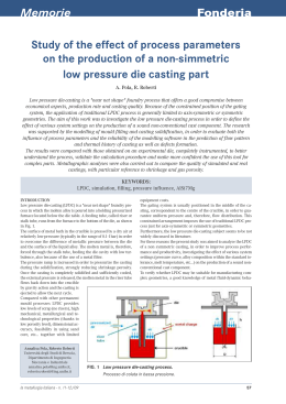
AMERICANt MUSEUM
Norntates
PUBLISHED BY THE AMERICAN MUSEUM OF NATURAL HISTORY
CENTRAL PARK WEST AT 79TH STREET, NEW YORK, NY 10024
Number 3256, 59 pp., 25 figures, 11 tables
March 4, 1999
Amerigo Vespucci and the Rat of Fernando de
Noronha: a New Genus and Species of Rodentia
(Muridae: Sigmodontinae) from a Volcanic Island
Off Brazil's Continental Shelf
MICHAEL D. CARLETON1 AND STORRS L. OLSON2
CONTENTS
Abstract ...................
.......................................
Introduction .
..........................................................
Materials and Methods ........................................................
Acknowledgments ..........................................................
Geology and Physiography of Fernando de Noronha .........
.......................
The Extinct Sigmodontine Rodent .
...............................................
Noronhomys, new genus .......................................................
Noronhomys vespuccii, new species ...............
............................
Comparisons with Holochilus and Lundomys ...........
..........................
Skeletal and Dental Anatomy .....................
...........................
Morphometric Analyses ......................................................
Phyletic Inferences ..........................................................
Character Definitions ........................................................
Results .
..........................................................
Discussion .
..........................................................
.................
Amerigo Vespucci and the Rat of Fernando de Noronha .......
Phylogenetic Relationships .
...................................................
...........................
Ecological and Functional Considerations ...........
...........................
Biogeography of Fernando de Noronha .............
References .
..........................................................
2
2
2
5
5
9
9
10
16
17
25
32
33
42
44
44
49
51
55
56
' Research Associate, Department of Mammalogy, American Museum of Natural History; Curator of Mammals,
Department of Vertebrate Zoology, National Museum of Natural History, Smithsonian Institution, Washington, D.C.
2Curator of Birds, Department of Vertebrate Zoology, National Museum of Natural History, Smithsonian Institution,
Washington, D.C.
Copyright C American Museum of Natural History 1999
ISSN 0003-0082 / Price $7.00
2
AMERICAN MUSEUM NOVITATES
NO. 3256
ABSTRACT
Noronhomys vespuccii, a new genus and
species (Muridae: Sigmodontinae), is described from Ilha Fernando de Noronha, a
small volcanic island located 345 km northeast of Cabo de Sao Roque, Brazil. The
abundant cranial and postcranial material of
the fossil rodent was recovered from old
beach dunes that are late Quaternary in age
(probably late Holocene). Noronhomys vespuccii retains incomplete mesolophs on its
moderately hypsodont molars and is compared with other tetralophodont oryzomyines
(sensu Voss and Carleton, 1993), especially
Lundomys molitor and species of Holochilus.
Morphometric analyses conducted separately
on craniodental, mandibular, and femoral
measurements reveal the sample of Noronhomys to be equally differentiated from those
of Lundomys and Holochilus. Based on the
criterion of parsimony, phylogenetic analyses
of 35 craniodental characters strongly support the recent common ancestry of the new
form and Holochilus. The clade (Pseudoryzomys (Lundomys (Holochilus-Noronhomys))) appears to represent a lineage of semiaquatic rodents that differentiated from an
oryzomyine ancestry in savanna landscapes
of southern South America. Morphometric
results and anatomical details of the pelvic
limb, however, suggest that Noronhomys was
not a semiaquatic form. It is hypothesized
that such aquatic skeletomuscular adaptations were lost (reversed) when the progenitor of Noronhomys became stranded on a
small oceanic island where palustrine habitats were scarce or absent. The specific patronym of the extinct rodent refers to Amerigo Vespucci's disputed landfall on Fernando
de Noronha in 1503 and his possible sighting
of "very large rats" on that island. If the
species Noronhomys vespuccii were alive in
1503, it became extinct shortly thereafter due
to the usual anthropogenic causes that have
extirpated so many vertebrate species on islands.
INTRODUCTION
On August 10, 1503, during his putative
ronha. Among the vertebrate material recovfourth voyage to the New World, Amerigo
ered were abundant, well-preserved bones of
a new sigmodontine rodent that was moderVespucci claimed to have landed on a small
island just three degrees south of the equator:
ately large in size and belongs to the diverse
radiation
of South American Oryzomyini.
[The] island we found uninhabited, and it contained
This extinct rodent is diagnosed and deplenty of trees, and so many birds, both marine and
scribed as a new genus and species. In forland, that they were without number ... and we saw
no other animals except very big rats and lizards with
mally recording its scientific name, we also
two tails, and some snakes. (Emphasis ours; translacontrast the new form with Lundomys and
tion of the original Italian text from Branner [1888:
its nearest probable relatives on
Holochilus,
869].)
continental South America, evaluate its phyGeographers and early naturalists have in- logenetic relationship to those taxa and to
other oryzomyine rodents, and discuss its
terpreted the small island mentioned by Vespucci as Ilha Fernando de Noronha (Branner,
biogeographic origin in the context of other
vertebrates indigenous to Ilha Fernando de
1888; Ridley, 1888), an oceanic island locatNoronha. The very recent existence of an ened off easternmost Brazil. Although Vespucdemic rodent on Fernando de Noronha proci's fourth journey to the New World, and by
vides corroboration of another biological deextension his landfall on Fernando de Notail in the account of Vespucci's fourth voyronha, has been questioned by some historiage, which may help in determining its auans (for example, Formisano, 1992), the forthenticity.
mer presence of "very big rats" on that
small island can now be empirically docuMATERIALS AND METHODS
mented. During August 1973, Olson discovered vertebrate fossils in old beach dunes at
SPECIMENS AND ANALYTICAL SAMPLES: The
fossil sample originated in late Quaternary
the eastern corner of Ilha Fernando de No-
1999
CARLETON AND OLSON: NEW FOSSIL RAT
beach sands found on Ilha Fernando de Noronha, Brazil. The holotype, along with representative crania, mandibles, and postcranial
material, has been deposited in the Museu de
Ciencias, Pontificia Universidade Catolica da
Rio Grande do Sul (MCP-PV), Porto Alegre,
Brazil. Comparable series have been retained
in the vertebrate paleontological collection,
Department of Paleobiology, National Museum of Natural History (USNM).
Museum specimens (skulls, mandibles,
and postcranial skeletons) of Holochilus and
Lundomys used for the various statistical tabulations and morphometric analyses are listed below and contained in the following collections of recent mammals: American Museum of Natural History, New York City
(AMNH); Field Museum of Natural History,
Chicago (FMNH); National Museum of Natural History, Smithsonian Institution, Washington, D.C. (USNM).
Holochilus brasiliensis: Argentina, Buenos Aires,
Carhue (USNM 236318-236320). Uruguay, Ca.nelones, Baniado de Tropa Vieja (AMNH
206362); Soriano, 3 km E Cardona (AMNH
206369, 206372, 206374-206379, 206383);
Tacuarembo, 16 km NNW San Jorge, Rio Negro, Isla Sanchez Chica (AMNH 206390).
Holochilus sciureus: Bolivia, Beni, mouth of Rio
Baures (AMNH 210218, 210221, 210226,
210228, 210232, 210233, 210235, 210237,
210239, 210241, 210244, 210249, 210255);
Mamore, San Joaquin (USNM 364750-364753,
364757-364759, 390248, 461031461035); Rio
Itenez, Costa Marques (AMNH 210268,
210271); Itenez, Lago Victoria (USNM
390767-390769). Brazil, Para, Utinga (USNM
394720-394726). Suriname, Commewijne,
Marienburg (USNM 312891-5, 319976319983). Venezuela, Trujillo, 30 km NW Valera, El Dividive (USNM 372652-372665).
Lundomys molitor: Uruguay, Canelones, Bafiado
de Tropa Vieja (AMNH 206363-206366); Minas, Pasos de Averias, Rio Cebollati (FMNH
29255, 29257, 29258; USNM 259641); Sonano, 3 km E Cardona (AMNH 206368, 206380,
206381); Trienta y Tres, 8 mi E Trienta y Tres
(FMNH 29260, 29261, 29263); 25 km WSW
Trienta y Tres, Rfo Olimar Chico (AMNH
206392, 206393).
For summary statistics and multivariate
comparisons, the specimens were grouped
into seven composite or operational taxonomic units (OTUs) as here defined:
3
OTU 1, Fernando de Noronha taxon;
OTU 2, all Lundomys molitor;
OTU 3, all Holochilus brasiliensis;
OTU 4, H. sciureus from Bolivia;
OTU 5, H. sciureus from Brazil;
OTU 6, H. sciureus from Venezuela.
Sample sizes for OTUs are presented in the
tables and varied according to the analysis
performed and the skeletal element compared. In particular, the relative rarity and
fragmentary nature of preserved crania limited sample sizes for morphometric characterization of the Noronha fossil.
Specimens in the USNM of five additional
oryzomyine taxa-Microryzomys minutus,
Oryzomys palustris, 0. subflavus, Pseudoryzomys simplex, and Zygodontomys brevicauda cherrei-were broadly surveyed to assist
evaluation of qualitative character variation
and to provide outgroup comparisons in the
phylogenetic analyses. Rationale for inclusion of these particular taxa is discussed under Phyletic Inferences.
MEASUREMENTS: Linear measurements
were taken from crania, mandibles, and femora as a basis for assessing covariation patterns and taxonomic discrimination. Skulls
were viewed under a dissecting microscope
when measuring the 13 cranial and 2 dental
variables to 0.01 mm by handheld digital calipers accurate to 0.03 mm. Fifteen dimensions of the mandible and its dentition were
also quantified in view of its greater abundance among the fossil material. All mandibular measurements were taken to 0.01 mm
by use of digital calipers mounted on the
movable stage of a binocular microscope set
at 45X magnification. Of the postcranial elements recovered for the Noronha fossil,
only the femur was preserved intact in sufficient numbers to permit numerical comparisons with living species. Thirteen femoral
variables were quantified using handheld calipers. All measurements, their abbreviations,
and definitions of landmarks where necessary, are enumerated below.
Cranial variables: Occipitonasal length
(ONL); zygomatic breadth (ZB); breadth of
braincase (BBC); least interorbital breadth
(IOB); length of nasals (LN); postpalatal
length (PPL); length of bony palate (BPL);
length of upper diastema (LD); length of in-
4
AMERICAN MUSEUM NOVITATES
cisive foramen (LIF); breadth of bony palate
across upper first molars (BM 1 s)-transverse
distance between the labial edges of the Mls;
posterior breadth of the bony palate (PPB)taken at the constriction of the maxillary
bones immediately posterior to the M3s;
breadth of zygomatic plate (BZP); depth of
auditory bulla (DAB)-an oblique distance,
from the dorsal rim of the auditory meatus
to the ventralmost curvature of the bullar
capsule; coronal length of maxillary toothrow (CLM); and greatest width of upper first
molar (WM 1).
Mandibular variables: Depth of mandible
below ml (DMml)-on the buccal side,
from the alveolar rim at the middle of ml to
the ventralmost projection of the symphysis;
depth of mandible below m3 (DMm3)-on
the lingual side, shortest distance from the
shelf behind m3 to the concave ventral margin of the dentary; length of the masseteric
crest (LMC)-from the point where the superior and inferior masseteric crests meet to
the anterior end of their conjoined ridge; coronal length of mandibular toothrow (CLm);
length of ml (Lml); length of m2 (Lm2);
length of m3 (Lm3); anterior width of ml
(AWml)-measured across the lateral apices
of the anteroconid; posterior width of ml
(PWml)-between the margins of the hypoconid and entoconid; anterior width of m2
(AWm2)-between the margins of the protoconid and metaconid; posterior width of
m2 (PWm2)-between the margins of the
hypoconid and entoconid; greatest width of
m3 (Wm3)-between the margins of the protoconid and metaconid; height of ml
(Hml)-on the lingual side, from the enamelcementum junction at the coronal base to the
apex of the metaconid; width of lower incisor
(Wi)-at the anterior surface just beyond the
emergence of the incisor from the dentary;
and depth of lower incisor (Di)-measured at
the proximal edge of the wear facet.
Femoral measurements: Femoral length
(FL)-dorsal margin of the head to the distalmost curvature of the medial condyle; distal width of the femur (DW)-width across
base of the shaft (diaphysis) just above the
condyles; middle width of the femur
(MW)-narrowest distance across the shaft,
measured slightly below the third trochanter;
depth of the femur (DF)-anterior-posterior
NO. 3256
thickness of the shaft taken at the same position as MW; diameter of the head (DH)
width across the articular ball; diameter of
the neck (DN)-narrowest constriction, measured along the anterior-posterior axis, below
the head; intertrochanteric distance (ITD)
an oblique distance, from the craniodorsal
rim of the greater trochanter to the posteromedial projection of the lesser trochanter;
breadth across the third trochanter (BTT)greatest expanse between the lateral margin
of the third trochanter (lateral crest) and an
opposite point on the medial edge of the
shaft; length of the third trochanter (LTT)distance between the dorsal edge of the
greater trochanter to the end of the scar for
insertion of the gluteus maximus on the third
trochanter; condylar breadth (CB)-transverse distance between the medial and lateral
edges of the articular condyles; condylar
depth (CD)-anterior-posterior thickness
measured from the dorsal lip of the trochlear
fossa to the caudal rim of- the medial condyle; length of the patellar fossa (LPF)from the dorsal lip of the patellar fossa to its
distal termination medial to the articular condyles; and width of the patellar fossa (WPF)distance between the lateral and medial edges
of the fossa at its midsection.
ANATOMICAL TERMINOLOGY AND MORPHOMETRIC AND PHYLOGENETIC ANALYSES: De-
scriptive features of the oryzomyine skull are
generally those used by Carleton and Musser
(1989) and Voss and Carleton (1993). Names
for enamel crests and reentrant folds of the
molars follow Reig (1977), as adapted and
illustrated for the oryzomyine dentition by
Carleton and Musser (1989). Positional abbreviations of the molars follow the common
alphanumeric convention of using uppercase
versus lowercase letters to identify maxillary
or mandibular teeth and numbers to indicate
their placement in the toothrow (for example,
Ml and ml). The myological studies of
Howell (1926) and Rinker (1954) are the
principal sources for names of muscles and
interpretations of their origins and insertions.
Standard descriptive statistics (mean,
range, standard deviation, coefficient of variation) were derived for the OTUs. Principal
components (PCs) and canonical variates
(CVs) were extracted from the variance-covariance or correlation matrix and computed
1999
CARLETON AND OLSON: NEW FOSSIL RAT
using natural logarithmic transformations of
the craniodental, mandibular, and femoral
variables. Loadings are expressed as Pearson
product-moment correlation coefficients between the principal components and the original skeletal and dental variables. Mahalanobis distances between OTU centroids were
clustered based on the unweighted pair-group
method using arithmetic averages (UPGMA).
All univariate and multivariate computations
were generated using Systat (version 5.0,
1992), a series of statistical routines programmed for microcomputers.
Phylogenetic relationships were explored
using PAUP (version 3.1.1; Swofford, 1993)
to find trees of minimal length (branch-andbound option) by the criterion of Wagner
parsimony. Both accelerated and delayed
transformation routines were used to optimize intermediate character state transformations, but only delayed transformation results are illustrated on trees. Characters were
not weighted, and trees were rooted using either a composite hypothetical ancestor (HA)
or a designated outgroup OTU. Both bootstrap percentages (1000 iterations, branchandbound search) and Bremer support indices are provided as measures of cladistic support; for the latter, the congruent cladistic
structure in mutually parsimonious trees is
summarized by the strict consensus method.
ACKNOWLEDGMENTS
Support for fieldwork by Olson was provided by a grant from the National Geographic Society (number 1105) and supplementary funding from the International
Council for Bird Preservation. The Conselho
Nacional de Pesquisas of Brazil authorized
the expedition to Fernando de Noronha and
facilitated various aspects of travel, as did
staff members of the American Consulate
General in Rio de Janeiro and Recife. Olson
was aided in fieldwork and collecting by 0.
A. Roppa, whose assistance was provided
through the Museu Nacional de Rio de Janeiro, I. A. Cruz, and other helpful individuals stationed on the island at the time. For
loans of recent specimens, we thank B. D.
Patterson (FMNH) and G. G. Musser and R.
S. Voss (AMNH). Dra. Maria Claudia L.
Malabarba of the Museu de Ciencias in Porto
5
Alegre, Brazil, kindly helped with the cataloging of material to be returned to her institution. Ralph Chapman, Morphometrics
Lab, USNM, provided advice on the use of
certain Systat routines. Documentation and
comparison of the new fossil were made possible through the creative talents of David F
Schmidt, who undertook all specimen photography and most line drawings; Karolyn
Darrow, who drew the postcranial figures;
and Janine Higgins, who illustrated the cranium and jaw of the holotype. Finally, we
appreciate the helpful manuscript reviews offered by Guy G. Musser, Scott J. Steppan,
and Robert S. Voss.
GEOLOGY AND PHYSIOGRAPHY
OF FERNANDO DE NORONHA
GENERAL: Fernando de Noronha is somewhat generously described as an archipelago
because of the 12 smaller islets that extend
from the northeast corner of the principal island. It lies just off the rim of the continental
shelf (latitude 3°50'S and longitude
32015'W), 345 km (215 mi) northeast of the
nearest Brazilian mainland at Cabo Sao
Roque (fig. 1). The total land area of the archipelago is 18.4 km2. of which Fernando de
Noronha itself, 10 km long by 3.5 km at its
greatest width, composes 16.9 km2 (fig. 2).
The highest point is the columnar igneous
plug known as Morro do Pico (321 m), but
the remainder of the island is, for the most
part, considerably lower and less rugged. A
rolling plain averaging 45 m elevation dominates the west-central part of the island, and
an irregular plateau averaging 70 m the eastcentral portion. Streams are small and seasonally intermittent, and nowhere have they
cut valleys of any significance.
The geology and geochemistry of the
small island chain have been treated in detail
by Branner (1889, 1890), de Almeida (1958),
Mitchell-Thome (1970), and Gunn and Watkins (1976). Fernando de Noronha is volcanic in origin, formed by a submarine mountain that rises abruptly 4000 m from the
ocean's floor as a classical conical seamount,
of which only the uppermost tip breaks surface to create the archipelago. Miocene phonolitic monoliths and domes form the high
points (160 to 320 m) that rim the main is-
6
AMERICAN MUSEUM NOVITATES
NO. 3256
Fig. 1. Position of Fernando de Noronha in relation to South America and its continental shelf
(stippled area approximates limit of 100 fathoms [180 m]). Distributional limits of Holochilus and
Lundomys (type locality, Lagoa Santa), rodent genera related to the new genus on Ilha Fernando de
Noronha, are also depicted. Numbers designate general areas of analytical samples of H. brasiliensis
(OTU 3) and H. sciureus (OTUs 4-7); see Materials and Methods for precise collecting localities and
sample sizes.
land, and Pliocene ankaramite lava flows underlie the central undulating plains. Potassium-argon dating indicates the greatest age of
subaerial rocks as 11.8 ± 0.4 million years
(Cordani, 1967), and active vulcanism is believed to have ceased by the late Pliocene to
very early Pleistocene (de Almeida, 1958).
The few sedimentary formations, products of
Pleistocene and Recent age, consist of calcareous sandstones of aeolian origin, some
marine limestone, and insubstantial alluvial
deposits. Old marine terraces record several
eustatic oscillations from 40 m above to 6 m
below current sea level.
Re~mdios
CARLETON AND OLSON: NEW FOSSIL RAT
1999
32 28
32 26
3 48
ARQUIPELAGO de
FERNANDO de NORONHA
7
32 24
3 48 -
rliha Rata
lihado Mo
liha
Sao Jose
500
-3 50
0
500 1000
liha Sela Gineta
e&Y
I2~'llha
Bahia do
Santo Antoni
Meters
.
Rasa
Ponta de
Santo Antonio
3 50-
t
liha Fernando de Noronha
3 52
L
3 52-
32128
32126
32124
Fig. 2. Islands composing the Arquipelago de Fernando de Noronha, located approximately 345 km
east of Cabo de So Rocque, Brazil. Fossil material of the new sigmodontine rodent was recovered from
late Quaternary beach dunes situated at the neck of Ponta de Santo Antonio, Ilha Fernando de Noronha.
Fernando de Noronha is actually the outermost of a series of volcanic peaks, all except Atoll das Rocas submerged, that extend
between the archipelago and the continental
shelf of Brazil. However, no geological evidence suggests that these peaks ever formed
continuous land, the depths between them
being too great-in some cases over 5000 m.
Mitchell-Thome (1970) likened the climate of Fernando de Noronha to that of Rio
Grande do Norte in eastermmost Brazil. The
climate is mild and marked by a pronounced
dry season (August to January) and distinct
rainy period (February to July); October is
the driest month and April the wettest. Annual rainfall averages 1318 mm, 60% falling
in the three wettest months. The average an-
nual temperature is 25.4°C, the minimum recorded temperature 18°C and the maximum
31°C; August is the coolest month and March
the warmest. Yearly relative humidity remains fairly uniform, ranging between an average of 81% in the dry season and 87% in
the wet season. Trade winds are strong and
constant, generally from the southeast. The
southern exposure of the island is thus the
weathered side, its shoreline mostly inaccessible and characterized by steep, rugged
cliffs; in contrast, the northern side boasts
several beaches of unsurpassed beauty and
tranquility.
THE FOSSIL LOCALITY: During three weeks
of searching in 1973, the only productive
fossiliferous site discovered was an area of
8
AMERICAN MUSEUM NOVITATES
NO. 3256
Fig. 3. Two views, looking westward, of the elevated Quaternary beach on Ilha Fernando de Noronha
where skeletal remains of Noronhomys vespuccii were discovered. Top: General overview of beach dunes
that straddle the neck of the Ponta de Santo Antonio (also see fig. 2); the phonolitic plug Morro do Pico,
"The Peak" (about 2.5 km distant in the right background) forms a conspicuous landmark (321 m) that
dominates the otherwise low relief of the island. Bottom: Southern portion of beach dunes with arrow
indicating the indurated nodular layer around which most surface material was recovered; note the presentday ocean level and active beach in lower left of photo. Photographs by Storrs L. Olson.
dunes that extend nearly across the narrow
neck of Ponta de Santo Antonio, which
forms the northeastern corner of the island
(fig. 2). Here, loose sands reach inland from
the southern beach at Bahia de Caleira and
extend almost to Bahia de Santo Antonio on
the northern side of the peninsula (fig. 3).
Just east of the dunes, on the south side of
the peninsula, there are also low outcrops of
indurated sandstone (calcareous arenites)
into which small caves have been weathered.
Similar consolidated aeolianites occur elsewhere on the main island and various islets
(Branner, 1890; de Almeida, 1958). No vertebrate fossils were found in the indurated
sandstone caves except one fragmented bone
of a frigate bird (Fregata).
Both the unconsolidated and indurated
sands are largely calcareous, composed of
polished grains representing the comminuted
organic remains of corals, nullipores, mollusks, and foraminifera. Analyses of dune
1999
CARLETON AND OLSON: NEW FOSSIL RAT
samples have accordingly yielded high percentages, 70 to 97%, of calcium carbonate
and small traces of silicon dioxide (Branner,
1890; Mitchell-Thome, 1970). We lack radiometric or other dates for these dune deposits, but the bulk of the sands may have
been blown inland during a glacial period of
lower sea level, when more reefs would have
been exposed and winds were stronger. Such
a taphonomic environment resembles that
hypothesized for the fossil-rich sands on St.
Helena and the Hawaiian Islands (Olson,
1975; Olson and James, 1982). Of course,
vertebrate remains could have been intermixed with the shifting sands at any time
afterwards, creating an unstructured stratigraphic context that leaves the age of the fossils as indeterminate. Some bones are obviously much older than others, being heavily
encrusted with indurated sand and appearing
well mineralized. Others, such as the remains
of domestic Felis, Rattus, and Mus, are much
more recent and display no signs of mineralization.
Vertebrate fossils recovered from the
dunes include various reptiles and seabirds,
the dove Zenaida auriculata, and an extinct
flightless rail (Olson, 1981), as well as the
sigmodontine rodent described below. The
vertebrate bones occurred mainly in three or
four localized patches characterized by an
abundance of land snail shells and small
white nodules, possibly conglomerates
formed from the phosphatic residue of guano. Sand removal from a small quarry had
exposed a semiconsolidated layer about 8-12
cm thick that was composed mainly of these
nodules; here, the terrestrial snail shells were
most prevalent. This layer ranged from 45
cm below the surface of the dunes to much
deeper and appeared to be the source of most
of the fossils collected. The identifiable land
snails, as determined by Joseph Rosewater
(late curator of mollusks at USNM), are Hyperaulax (Hyperaulax) ridleyi, H. (Bonnanius) ramagei, Ridleyi quinquelirata, and Lamellaxis micra. The first three are endemic
to the island.
THE EXTINCT SIGMODONTINE
RODENT
Olson and assistants collected vertebrate
fossils on nine days between 27 July and 16
9
August 1973. These were found mostly by
surface picking, supplemented by sand
screening in the more productive areas (see
fig. 3). The preserved material of the extinct
rodent, rich both in the skeletal parts and
number of individuals represented, consists
of several crania, many mandibles with intact
or partial toothrows, and numerous postcranial elements. Most bones recovered were
isolated, but the burial context of some, including the skull with associated mandibles
selected as the holotype, suggests that they
came from a single individual.
Noronhomys, new genus
TYPE SPECIES: Noronhomys vespuccii, new
species.
DIAGNOSIS: A genus of sigmodontine rodent, tribe Oryzomyini (sensu Voss and
Carleton, 1993), characterized by robust cranial proportions and moderately large size
(ONL = 38-40 mm), smaller than Lundomys
molitor (ONL = 42-46 mm) and Holochilus
brasiliensis (ONL = 40-44 mm) but equivalent to larger-bodied examples of H. sciureus (see tables 1, 2). Interorbital region relatively broad and hourglass shaped without
dorsally raised beads (present in mature
Holochilus), temporal ridging over braincase
low in relief (well developed in adult Holochilus and Lundomys); zygomatic plate comparatively narrow, notch shallow, lacking
spine on anterodorsal rim (plate broader,
notch well inscribed, and anterodorsal edge
produced as moderate to strong spine in Lundomys and Holochilus); bony palate relatively short, surface broad and flat, mesopterygoid fossa projecting conspicuously between
M3s (palate narrower and corrugated, extending even with or beyond the end of M3
in Lundomys and Holochilus). Molars moderately hypsodont with planar occlusal surface (cuspidate and terraced in Lundomys);
short mesolophs consistently present on upper MI and M2 (absent in most Holochilus);
Ml and M2 posteroloph absent (present on
little-worn molars of Lundomys and Holochilus); m2 posteroflexid and m3 entoflexid
closed off as enamel islands, m3 anterolabial
cingulum and protoflexid absent (folds confluent with lingual margin, small cingulum
10
AMERICAN MUSEUM NOVITATES
NO. 3256
Fig. 4. Dorsal and ventral cranial views of the holotype (MCP 3460-PV) of Noronhomys vespuccii
(occipitonasal length = 39.2 mm).
and protoflexid present in Lundomys and
Holochilus).
ETYMOLOGY: Named for Ilha Fernando de
Noronha, where the extinct fossil species was
discovered and to which Noronhomys was
probably endemic. In Portuguese, the nh in
"Noronha" is pronounced like the Spanish
n or the ny in "canyon."
Noronhomys vespuccii, new species
Figures 4-15; Tables 1-3
HOLOTYPE: MCP 3460-PV, a nearly complete cranium with associated lower jaws,
collected by S. L. Olson and 0. A. Roppa,
on 16 August 1973.
The preservation of the type specimen is
remarkably fine, and its moderately worn
molars suggest a fully adult animal. The cranium is nearly intact, lacking only the left
zygomatic arch and the delicate pterygoid
and paroccipital processes (figs. 4-7). There
is some breakage of the nasolacrimal capsules, sphenopalatine vacuities, and basal alisphenoid region. Both otic capsules are present though slightly askew from their normal
position. Embedding matrix occludes most
cranial formina and cements the paired lower
jaws found in close physical association; otherwise, the skull is well cleaned and anatomical detail can be readily observed.
Selected dimensions (in mm) of the holotype are ONL, 39.2; ZB. 20.1; BBC, 13.4;
IOB, 5.5; PPL, 14.9; BPL, 7.9; LD, 11.2;
LIF, 7.1; CLM, 7.28; WM1, 2.24; DMml,
1999
CARLETON AND OLSON: NEW FOSSIL RAT
11
Fig. 5. Lateral view of the cranium and associated left mandible of the holotype (MCP 3460-PV)
of Noronhomys vespuccii.
7.6; CLm, 8.07; and PWml, 1.95. See tables
1 and 2 for descriptive cranial and mandibular statistics of the type series and mensural
comparisons with samples of Lundomys and
Holochilus.
TYPE LOCALITY: Brazil, Territorio de Fernando de Noronha, Ilha Fernando de Noronha, late Quaternary beach dunes located
near Ponta de Santo Antonio (figs. 2, 3); approximately 3°50'S, 32°24'W.
DIAGNOSIS: As for the genus.
REFERRED MATERIAL: Skull, partially invested with matrix, with attached mandibles
(USNM 490263); fragmented cranium with
mandibles (MCP 3578-PV); cranium with incomplete zygoma (USNM 490264); rostral
portion of crania with maxillary toothrows
(MCP 3579-PV; USNM 490265); left and
right dentaries with intact or partial denti-
tions (MCP 3580-3610-PV; USNM 490266490297); maxillaries with intact or partial
molar rows (MCP 3611-3613-PV; USNM
490297-490299); left femur (USNM
490300); left tibia (USNM 490301); right ilium (USNM 490302); and miscellaneous uncataloged and mostly fragmentary postcranial elements (MCP-PV, USNM).
ETYMOLOGY: The specific epithet is the
Latin genitive singular of the surname of
Amerigo Vespucci, Florentine navigator of
the late 15th and early 16th centuries. Vespucci may have been one of the few humans
to have seen this rodent alive, although his
account of landing on Fernando de Noronha
in 1503 and there finding "very big rats" has
been tainted by controversies surrounding the
veracity of his fourth voyage (Pohl, 1944;
Formisano, 1992). Whether his observations
12
-t. ,rz
%.s
s:w[ _s
.
_.
4f-
'";
t
f
*i}
z
I;
-Xt
i
. S; X
54S,
^
,,
9
AMERICAN MUSEUM NOVITATES
I
....
/
NO. 3256
IA
I'TI
)
s Ph,,
.4N,,',.
a+'?
Wz
aw
ii
b
~
~
r
.-,;,*_
r K
t'd'-
rll,..s
S'-11
';.
.
Fig. 6. Dorsal and ventral views of skulls (about 1.5x) of adult Lundomys, Noronhomys, and Holochilus: A, L. molitor (AMNH 206393); B, N. vespuccii (MCP 3460-PV); C, H. brasiliensis (AMNH
206369); and D, H. sciureus (AMNH 210263).
were personal or purloined from another
mariner's log, the 1504 account of four voyages attributed to Vespucci is the first and
only reference known that suggests the existence of an indigenous rodent on the island
(also see Discussion).
DESCRIPTION: Skull stoutly constructed and
moderately large (ONL = 38-40 mm), similar in general size and proportions to largebodied populations of H. sciureus (table 1).
Dorsal profile flattened (fig. 5), the highest
point of the cranial vault situated near the
middle of the parietals and sloping in a nearly straight line from the frontoparietal suture
to the nasal tips. Rostrum short and broad;
anterior tips of nasals apparently bluntly
rounded (edges damaged in all specimens),
posterior borders squared off, terminating
even with the rostral process of the premaxillary. Interorbital constriction broad relative
to expanse of the zygoma; dorsolateral margins of interorbit hourglass shaped, with the
CARLETON AND OLSON: NEW FOSSIL RAT
1999
-
,) j
-- .--
.41
N
I
#Of
-.
,&a
t.1%'.w
I
-
ZJM4.
Fig. 7. Lateral view of skulls (about 1.5X) of
adult Lundomys, Noronhomys, and Holochilus: A,
L. molitor (AMNH 206393); B, N. vespuccii
(MCP 3460-PV); C, H. brasiliensis (AMNH
206369); and D, H. sciureus (AMNH 210263).
frontal edges sharp, not rounded, and produced as a slight supraorbital shelf at the rear
of the orbits. Vertical bony ridge present on
orbital wall, extending from a point just below the rear of the supraorbital shelf to one
just above the zygomatic root of the squamosal, partially obscuring the frontosquamosal suture from lateral view. Shape of
braincase somewhat rectangular as seen from
13
above, an effect heightened by the placement
of the postorbital vertical ridges (fig. 4);
frontoparietal suture defining a broad curve,
angle formed at midsagittal junction obtuse;
temporal and lambdoidal ridging present but
weakly pronounced, even in the oldest specimens available. Interparietal bone short and
wide, its lateral apices not reaching the squamosals.
Zygomatic arches heavy and laterally
flared, widest across their squamosal roots
and gradually convergent toward the zygomatic plates. Jugal very small and the maxillary and squamosal processes of the arch in
contact. Anterior rim of zygomatic plate
more or less straight, oriented vertically,
lacking a spinous process on its free anterodorsal edge; zygomatic notch moderately incised; as seen in lateral view, posterior margin of plate aligned approximately with the
anterior root of Ml.
Incisive foramina medium in length (LIF
about 62% of LD), their posterior ends terminating in front of the molar alveoli (fig.
4); foraminal openings nearly parallel
throughout their length, without notable medial expansion, and bluntly tapered at both
ends, not sharply pointed. Palatal bridge relatively wide and short, its posterior border
positioned about the middle of the third molars (mesopterygoid fossa conspicuously projecting between the M3s); bridge relatively
flat, unmarked by well-defined palatal gutters
or median longitudinal ridge. Posterior palatine foramina exiting within the maxillarypalatine suture, just medial to the lingual root
of the M2s; posterolateral palatal pits not defined as such, but simple unrecessed foramina occurring just in front of the parapterygoid fossae. Anterior margin of mesopterygoid fossa evenly convex, the palatines lacking a posterior extension or spine; walls of
mesopterygoid fossa seemingly well ossified
but presence or extent of sphenopalatine vacuities indeterminate with available specimens; parapterygoid fossae each about as
wide as mesopterygoid fossa and moderately
excavated above the level of the bony palate.
Robust strut of the alisphenoid bone dividing the masticatory-buccinator foramen from
the foramen ovale accessorius. Stapedial and
sphenofrontal foramina absent; no vascular
grooves occurring on the inner surface of the
14
AMERICAN MUSEUM NOVITATES
TABLE 1
TABLE 1-(Continued)
Selected Cranial Dimensions (in mm) of Noronhomys vespuccii, Lundomys molitor, Holochilus
brasiliensis, and H. sciureus
Species
(and OTUa)
N
Mean
Range
SD
38.0-39.2
40.2-48.3
38.3-45.6
30.0-38.7
34.2-45.6
0.6
2.6
2.3
3.1
3.1
Breadth
20.1
24.9
22.5-27.0
24.1
22.2-24.1
19.3
16.8-21.1
21.8
19.4-24.2
1.4
1.3
1.5
1.4
Occipitonasal Length
N. vespuccii
L. molitor
H. brasiliensis
H. sciureus (OTtU 4)
H. sciureus (OTtU 7)
3
11
13
10
14
Zygomatic
N. vespuccii
1
L. molitor
10
H. brasiliensis
14
H. sciureus (OTtU 4) 10
H. sciureus (OTtU 7) 14
38.5
44.3
41.8
35.6
40.1
NO. 3256
Breadth of Braincase
3 14.1
13.4-14.8 0.7
5 16.7
15.7-17.5 0.6
6 15.5
15.0-15.9 0.4
10 13.9
12.8-14.8 0.6
9 14.2
13.6-14.9 0.5
Interorbital Breadth
N. vespuccii
4
5.7
5.4--6.0 0.3
L. molitor
11
5.1
4.7-5.4
0.2
H. brasiliensis
14
4.7
4.5-5.5
0.3
H. sciureus (OTU 4) 10
4.6
4.3-4.9 0.3
H. sciureus (OTU 7)
14
4.9
4.4-5.5
0.3
Breadth of Zygomatic Plate
N. vespuccii
4.0-4.5
5
4.3
0.2
L. molitor
4.8
5
4.4-5.1
0.3
H. brasiliensis
5.2
4.2-5.7
0.7
5
H. sciureus (OTU 4)
10
4.3
3.4-4.9 0.5
H. sciureus (OTU 7)
9
4.7
3.8-5.3
0.5
N. vespuccii
L. molitor
H. brasiliensis
H. sciureus (OTU 4)
H. sciureus (OTU 7)
Postpalatal Length
N. vespuccii
1 14.9
L. molitor
11 14.3
12.5-16.1
H. brasiliensis
12 14.2
12.7-16.8
H. sciureus (0'TU 4)
8.9-12.5
10 11.3
H. sciureus (0' Tu 7)
14 13.1
11.2-14.4
1.2
1.3
1.2
1.0
Length
N. vespuccii
L. molitor
H. brasiliensis
H. sciureus (0'TU 4)
H. sciureus (0'TU 7)
0.5
0.7
0.7
0.8
0.8
of Bony Palate
4
8.0
7.7-8.7
11 10.0
8.4-10.7
14
9.6
8.7-11.2
7.9
6.4-9.2
10
14
8.9
7.6-10.8
Species
(and OTUa)
N
Mean
Range
Length of Diastema
N. vespuccii
5 11.4
11.2-12.2
L. molitor
8 12.3
10.2-13.4
H. brasiliensis
9 13.3
11.7-15.0
H. sciureus (OTU 4) 10 10.4
8.0-11.8
H. sciureus (OTU 7)
9 12.0
9.3-14.4
Length of Incisive Foramen
7
7.1
6.7-7.7
11
8.9
8.1-9.9
14
8.0
7.0-9.0
10
7.1
6.0-8.3
14
7.9
6.7-8.9
Palatal Breadth across Mls
4
N. vespuccii
8.4
8.1-8.9
8
L. molitor
9.0
7.6-9.7
H. brasiliensis
9
9.0
8.3-9.6
H. sciureus (OTU 4)
10
7.0
6.1-7.8
H. sciureus (OTU 7)
9
7.6
6.8-8.0
Depth of Auditory Bulla
N. vespuccii
2
5.2
5.1, 5.3
L. molitor
11
6.7
6.3-7.1
H. brasiliensis
6.7
6.4-7.0
13
H. sciureus (OTU 4) 10
5.5
5.0-5.9
H. sciureus (OTU 7) 14
6.2
5.7-6.6
N. vespuccii
L. molitor
H. brasiliensis
H. sciureus (OTU 4)
H. sciureus (OTU 7)
Length of Maxillary Toothrow
3
N. vespuccii
7.37 7.15-7.60
L. molitor
15
8.27 7.95-8.75
14
H. brasiliensis
7.73 7.05-8.15
H. sciureus (OTU 4) 10
6.61 6.10-7.13
H. sciureus (OTU 7) 14
7.31 7.00-7.60
N. vespuccii
L. molitor
H. brasiliensis
H. sciureus (OTU 4)
H. sciureus (OTU 7)
Width of Ml
7
2.25
2.53
15
14
10
14
2.47
2.01
2.24
2.16-2.33
2.4-2.65
2.30-2.60
1.80-2.24
2.15-2.40
SD
0.4
1.2
1.1
1.3
1.4
0.4
0.7
0.6
0.8
0.6
0.3
0.7
0.5
0.5
0.4
0.2
0.2
0.3
0.2
0.23
0.19
0.29
0.27
0.16
0.06
0.09
0.10
0.12
0.07
a
Operational taxonomic unit (OTU) 4 represents specimens from Bolivia and OTU 7 those from Venezuela.
squamosal and alisphenoid bones or across
the posterolateral angle of the parapterygoid
fossa (carotid cirulation derived, lacking supraorbital and infraorbital branchs of the stapedial artery). Carotid canal of moderate
size, its perimeter delimited by the posteromedial edge of the bony eustachian tube and
anterolateral edge of the basioccipital.
1999
15
CARLETON AND OLSON: NEW FOSSIL RAT
TABLE 2-(Continued)
TABLE 2
Selected Mandibular Dimensions (in mm) of
Noronhomys vespuccii, Lundomys molitor, Holochilus brasiliensis, and H. sciureus
Species
(and OTUa)
N
Mean
Range
SD
Depth of Ramus below ml
N. vespuccii
50 7.68 5.65-9.80
L. molitor
9 7.79 6.95-8.80
H. brasiliensis
12 8.56 6.90-9.80
H. sciureus (OTU 4)
16 7.37 5.40-8.40
H. sciureus (OTU 6)
12 7.22 6.85-7.48
H. sciureus (OTU 7)
14 7.69 7.52-7.99
0.76
0.65
0.76
0.82
0.20
0.13
Length of Masseteric
N. vespuccii
56 3.86
L. molitor
9 2.61
H. brasiliensis
12 4.00
H. sciureus (OTU 4)
16 2.05
H. sciureus (OTU 6)
12 1.84
H. sciureus (OTU 7)
14 2.54
Crest
3.04-4.60
2.12-3.18
2.79-4.91
1.10-2.71
1.03-2.99
1.46-3.32
0.35
0.40
0.69
0.45
0.66
0.48
of Mandibular Toothrow
39 8.00 7.57-8.29
9 8.95 8.74-9.14
12 8.61 8.24-8.99
15 6.93 6.40-7.29
4)
12 7.22 6.85-7.48
6)
14 7.69 7.52-7.99
7)
0.18
0.14
0.26
0.22
0.20
0.13
Length
N. vespuccii
L. molitor
H. brasiliensis
H. sciureus (OTU
H. sciureus (OTU
H. sciureus (OTU
Length
N. vespuccii
53
L. molitor
9
H. brasiliensis
12
H. sciureus (OTU 4)
16
H. sciureus (OTU 6)
12
H. sciureus (OTU 7)
14
of ml
3.15
3.63
3.43
2.75
2.91
3.00
Length of m2
44 2.27
9 2.54
12 2.34
15 1.92
12 2.01
N. vespuccii
L. molitor
H. brasiliensis
H. sciureus (OTU 4)
H. sciureus (OTU 6)
H. sciureus (OTU 7)
14
2.29
2.94-3.32
3.56-3.82
3.24-3.73
2.45-3.02
2.70-3.08
2.86-3.18
0.09
0.08
0.16
0.13
0.11
0.11
2.00-2.42
2.42-2.78
2.14-2.43
1.82-1.99
1.91-2.15
2.14-2.73
0.08
0.12
0.10
0.05
0.07
0.18
Length of m3
38 2.51 2.32-2.75
N. vespuccii
L. molitor
H. brasiliensis
H. sciureus (OTU 4)
H. sciureus (OTU 6)
H. sciureus (OTU 7)
9
12
15
12
14
2.95
2.97
2.38
2.43
2.55
2.81-3.20
2.64-3.21
2.17-2.68
2.23-2.56
2.22-2.80
0.12
0.12
0.19
0.14
0.09
0.17
Species
(and OTUa)
N
Mean
Range
SD
Posterior Width of ml
N. vespuccii
48 1.93 1.80-2.08
L. molitor
9 2.18 2.04-2.26
H. brasiliensis
12 2.22 2.12-2.38
H. sciureus (OTU 4)
16 1.82 1.62-1.92
H. sciureus (OTU 6)
12 1.93 1.85-2.10
H. sciureus (OTU 7)
14 2.07 1.96-2.19
0.06
0.07
0.07
0.08
0.07
0.06
Anterior Width of m2
N. vespuccii
40 2.00 1.90-2.13
9 2.29 2.18-2.36
L. molitor
H. brasiliensis
11 2.28 2.16-2.40
H. sciureus (OTU 4)
15 1.98 1.84-2.09
H. sciureus (OTU 6)
12 2.07 1.98-2.23
H. sciureus (OTU 7)
14 2.28 2.17-2.44
0.06
0.05
0.08
0.07
0.07
0.08
Width of m3
34 1.88 1.74-2.01
N. vespuccii
9 2.12 1.99-2.31
L. molitor
H. brasiliensis
11 2.17 2.09-2.30
H. sciureus (OTU 4)
13 1.84 1.68-1.96
12 1.95 1.83-2.07
H. sciureus (OTU 6)
14 2.18 2.09-2.43
H. sciureus (OTU 7)
0.07
0.10
0.05
0.10
0.09
0.09
Width of Incisor
38 1.32 0.88-1.70
9 1.74 1.28-2.08
11 1.84 1.53-2.11
16 1.52 1.12-1.81
12 1.63 1.10-1.85
14 1.82 1.37-2.19
0.18
0.24
0.18
0.19
0.14
0.23
N. vespuccii
L. molitor
H. brasiliensis
H. sciureus (OTU 4)
H. sciureus (OTU 6)
H. sciureus (OTU 7)
Depth of
N. vespuccii
38
9
L. molitor
H. brasiliensis
11
H. sciureus (OTU 4)
16
12
H. sciureus (OTU 6)
14
H. sciureus (OTU 7)
Incisor
2.03
2.06
2.28
1.81
1.79
1.99
1.48-2.35
1.69-2.29
1.88-2.72
1.27-2.06
1.50-2.10
1.56-2.43
Height of ml Metaconid
64 0.81 0.31-1.26
N. vespuccii
L. molitor
H. brasiliensis
H. sciureus (OTU 4)
H. sciureus (OTU 6)
H. sciureus (OTU 7)
9
12
16
12
14
1.12
0.92
0.73
0.81
0.83
0.74-1.44
0.41-1.25
0.25-1.04
0.59-1.00
0.58-1.06
0.21
0.16
0.28
0.24
0.18
0.25
0.24
0.26
0.28
0.21
0.13
0.14
a OTU 4 represents specimens from Bolivia, OTU 6
those from Suriname, and OTU 7 those from Venezuela.
16
AMERICAN MUSEUM NOVITATES
Auditory bulla globoid, eustachian tube
short and wide, bullar inflation moderate. Terete spine (stapedial process sensu Steppan,
1995) formed on anterolateral wall of bulla,
braced against posterolateral margin of the
alisphenoid. Tegmen tympani visible within
the postglenoid space, apparently not contacting the ventrolateral edge of the squamosal (anatomical orientation uncertain due
to investing matrix and probable dislocation
of bullae on available crania). Subsquamosal
fenestra present as a short, narrow cleft; area
of postglenoid foramen greater than subsquamosal fenestra, but not so spacious as observed in Holochilus; hamular process of
squamosal correspondingly short and stout.
Mandible deep and stocky; ascending ramus broad, its leading edge nearly vertical
and terminating dorsad in a sweeping, crescent-shaped coronoid process; dorsalmost
extent of the condyloid and coronoid processes equal, the two circumscribing a deep,
semioval sigmoid notch; angular process
short and bluntly rounded, angular notch a
shallow emargination along the posterior
edge of ramus; superior masseteric ridge intersects inferior ridge at right angle, the two
conjoined as a strong crest that continues anteriad to a point below the anterior root of
ml (fig. 5); terminus of incisor alveolus
marked by raised capsular process, situated
below the ventral rim of the sigmoid notch;
posterior end of symphysis forming rugose,
sharply angled projection that accentuates
depth of mandible; mental foramen positioned on lateral surface of mandibular diastema, set apart from the end of the masseteric crest.
Upper incisors moderately wide and
heavy, asulcate, opisthodont in form; enamel
bands pale to deep orange. Lower incisors
somewhat narrow relative to the stout appearance of the mandible.
Molar teeth moderately hypsodont, forming planar occlusal surfaces; margin of lingual and labial cusps bluntly rounded (figs.
8, 9); major cusps positioned nearly opposite
one another in upper molars, more alternate
in lowers. First molars longest of cheek teeth
(LM1 about 40% of CLM); upper and lower
third molars slightly longer than contiguous
second molars (LM3 about 107% of LM2;
Lm3 about 110 % of Lm2) but narrower. An-
NO. 3256
terocone of M1 almost as broad as greatest
width of tooth, without indication of anteromedian flexus or fossettus for any wear
stage of specimens at hand; ml anteroconid
similarly wide, enclosing large enamel pit
(anteromedian fossettid?) that persists until
late wear stages. Short mesolophs, typically
not reaching buccal margin of tooth, uniformly present on Mls and M2s and contributing to planar chewing surface (fig. 8); mesoloph better defined on elongate M3; mesolophids lacking on all lower molars. Anteroloph and anterolophid not developed;
protoflexus undefined on M2 and M3; posteroloph of M1 and M2 absent, residual
enamel fold or island never observed on the
least worn molars. Posteroflexid of m2 and
entoflexid of m3 closed off from lingual margin of tooth, represented as fossettids in
youngest wear stages in sample; other lingual
and labial enamel folds on both upper and
lower molars open to basal margins of tooth.
Anterolabial cingulum weakly defined on
m2, never defined on m3.
Ml with large labial root in addition to
usual complement of anterior, posterior, and
lingual roots (total of four roots), M2 and M3
with three roots each; ml with accessory labial root (three roots) and usually with lingual accessory root (four roots), m2 and m3
each with two roots.
Entepicondylar foramen of humerus absent; trochlear process of calcaneum positioned distally relative to posterior articular
facet (see character 40 of Carleton, 1980);
iliac blade of innominate and limb bones
generally stout in proportions (see below for
comparisons with Holochilus). Meristic traits
of vertebral column indeterminate due to disarticulated condition of fossil samples.
COMPARISONS WITH HOLOCHILUS
AND LUNDOMYS
As underscored by our diagnosis, specimens of Noronhomys resemble those of
Holochilus and Lundomys in many morphological features. The cranial similarities to
Holochilus are many and include the comparatively large skulls with stoutly constructed zygomatic arches and reduced jugals; the
relatively narrow constriction and amphorallike shape of the interorbit; the possession of
1999
CARLETON AND OLSON: NEW FOSSIL RAT
6h,
17
L
-Al
Fig. 8. Occlusal view of right upper molars (about 12X) of adult Lundomys, Noronhomys, and
Holochilus: A, L. molitor (AMNH 206388); B, N. vespuccii (MCP 3460-PV); C, H. brasiliensis (AMNH
206372); and D, H. sciureus (AMNH 134703).
vertically elongate postorbital ridges; the
short, broad interparietal; the relatively short
incisive foramina that terminate in front of
the molar rows; the deeply recessed parapterygoid fossae; the absence of an alisphenoid strut; a derived carotid circulation and
corresponding loss or occlusion of cranial foramina and vascular traces; and the conformation of the mandible and masseteric crests.
Dental resemblances between Noronhomys
and Holochilus are equally noteworthy: occlusal surfaces that are planar; pronounced
involution of the enamel folds; reduction or
absence of the mesoloph and loss of the mesolophid; broad anterocone lacking an anteromedian crease; presence of an internal
enamel pit on ml; sigmodont form of the
m3s; and presence of satellite roots on the
upper and lower first molars.
Considered together, the shared craniodental traits are sufficiently impressive to advance preliminary hypotheses of close phylogenetic relationship between Noronhomys
and Holochilus and the more distant kinship
of these two genera to Lundomys, hypotheses
explored in broader phylogentic context in
the next section. The weight of such similarities notwithstanding, substantial differences,
both discrete and proportional, exist among
these genera and warrant more detailed contrast.
SKELETAL
AND
DENTAL ANATOMY
CRANIAL VAULT AND ORBITAL REGION: The
skull of Noronhomys, although as robust as
in Holochilus and Lundomys, has a flatter
dorsal profile. The apex of the cranial vault
occurs at about the middle of the parietals
(with the molar rows positioned horizontally
as reference) and slopes gently as a nearly
straight line over the interorbit and rostrum.
In contrast, the dorsal contour of the latter
genera is evenly arched with the apex centered over the interorbital region (figs. 7, 10).
The stronger development of the dorsally di-
AMERICAN MUSEUM NOVITATES
18
NO. 3256
I
I
Fig. 9. Occlusal view of right lower molars (about 12x) of adult Lundomys, Noronhomys, and
Holochilus: A, L. molitor (AMNH 206388); B, N. vespuccii (USNM 490287); C, H. brasiliensis (AMNH
206372); and D, H. sciureus (AMNH 134703).
rected supraorbital ridges of Holochilus perhaps exaggerates the dissimilarity in cranial
profiles, but the interorbital construction of
Lundomys more nearly resembles that of Noronhomys and still presents the same contrast
in shape.
The interorbital contour of all three genera
is basically hourglass shaped and appears
narrow relative to the expanse of the zygomatic arches (actually more attenuate in
Holochilus and Lundomys than in Noronhomys, see below). The dorsolateral margins of
the frontals are squarely edged, not rounded,
but do not form pronounced supraorbital
shelves; slight supraorbital ledges do occur
at the rear of the orbits in Holochilus and
may be found in very old Lundomys and Noronhomys. Adult and older specimens of
Holochilus possess a dorsally reflected supraorbital bead along the rear of the frontal,
generally continued as a prominent temporal
ridge over the squamosal and parietal. Even
old adult Lundomys and Noronhomys lack
such a raised bead but may exhibit temporal
ridging of low relief.
A rugose vertical crest or ridge, like that
seen in Holochilus (Voss and Carleton, 1993:
fig. 9), marks the rear orbital wall of Noronhomys. A postorbital ridge is not developed
in Lundomys.
ZYGOMATIC REGION: The conformation of
the maxillary root of the zygoma sets Noronhomys apart from both Holochilus and
Lundomys. No anterodorsal spine is apparent
in any of the six fossils that have the zygomatic plate suitably preserved. In each, the
zygomatic notch appears relatively shallow,
and the rostral border of the plate is nearly
straight (figs. 5, 11). Individuals of Holochilus and Lundomys, on the other hand, typically possess a protuberant anterodorsal
spine, a feature that accentuates the depth of
the zygomatic notch and the concavity along
the forward margin of the plate (fig. 1 1). The
position of the inferior root of the zygomatic
arch, as observed relative to the molar rows
CARLETON AND OLSON: NEW FOSSIL RAT
1999
A
I
B
19
I
Fig. 10. Lateral cranial profiles of adult representatives of A, Noronhomys vespuccii (MCP 3460PV); B, Lundomys molitor (AMNH 206368); C, Holochilus sciureus (USNM 394720); and D, H. brasiliensis (AMNH 206379). The heavy vertical line approximates the position of the apex of the cranial
vault when skulls are coaligned on a plane horizontal with their molar occlusal surfaces.
and to the premaxillary-maxillary juncture,
also differs. In Holochilus and Lundomys, the
caudal border of the plate is set in front of
the anterior root of the Mls, and its rostral
border is situated well posterior to the suture
of the premaxillary-maxillary bones. In Noronhomys, the rearward edge is situated more
or less over the anterior root of Ml and the
forward edge originates just behind the suture (fig. 11).
As judged by the holotype of Noronhomys, which is the only specimen with a nearly complete arch, the jugal is small and irregularly defined, with the maxillary and
squamosal processes of the arch being in
contact. This condition, if representative of
the taxon, agrees with that of Holochilus and
departs from the consistently formed, larger
jugal observed in Lundomys.
BONY PALATE AND PALATAL FORAMINA:
The construction of the bony palate of Lundomys conforms to the long type (sensu
Hershkovitz, 1962), in which the posterior
margin clearly extends behind the M3s. The
long palate also characterizes adult specimens of H. brasiliensis, but in those of H.
sciureus, it terminates more or less equal
with the posterior margin of the M3s (Voss
and Carleton, 1993). In those examples of
Noronhomys that have the palatal region in-
tact, the hard palate reaches to only the middle of the M3s, and the mesopterygoid fossa
correspondingly protrudes between them
(figs. 4, 6). Furthermore, the intermolar area
of Noronhomys is relatively broad, without
notable palatal gutters or a median longitudinal ridge as observed in mature examples
of Lundomys and Holochilus.
Variation in development of the posterolateral palatal pits may correlate with the
posterior extent of the hard palate. These
vascular foramina occur as highly perforated
palatal recesses in both Holochilus and Lundomys; in Noronhomys, apparently homologous foramina penetrate the anterolateral
walls of the mesopterygoid fossa but are not
set within a conspicuous depression.
ALISPHENOID REGION, OTIC CAPSULES, AND
CRANIAL FORAMINA: A sturdy alisphenoid
strut, separating the masticatory-buccinator
and accessory oval foramina, can be verifed
on several crania of Noronhomys. The strut
is likewise present in members of Holochilus
but is uniformly lacking in Lundomys (Voss
and Carleton, 1993: fig. 10).
The auditory bullae of Noronhomys are
moderately bulbous, shaped like those of
Holochilus and Lundomys but less inflated.
Their ectotympanic volume approximates
20
AMERICAN MUSEUM NOVITATES
NO. 3256
B
C A e~ ~ ~ ~ ~ ~ ~
Zn~~~~a
pro~~~~~~~~~~~~~~~~~~~~~~~~~~~~~~f
Fig. 11. Architecture of the anterior zygomatic region (left, lateral
adult representatives of A, Noronhomys vespuccii (USNM 490265);
206368); and C, Holochilus sciureus (USNM 394724). Abbreviations
nc, nasolacrimal capsule; nas, nasal; pre, premaxillary; zn, zygomatic
zs, zygomatic spine.
certain Oryzomys sensu stricto, such as 0.
palustris and 0. subflavus.
The apparently unequal size of the postglenoid foramen and subsquamosal fenestra
and the short thick hamular process in No-
view, and right, dorsal view) in
B, Lundomys molitor (AMNH
are fr, frontal; max, maxillary;
notch; zp, zygomatic plate; and
ronhomys recall the condition in Holochilus.
In Lundomys, equally large temporal openings perforate the lateral wall of the braincase and define a thin, elongate hamular process (Voss and Carleton, 1993: fig. 4). The
1999
I~ ~ gf
CARLETON AND OLSON: NEW FOSSIL RAT
damaged or partially concealed condition of
the temporal region in the available fossil
sample advises some caution in accepting
these contrasts as typical of Noronhomys. In
like manner, the slight dislocation of the bullae and their retention in so few specimens
obscure the configuration of the tegmen tympani. The unmodified posteroventral edge of
the squamosal nonetheless intimates that
overlap and fitted contact are absent in Noronhomys, as has been described for other
oryzomyines including Holochilus and Lundomys (Voss, 1993; Voss and Carleton,
1993).
Several fossil specimens with undamaged
otic capsules have a pinhole-sized stapedial
foramen that pierces the petrotympanic fissure. The reduced size of the stapedial foramen, together with the lack of a sphenofrontal foramen and squamosal-alisphenoid vascular groove and occlusion of the posterior
opening to the alisphenoid canal, suggest that
the orbitofacial circulation of Noronhomys
originates mainly from the internal carotid
instead of the stapedial artery (Bugge, 1970;
Carleton, 1980; Voss, 1988; Carleton and
Musser, 1989). Such a tiny stapedial aperture
occurs irregularly in individuals of Holochilus and Lundomys, genera that also have derived cephalic circulatory patterns (Voss and
Carleton, 1993).
POSTCRANIAL SKELETON: Comparisons here
emphasize aspects of the pelvic girdle, both
because hind-limb bones were more frequently recovered as nearly intact or whole
elements and because their morphology significantly relates to locomotory adaptations
considered below. Although Noronhomys is
contrasted with only H. brasiliensis in the
illustrations (the two average nearly the same
in femoral length), the differences noted are
more exaggerated in the larger-bodied Lundomys and are generally less conspicuous in
the smaller-bodied H. sciureus.
The pelvis of Noronhomys is heavily constructed with a broad neck between the acetabulum and iliac blade. The greater circumference of its acetabulum (fig. 12, top), relative to those of Holochilus and Lundomys,
is immediately obvious and predictably conforms to the wider diameter of the articular
head of the femur (table 3). In Noronhomys,
the femoral tubercle, a rugosity near the an-
21
sgf
Fig. 12. Dorsolateral views of anterior portion
of the right innominate of top, Noronhomys vespuccii (USNM 490302) and (bottom), Holochilus
brasiliensis (AMNH 206383). Note the wider diameter of the acetabulum of Noronhomys and
conspicuous projection of the femoral tubercle in
Holochilus. Abbreviations are ac, acetabulum; ft,
femoral tubercle; ib, blade of ilium; igf, inferior
gluteal fossa; Ir, lateral ridge; and sgf, superior
gluteal fossa.
terior lip of the acetabulum that provides origin for the rectus femoris muscle, is a small
irregular mound; in both Holochilus and
Lundomys, the tubercle forms a well-defined,
conical eminence positioned farther craniad
from the acetabulum (fig. 12, bottom). The
lateral ridge in Noronhomys is gently rounded, the superior gluteal fossa broad and flat;
the inferior gluteal fossa appears much
smaller in area and faces more ventrad. In
contrast, the innominates of Holochilus and
Lundomys possess a more acute lateral ridge
that scallops the lateral surface of the ilium,
clearly demarcating the superior and inferior
gluteal fossae; the inferior gluteal fossa is
oriented more ventrolaterally, and its area is
only slightly smaller than that of the superior
fossa (fig. 12, bottom). The iliac crest in
Holochilus and Lundomys also bends more
conspicuously laterad and accentuates the
curvature of the iliac blade.
In general aspect, the long limb bones of
Noronhomys are robust compared with the
appendicular skeletons of Holochilus and
Lundomys. This contrast is best conveyed by
dimensions of the femur, the most abundant
long bone recovered as intact fossils. Measurements of the head, neck, and shaft of the
AMERICAN MUSEUM NOVITATES
22
TABLE 3
NO. 3256
TABLE 3-(Continued)
Selected Femoral Dimensions (in mm)of NoSpecies
(and OTUa)
ronhomys vespuccii Lundomys molitor, Holochilus brasiliensis, and H. sciureus
Species
(and OTUa)
N
Mean
Range
Length of Femur
N. vespuccii
9 36.5
L. molitor
6 38.8
H. brasiliensis
6 37.6
H. sciureus (OTU 4) 15 30.8
32.5-41.0
30.3-44.5
33.8-41.0
25.2-35.8
Distal Width of Shaft
N. vespuccii
9
6.68 5.17-7.77
L. molitor
6 5.91 5.31-6.40
H. brasiliensis
6 5.71 5.22-6.19
H. sciureus (OTU 4) 15
5.01 4.10-5.92
Middle Width of Shaft
N. vespuccii
9 4.39 3.01-5.72
L. molitor
6 4.09 3.17-4.84
H. brasiliensis
6 3.66 3.47-3.96
H. sciureus (OTU 4) 15
3.26 2.69-3.96
Depth of Shaft at Middle
N. vespuccii
9
3.20 2.55-3.83
L. molitor
6 3.33 2.81-3.57
H. brasiliensis
6 3.15 2.82-3.32
H. sciureus (OTU 4) 15
2.70 2.23-3.17
Diameter of Head
N. vespuccii
9
3.99 3.53-4.59
L. molitor
6 3.90 3.61-4.07
H. brasiliensis
6
3.56 3.30-3.86
H. sciureus (OTU 4) 15
3.13 2.60-3.46
Diameter of Neck
N. vespuccii
9 2.28 1.78-2.71
L. molitor
6
1.94 1.74-2.10
H. brasiliensis
6
1.79 1.67-1.87
H. sciureus (OTU 4)
15
1.57 1.33-1.85
Consdylar Breadth
N. vespuccii
9 7.02 6.30-7.75
L. molitor
6 7.70 7.11-8.14
H. brasiliensis
6 6.71 6.34-7.12
H. sciureus (OTU 4) 15
5.81 5.35-6.22
SD
3.23
5.44
2.66
3.22
0.92
0.39
0.34
0.57
1.01
0.63
0.17
0.45
0.45
0.33
0.20
0.28
0.37
0.20
0.20
0.27
0.33
0.13
0.07
0.17
0.54
0.37
0.26
0.30
Condylar Depth
N. vespuccii
L. molitor
H. brasiliensis
H. sciureus (OTU 4)
9
7.75 6.92-8.49
8.95 7.90-9.50
6
6
8.54 8.18-8.89
15
6.94 6.10-7.80
Length of Patellar Fossa
9
6.15 5.44-6.68
N. vespuccii
6
7.30 6.01-8.17
L. molitor
H. brasiliensis
6 7.05 6.71-7.55
5.80 5.08-6.51
H. sciureus (OTU 4) 15
0.59
0.59
0.24
0.49
0.45
0.76
0.29
0.46
N
Mean
Range
Width of Patellar Fossa
N. vespuccii
9
2.99 2.35-3.50
L. molitor
6
2.96 2.68-3.22
H. brasiliensis
2.72 2.52-2.91
6
H. sciureus (OTU 4) 15
2.30 2.05-2.58
a
SD
0.42
0.20
0.13
0.19
OTU 4 represents specimens from Bolivia.
femur of Noronhomys typically exceed even
those of H. brasiliensis or L. molitor (table
3), species that are considerably larger based
on average femur length and craniodental
size (table 1). The patellar fossa (or trochlea),
the trough in which the patella slides, is shallow, short, and wide in Noronhomys but
deeper, longer, and relatively narrow in Holochilus and Lundomys (fig. 13). The greater
and lesser trochanters are developed similarly in all three genera; however, the lateral
femoral crest (third trochanter) of Noronhomys is scored by a shallow flute along its free
edge, a feature not observed in Holochilus or
Lundomys. The distal articular condyles are
less rounded in Noronhomys, the breadth
across their lateralmost edges being about
equal to their anterior-posterior depth; the
condyles of Holochilus and Lundomys are
more bulbous and pulleylike in contour, their
depth greater than the greatest width and the
intercondyloid notch correspondingly deeper.
The shape discrimination offered by these
and other univariate differences are more
readily appreciated in the multivariate analyses reported in the following section.
In Noronhomys, the tibial crest forms a
low, curved ridge that approximately bisects
the anterior face of the upper tibia; the lateral
tibial fossa is nearly flat and fully exposed in
frontal view; and the crest is marked by a
raised scar (insertion of the semitendinosus?)
at its distal end where it merges with the
shaft (fig. 14A). In Holochilus and Lundomys, the tibial crest is straighter and displaced laterally, its anteromedial surface thus
broad and smoothly convex; the lateral fossa
is deeply concave and hence largely obscured in frontal view (fig. 14B); and no rugosity marks the distal end of the anterior
crest. The medial and lateral ridges of No-
1999
CARLETON AND OLSON: NEW FOSSIL RAT
23
A
B
nf
mc
Fig. 13. Full frontal comparisons of the left femur of A, Noronhomys vespuccii (USNM 490300,
femoral length = 37.4 mm) and B, Holochilus brasiliensis (AMNH 206362, femoral length = 38.4
mm). Middle views portray medial (top) and frontal (bottom) enlargements of the distal articular condyles of N. vespuccii (left pair) and H. brasiliensis (right pair). Note the greater circumference of the
medial articular condyle and longer patellar fossa that characterize H. brasiliensis as well as Lundomys
molitor. Abbreviations are gt, greater trochanter; hf, head of femur; Ic, lateral condyle; It, lesser trochanter; mc, medial condyle; nf, neck of femur; pf, patellar fossa; and tt, third trochanter.
ronhomys are short and low, defining a shallow posterior tibial fossa; in contrast, the
posterior ridges of Holochilus and Lundomys
are longer and project above the rear tibial
surface, enfolding a cavernous caudal fossa
(fig. 14C). The proximal articular facets appear more convex on the tibiae of Holochilus
and Lundomys, especially as observed in the
posterior overhang of the lateral tuberosity.
DENTITION: Voss and Carleton (1993: fig.
12) drew attention to the beveled edge and
angular surface that mark the enamel face of
the upper incisors in species of Holochilus.
This faceted condition is more distinct in H.
sciureus than in H. brasiliensis or Noronhomys. Moreover, the lower incisors in H. sci-
ureus exhibit complementary development
of an enamel facet and possess a wide, flattened lateral edge set off from a narrower
medial bevel. Examples of H. brasiliensis
and Noronhomys possess evenly curved
enamel bands on the lower incisors, in contrast to the angularization that characterizes
those of H. sciureus. Description of this subtle enamel variation and delineation of character states would profit from careful scanning electron microscopy and histological
examination.
The cuspidate molars of Lundomys differ
strikingly from those of Noronhomys, in basically the same ways as it does from Holochilus (Voss and Carleton, 1993: 19-20).
AMERICAN MUSEUM NOVITATES
24
NO. 3256
C
B
A
It
mt
Fig. 14. Frontal (A), lateral (B), and caudal (C) views of the proximal half of the left tibia of
Noronhomys vespuccii (USNM 490301, left member in each pair) and Holochilus brasiliensis (AMNH
206362, right member). Note the pronounced excavation of the lateral and posterior tibial fossae in H.
brasiliensis and the presence of the rugosity on the tibial crest of N. vespuccii. Abbreviations are It,
lateral tuberosity; Itf, lateral tibial fossa; mt, medial tuberosity; ptf, posterior tibial fossa; and tc, tibial
crest.
The moderately hypsodont, planar molars
of Noronhomys and Holochilus are generally
similar but contrast subtly in formation of
certain enamel folds and their corresponding
ridges. All preserved Ml and M2s of NoronA
homys have truncated but well-formed mesolophs that contribute to the planar occlusal
surface (fig. 8). The short mesolophs, in lateralmost projection like those found in Lundomys, are consistently defined, unlike the
B
-
_7
1)
I
"
Fig. 15. Dorsal (occlusal) and medial (lingual) views of the lower right toothrow of A, Holochilus
sciureus (USNM 390249) and B, Noronhomys vespuccii (USNM 490287). Abbreviations are amf, anteromedian fossettid; ef, entoflexid; efd, entofossettid; mf, metaflexid; pf, posteroflexid; pfd, posterofossettid; and prf, protoflexid. Note the isolation of certain medial folds as enamel islands (fossettids)
on the second and third molars of Noronhomys as compared with Holochilus, as well as the lack of an
anterolabial cingulum and protoflexid on the third molar of Noronhomys.
1999
CARLETON AND OLSON: NEW FOSSIL RAT
occasional spur that arises from the central
mure of some H. brasiliensis. Moreover, in
such instances in the latter species, the tip of
the mesoloph recedes into the metaflexus and
does not form part of the triturative surface
as in Noronhomys. Upper first and second
molars of H. sciureus uniformly lack a mesoloph. In both species of Holochilus, a remnant of the posteroflexus persists as an enamel island (posterofossetus) at the posterior
rim of the Ml and M2s until moderately
worn. This fold and the adjoining posteroloph are not evident on any molars of Noronhomys, apparently being wholly lost or
undefinably incorporated into the laminated
union of the hypocone and metacone across
the rear of the tooth.
Lingual and labial folds of the lower molars of Holochilus broadly coalesce with the
basal margins of the crown. On the lowers
of Noronhomys, the posteroflexid of m2 and
entoflexid of m3 are sequestered as enamel
pits (fig. 15). Accordingly, the medial borders of the m2 and m3 are not dissected by
emergent valleys corresponding to those visible on molars of Holochilus. Incipient isolation of the ml posteroflexus in Noronhomys is also suggested by its relatively shallow indentation along the medial wall. These
contrasts hold even on the youngest, least
abraded specimens of Noronhomys in our
sample, which compare favorably in wear
stage to young adult Holochilus. The dissimilarity in closure of the lingual folds may relate to the greater coronal hypsodonty seemingly characteristic of Noronhomys, an impression that we cannot easily quantify, however. The m3 of Noronhomys lacks a
cingulum and enamel crease (protoflexid) on
its anterolabial corner (fig. 15); however,
both features are typically present on m3s of
Holochilus and Lundomys, although small
and weakly defined on some individuals.
Examples of Noronhomys possess stout labial roots on the upper and lower first molars,
like those found in Holochilus (Voss and
Carleton, 1993: fig. 13). Also like Holochilus
and Lundomys, a smaller lingual root usually
anchors the middle of ml of Noronhomysamong 44 specimens in which the crown
base is suitably exposed, a lingual root is present and well developed on 9, present but
25
weakly to moderately defined on 25, and apparently not formed on 10.
MORPHOMETRIC ANALYSES
The five population samples of Holochilus
used in the numerical analyses are geographically scattered but fairly circumscribe the
range of craniodental divergence in this
widely distributed sigmodontine (fig. 1). Although usage of the nominal epithets H.
brasiliensis and H. sciureus conforms to recent classifications of Sigmodontinae (Massoia, 1981; Musser and Carleton, 1993), we
must emphasize the inadequate comprehension of specific limits among populations of
Holochilus, in particular those now grouped
under H. sciureus. Although insufficient to
clarify the alpha taxonomy of Holochilus,3
the coarse sampling design does furnish an
adequate backdrop to interpret variational
patterns within our sample of N. vespuccii
and to gauge its level of differentiation from
continental forms. The fossil sample, given
the disassociation of skeletal parts and their
usually broken condition, constrained the analytical design and scope of multivariate
analyses performed. Consequently, the following morphometric comparisons with living species were computed separately for
mensural data obtained from crania, mandibles, and femora.
SAMPLE VARIATION AND UNIVARIATE STATISTICS: Of the skeletal elements recovered
for Noronhomys, only the mandibles, in view
of their abundance, offer a suitable sample to
assess population variation and homogeneity.
Among the 15 measurements obtained from
the mandible and its dentition, two subsets of
highly intercorrelated variables are apparent-those representing the molars, which
once erupted do not change in size, and those
representing the dentary bone, which exhibits some age-related increase in size. Molar
3 Although sample sizes are small, we draw attention
to the consistent separation in our multivariate results of
the sample (OTU 7) drawn from the rim of the Maracaibo Basin, Venezuela. Northernmost populations, as
H. venezuelae, have been argued as specifically distinct
from H. sciureus based on chromosomal data (Aguilera
and P6rez-Zapata, 1989). These suggestive lines of ev-
idence should be integrated in order to illuminate fully
the taxonomic status and distributions of Holochilus
populations in Venezuela.
26
AMERICAN MUSEUM NOVITATES
NO. 3256
TABLE 4
Matrices of Pairwise Pearson Correlation Coefficients among Dental and Mandibular Measurements of Noronhomys vespuccii and Holochilus sciureus from Bolivia (OTU 4)
CLm
Lml
Lm3
Lm2
AWml
PWml
AWm2
0.57***
0.38*
0.43**
0.15
0.19
0.13
-0.15
-0.07
0.19
0.25
0.76***
0.71
0.40*
0.01
0.03
-0.03
0.15
0.13
0.27
0.81
0.58***
0.11
0.14
0.19
-0.03
0.07
0.23
0.81***
0.45
0.60*
0.28
0.08
-0.02
0.05
-0.02
0.17
-0.21
0.55*
0.66**
0.48
0.23
0.16
0.11
0.07
0.27
-0.23
0.81***
0.48
0.37
0.24
-0.24
-0.26
0.30
0.10
Noronhomys vespuccii
(N = 20-64)a
x
Lml
Lm2
Lm3
AWml
PWm1
AWm2
PWm2
Wm3
wi
Di
Hml
LMC
DMm1
DMm3
0.65***
0.67***
0.55***
0.44**
0.46**
0.62***
0.62***
0.55***
-0.43*
-0.14
0.13
0.02
-0.22
0.02
Lml
Lm2
Lm3
AWml
0.72**
0.70**
0.65**
0.77***
0.72**
0.26
0.41
0.47***
-0.06
0.44**
0.29*
0.29
0.34*
0.06
0.16
0.17
-0.37*
0.12
0.18
0.22
0.19
0.31 *
0.33
0.28
0.45**
0.34*
0.46**
0.42**
0.32
0.24
0.72***
-0.17
-0.26
-0.11
-0.05
-0.25
0.37*
-0.16
0.16
-0.13
-0.11
0.02
0.05
Holochilus sciureus
(N
PWml
AWm2
PWm2
Wm3
wi
Di
Hml
LMC
DMm1
DMm3
a
*
0.65*
0.11
0.01
0.02
0.07
0.33
-0.27
0.43
0.01
0.52*
0.55*
0.36
0.19
0.11
0.17
0.09
-0.11
-0.29
0.20
-0.24
0.44
0.65**
0.61*
0.08
0.35
0.18
-0.15
-0.21
0.53*
-0.04
-0.13
-0.61*
=
16)
0.43
0.43
0.04
0.34
0.73**
-0.09
-0.16
0.28
0.58*
0.05
-0.20
Due to the variable preservation of recovered dentaries, not all measurements could be recorded for all specimens.
P - 0.05; ** P - 0.01; *** P < 0.001.
dimensions are, in general, positively and
strongly correlated (table 4); correlation values of length and width measurements of an
individual tooth typically surpass those recorded between different teeth. The other
consistent association involves dimensions
measured on the mandible (LMC, DMml,
DMm3) and lower incisor (Wi, Di), most of
which also correlate positively with one another (table 4). The height of the ml, which
incidentally decreases with advancing age, is
inversely associated with this dentary subset,
and significantly so except for length of the
masseteric crest. Although correlation coef-
ficients tend to be large and positive among
variables within each subset, they are smaller, commonly negative, and usually insignificant between the two. The same two suites
of intercorrelated mandibular variables also
characterize the series of H. sciureus from
Bolivia (OTU 4), although coefficients tend
to be smaller than those computed for the
larger sample of Noronhomys (table 4).
Coefficients of variation (CV) impart a
similar contrast among variables recorded
from the mandible and lower dentition. Values of sample variation are typically small
(CV = 2.3-4.6) for all molar dimensions of
1999
CARLETON AND OLSON: NEW FOSSIL RAT
27
TABLE 4-(Extended)
PWm2
Wm3
Wi
Hml
Di
LMC
DMm1
0.53***
0.50***
0.91***
0.07
0.11
0.78***
Noronhomys vespuccii (extended)
(N = 20-64)-
0.48**
-0.13
-0.08
0.13
-0.19
-0.10
0.08
-0.14
0.08
0.37*
-0.05
-0.07
0.02
0.91***
-0.45**
0.53**
0.89***
0.84***
0.43
0.00
-0.15
0.11
0.02
0.01
-0.22
0.21
0.18
-0.12
0.10
0.38
0.13
0.95***
-0.70**
0.07
0.87***
0.75**
-0.41**
-0.02
0.57***
-0.42**
0.92***
0.87***
-0.22
Holochilus sciureus (extended)
(N = 16)
-0.67**
0.08
0.91***
0.79***
Noronhomys (except height of ml metaconid), whereas they are considerably larger
(CV = 9.2-13.5) for those dimensions measured on the dentary and its incisor (table 5).
Moreover, the magnitudes and patterns of
measurement variability closely parallel that
observed in population samples of H. brasiliensis and H. sciureus; these exhibit comparably small coefficients for molar size, intermediate values for incisor and mandibular
dimensions, and greatest variation for the
contingently age-related crown height (fig.
16).
That postweaning growth, or lack thereof,
explains some of the covariation among
these measurements is suggested by sample
0.16
-0.75***
-0.78***
means and analyses of variance derived from
qualitatively defined tooth wear classes,
which roughly approximate relative age of
specimens. The dentary and lower incisor of
Noronhomys exhibit relatively large, incre-
mental increases in mean size from young to
old-adult age classes; however, mean dimensions of the molar teeth vary little and erratically with respect to age. Correspondingly,
f-values for age-related effects are much
greater and significant for the incisive and
mandibular measurements (except LMC) but
small and negligible for all molar dimensions
(table 5). Again, metaconid height of the first
lower molar (Hml) is inversely related to
age, as indexed by tooth wear class, a factor
28
coi
AMERICAN MUSEUM NOVITATES
* Holochilus brasiliensis
35
30
c
.o
0
25
._
o
0
*O
a1)
C
20
15
a1)0
10
5
0
D
q
q
NO. 3256
O Holochilus sciureus
A Noronhomys
~ ~ t-
cm
q
E
E
cti
CM
E
E
E
0E
D-
3r.a
I
E
a
Mandibular Dimension
Fig. 16. Profiles of coefficients of variation (sample mean divided by sample standard deviation X
100) for dental and mandibular variables of Noronhomys vespuccii (N = 34-64), Holochilus brasiliensis
(N = 12), and three samples of H. sciureus (OTU 4, N = 16; OTU 6, N = 12; and OTU 7, N = 14).
See Materials and Methods for explanation of OTUs and variable abbreviations.
which logically accounts for most of its extensive variation.
Univariate ranges, correlation coefficients,
and measures of dispersion among mandibular and lower molar dimensions of Noronhomys are unremarkable in comparison with
those derived from population samples of extant Holochilus. Further, the intensity, direction, and pattern of these numerical associations conform to those reported for other muroid species, particularly in regard to age-related size increase of the mandible and
incisor and to age-invariance of fully erupted
molars (for example, see Carleton and Eshelman, 1979; Voss, 1988, 1991). Aside from
the clearly recent admixture of commensal
Rattus rattus and Mus musculus, these statistics support the specific homogeneity of the
fossil sample and supply no evidence for the
presence of some cryptic, morphologically
similar species within
our
sample of N. ves-
pUCCii.4
MULTIVARIATE COMPARISONS: Results from
both discriminant function and principal
component analyses reveal the cranial geometry of the island form to be distinctive.
The dispersion of individual specimens and
distances between centroids among OTUs of
4Although sex cannot be determined in the fossil
sample, no bimodal patterns are apparent in any of the
measurements of N. vespuccii with sufficient sample
size, nor do probablility plots or goodness-of-fit tests
supply persuasive evidence for departure from a lognormal distribution for most variables. Although slight
sexual dimporphism in size is demonstrable in balanced
series of known-aged muroid rodents (Voss et al., 1990),
such divergence is minor relative to that attributed to
age and geographic effects within sigmodontine species
and wholly negligible compared with taxonomic differences between them (for example, Carleton and Eshelman, 1979; Voss, 1988, 1991; Carleton and Musser,
1989, 1995).
CARLETON AND OLSON: NEW FOSSIL RAT
1999
TABLE 5
Mean Values of Dental and Mandibular Vari-
ables for Age Cohorts of Noronhomys
(Given are the mean values by age class, coefficients of variation [CV], and f values from
one-way ANOVAs for age effects. Age classes
are Y, young adult [N = 3-6]; A, full adult [N =
19-33]; and 0, old adult [N = 14-25].)
Age class
ANOVAs
Variable
Y
A
0
CV
CLm
Lml
Lm2
Lm3
AWml
7.96
3.08
8.06
3.15
2.27
2.54
1.54
1.95
2.01
2.08
1.91
1.30
2.00
0.91
3.91
7.56
4.92
7.93
3.15
2.27
2.45
1.51
1.91
1.99
2.03
1.86
1.39
2.12
0.60
3.85
8.02
4.92
2.3
2.8
3.6
4.6
4.3
3.4
2.8
3.4
3.8
13.5
10.2
29.2
9.2
9.9
10.8
PWml
AWm2
PWm2
Wm3
Wi
Di
Hml
LMC
DMml
DMm3
*
2.24
2.55
1.51
1.91
2.01
2.04
1.86
1.10
1.76
1.18
3.58
6.52
4.36
P ' 0.05;
**
P ' 0.01;
***
f (Age)
2.9
1.1
0.2
3.1
1.3
1.5
0.7
2.1
1.6
5.9**
6.9**
48.8***
1.9
7.4**
3.3*
P ' 0.001.
Lundomys and Holochilus are consistent with
our current understanding of their generic
and specific classification (fig. 17). The skull
of the holotype of N. vespuccii stands apart
from the samples of both Lundomys and
Holochilus. The extremal position of Noronhomys on canonical variate II reflects certain
breadth measurements (IOB, BMls, PPB)
and the long basicranium (PPL) and toothrow (CLM) relative to size of skull (fig. 17A;
table 1). On canonical variate III, the relatively short zygomatic plate (BZP), small ectotympanic bulla (DAB), and narrower molars (WMI) similarly influence the high
score of Noronhomys on this factor.
Based upon log-transformed means of
OTUs, the first two principal components extracted summarize most intersample variation
(93.4%). The large, positive correlations of
most dimensions with the first component
implicate general size as a primary explanation for the array of OTU scores (table 6).
Along this component, the type specimen of
N. vespuccii compares favorably with a large
example of H. sciureus, a numerical finding
29
which corroborates one's visual impression
when laying out skulls for comparison. As in
the canonical variate scatter plots, proportional contrasts in three variables (DAB,
IOB, PPL) largely account for the segregation of Noronhomys from examples of Lundomys and Holochilus on the second principal component (fig. 18; table 7).
Several of the mensural variables that contribute to the consistent morphometric separation of Noronhomys lend objectivity to
qualitative differences noted in the anatomical comparisons. For example, the narrower
constriction of the interorbit and broader zygomatic plates of Holochilus and Lundomys
agree with the patterns of variable loading
uncovered and their influence on the ordinal
segregation of Noronhomys, whether seen in
discriminant function or principal component
analysis (figs. 17, 18). However, the large effect divulged for postpalatal length was unexpected with regard to the divergence of
Noronhomys. The comparatively greater
postpalatal length, and by extrapolation the
basicranial segment this linear dimension
spans, conceivably relates to the flat cranial
profile characteristic of Noronhomys and certainly correlates inversely with the shortness
of its hard palate. In summary, while we acknowledge the minimal sample size of one
intact skull, the covariation patterns among
the 15 craniodental variables depict the fossil
form as something other than an insular variant of either Holochilus or Lundomys.
In view of the ample number of mandibles
recovered for Noronhomys, small sample
size cannot be advanced to qualify interpretation of multivariate analyses based on measurements of the dentary and lower dentition.
Thirty-eight of the 64 mandibles analyzed
possess complete molar rows, and 18 of
those are sufficiently intact to permit measurement of all 15 variables defined for the
lower jaw. Compared with the topographically complex, three-dimensional conformation of the rodent cranium, a lower discriminatory potential might be anticipated for linear variables obtained from the flat, basically
two-dimensional dentary.
Nevertheless, principal component analysis of these dimensions, as extracted from the
correlation matrix, still conveys the singular
morphology of Noronhomys (fig. 19, table
30
AMERICAN MUSEUM NOVITATES
NO. 3256
A
10
5
0
-10
B
.4
.5
I7
.3
.2
1
20
16
12
8
4
Mahalanobis D
Fig. 17. Results of discriminant function analysis performed on 15 log-transformed craniodental
variables as measured on 35 intact specimens representing samples of Holochilus (OTUs 3-5, 7), Lundomys, and Noronhomys (see table 6). A, Projection of specimen scores on first three canonical variates
(CVs) extracted. B, UPGMA phenogram based on Mahalanobis distances (D) between the six OTU
centroids. See Materials and Methods for explanation of OTUs.
31
CARLETON AND OLSON: NEW FOSSIL RAT
1999
TABLE 6
TABLE 7
Discriminant Function Analysis and One-Way
ANOVAs Based on Craniodental Variables
(Analyses performed on intact specimens [N =
35] representing six OTUs: Holochilus [Nos. 35, 7]a, Lundomys, and Noronhomys; see fig. 17.)
Principal Component Analysis Based on
Craniodental Variables
(Analysis performed on log-transformed means
representing six OTUs: Holochilus [OTUs 3-5,
and 7, as in table 6], Lundomys, and Noronhomys;
see fig. 18.)
Variable
ONL
ZB
BBC
IOB
LN
BZP
PPL
BPL
LD
LIF
BMIs
PPB
CAB
DLM
WM1
Canonical
correlation
Correlations
CVb I CV II CV III
0.71
0.76
0.89
0.41
0.72
0.34
0.60
0.74
0.48
0.71
0.71
0.83
0.76
0.83
0.81
0.37
0.36
0.12
0.48
0.17
0.40
0.54
0.31
0.37
0.51
0.51
0.42
0.32
0.49
0.42
-0.11
-0.26
-0.17
0.27
0.00
-0.32
-0.02
-0.27
-0.23
-0.21
-0.21
0.00
-0.43
-0.10
-0.29
0.98
0.97
0.95
ANOVAs
f (OTU)
10.4***
16.7***
25.9***
5.9**
9.2***
3.4*
10.3***
12.6***
4.5**
6.2**
21.3***
44.9***
27.8***
60.5***
39.1***
a OTU 3 represents all H. brasiliensis; OTU 4 represents H. sciureus from Bolivia, OTU 5 those from Brazil, and OTU 7 those from Venezuela.
b Canonical variate.
* P ' 0.05; ** P ' 0.01; *** P ' 0.001.
8). Most variables, but especially molar
lengths and widths, contribute strongly to
variance of the first principal component, an
association that is consistent with the separation of the two largest species, L. molitor
and H. brasiliensis, from all others on this
axis. No discrimination among taxa is apparent along the second component, which atypically emerged as an age factor as indicated
by the large positive coefficient for crown
height of ml and by the strong negative correlations disclosed for incisor girth and mandibular depth (table 8). The third factor extracted suggests the distinctiveness of examples of Noronhomys from those of Holochilus and Lundomys, an isolation influenced
by certain molar dimensions, the narrow
width of the incisor, and the long masseteric
crest. The somewhat disharmonious combination of these variable covariations portrays
Noronhomys as a rodent having a robust
mandible, a long toothrow consisting of rel-
Variable
ONL
ZB
BBC
IOB
LN
BZP
PPL
BPL
LD
LIF
BM1s
PPB
DAB
CLM
WM1
Eigenvalue
Percent variance
Correlations
PC II
PC I
0.99
0.98
0.93
0.40
0.85
0.86
0.80
0.96
0.92
0.90
0.93
0.89
0.82
0.98
0.99
0.12
79.1
0.04
0.21
0.16
-0.84
0.30
0.25
-0.59
0.27
0.01
0.32
-0.31
-0.38
0.52
-0.15
-0.06
0.02
14.3
atively narrow molars, and decidedly thin yet
deep incisors. This shape syndrome contrasts
with that observed in members of Holochilus, which also possess a robust dentary and
long toothrow but have wider molars and notably broader lower incisors (see table 2 for
mensural comparisons).
We necessarily limited morphometric analyses of the hind limb to the femur because
of its greater occurrence as an intact bone
within Noronhomys material. Discriminant
function analysis, using log-transformed values of the 13 femoral measurements, consistently isolated Noronhomys from the three
living taxa on the second canonical variate
extracted (fig. 20). Dimensions that influence
the Noronhomys separation include those that
reflect the greater girth of its femoral shaft
(MW, DW), its stout neck and articular ball
(DH, DN), and the area of the patellar fossa
(LPF, WPF). All hind-limb variables are significantly and positively associated with the
first canonical variate (table 9), which predictably sorts the specimens from smallest
(H. sciureus) to largest (Lundomys); on this
NO. 3256
AMERICAN MUSEUM NOVITATES
32
A Noronhomys
* Holochilus brasiliensis
0 Lundomys
0.2
O Holochilus sciureus
II
0.1
0
O
0.0
%%Ol
-0.1
a-
-0.2
A
-0.3
-0.4
IlI
I
-0.5
-0.4
-0.3
-0.2
-0.1
0.0
0.1
0.2
0.3
0.4
0.5
PC 1(79.1%)
Fig. 18. Projection of sample scores on first two principal components (PCs) extracted from analysis
of log-transformed means of 15 craniodental variables representing six OTUs of Holochilus (OTUs 35, 7), Lundomys, and Noronhomys (see table 7).
axis, examples of Noronhomys are inseparable from those of H. brasiliensis. These results underscore the long, slim appearance of
the femora possessed by Holochilus and Lundomys relative to the stocky conformation
characteristic of Noronhomys (fig. 13).
In order to better grasp shape differences
with respect to age influences and possible
allometric effects, ordination of the femoral
data was repeated for the three largest species (omitting H. sciureus), and individual
scores were plotted against actual femur
length (excluding FL from principal components analysis). Variable coefficients for
the first principal component extracted from
the covariance matrix again reveal the large
positive correlation of this axis of variation
with most measurements. For a long limb
bone like the femur, much of its variation in
greatest length plausibly represents growthrelated age differences, expressed as simultaneous increases in the girth of its shaft, the
size of its articular condyles, and the size of
for muscle attachment (table 10).
Taxonomic separation is more clearly evident
in the dispersion of scores along the second
component, which instead suggests certain
proportional contrasts in development of the
distal articular condyles and associated patellar fossa and in thickness of the neck (fig.
21).
processes
PHYLETIC INFERENCES
In assessing the phylogenetic relationships
of Noronhomys, we a priori accept the monophyly of the tribe Oryzomyini (Muridae: Sigmodontinae) and its generic contents as recently argued by Voss and Carleton (1993).
This hypothesis has been generally borne out
by subsequent cladistic studies surveying
more sigmodontine taxa and additional morphological characters (Steppan, 1995) and by
studies based on other kinds of data, such as
karyology (Baker et al., 1983, as reanalyzed
by Voss and Carleton, 1993) and DNA se-
33
CARLETON AND OLSON: NEW FOSSIL RAT
1999
2
0
0
-3.0
-1
-1.5
0.0
0
1.5
0.0
PC
-1.5
-3.0
3.
3.0
Fig. 19. Projection of individual scores on first three principal components (PCs) extracted from
analysis of 15 log-transformed mandibular variables as measured on 80 intact specimens representing
Holochilus (OTUs 3-7), Lundomys, and Noronhomys (see table 8). See Materials and Methods for
explanation of OTUs.
quencing (Myers et al., 1995; Patton and da
Silva, 1995). These tribal limits thus exclude
the short palate, pentalophodont thomasomyines, contra Reig (1980), but accord
closely with the narrower definition of oryzomyines as first outlined by Thomas (1906,
1917) and reiterated by Hershkovitz (1944,
1960) and Carleton and Musser (1989).
However, membership of Oryzomyini sensu
stricto does embrace certain tetralophodont
genera (namely Holochilus, Pseudoryzomys,
and Zygodontomys) that were formerly allocated to other tribes. Voss (1991), Voss and
Carleton (1993), and Steppan (1995) review
the taxonomic history of the Oryzomyini and
discuss the morphological evidence for these
a close kinship among Holochilus, Pseudoryzomys, and Zygodontomys (Voss, 1991;
Voss and Carleton, 1993; Steppan, 1995),
genera that also need to be considered in il-
generic reassociations.
Diagnosis of Noronhomys necessarily re-
In total, 40 ordered characters, most restricted to cranial and dental features for
comparability with available material of the
extinct Noronhomys, are described below for
nine species of Oryzomyini and a hypothetical sigmodontine ancestor (table 11). Char-
quired comparisons with Holochilus and
Lundomys, but other oryzomyine genera
were examined to provide a broader phylogenetic context. Prior studies have implicated
luminating the relationships of Noronhomys.
In addition, we included Oryzomys palustris,
the type species of the genus, 0. subflavus,
a South American relative of 0. palustris,
and Microryzomys, a genus which is endemic
to Andean wet montane forests and which
possesses many plesiomorphic traits within
the Oryzomyini (Carleton and Musser,
1989).
CHARACTER DEFINITIONS
34
AMERICAN MUSEUM NOVITATES
TABLE 8
Principal Component Analysis Based on
Mandibular Variables
(Analysis performed on intact specimens of H.
brasiliensis [N = 9], H. sciureus [N = 44], Lundomys [N = 9], and Noronhomys [N = 18]; see
fig. 19.)
Variable
CLm
Lml
Lm2
Lm3
AWml
PWml
AWm2
PWm2
Wm3
Wi
Di
Hml
LMC
DMml
DMm3
Eigenvalue
Percent variance
PC I
0.88
0.84
0.80
0.81
0.86
0.89
0.82
0.91
0.78
0.51
0.57
0.34
0.40
0.56
0.70
8.1
54.0
Correlations
PC II
PC III
0.19
0.15
0.23
0.29
0.18
0.21
0.09
0.09
0.10
-0.53
-0.68
0.76
-0.18
-0.73
-0.60
2.5
17.0
0.36
0.31
0.26
0.09
-0.13
-0.21
-0.49
-0.08
-0.49
-0.58
0.31
0.05
0.71
-0.16
0.27
1.9
12.4
acter definitions and rationale for the polarities adopted draw upon a now substantive
body of phylogenetic investigations on neotropical muroid rodents-for example,
Hershkovitz (1962), Carleton (1980), Voss
(1988, 1991, 1993), Carleton and Musser
(1989), Braun (1993), and Steppan (1995). In
their study of Lundomys and Holochilus,
Voss and Carleton (1993) elaborated upon
and figured many of the characters listed below, so their polarity arguments are not repeated here. Remarks are given where our
character-state treatment contradicts or clarifies aspects of the ongoing dialogue about
muroid character analysis. The alphabetic order of states within most characters represents the hypothesized linear sequence of
primitive to derived changes; the ancestral
state of several characters is acknowledged
as unknown and the condition of the five external variables (36-40) for Noronhomys as
missing.
Character 1. Development of zygomatic
plate and notch:
(a) plate narrow, notch indistinct;
NO. 3256
(b) plate broader with moderately deep
notch, anterodorsal margin smoothly rounded, without a sharp corner or spinous process;
(c) plate broad and notch conspicuous, anterodorsal margin produced as a sharp corner
or spinous process.
Character 2. Posterior margin of zygomatic plate:
(a) situated noticeably anterior to the alveolus of Ml, as observed in ventral view;
(b) approximately even with the alveolus
of Ml.
Remarks: Among the oryzomyines included, a more rostral placement of the zygomatic plate, as judged by the position of its
rear edge relative to the molar row, is the
usual condition (table 11). Steppan (1995)
provisionally interpreted the relatively forward orientation as plesiomorphic in view of
its slightly broader occurrence among his
outgroup taxa; on the other hand, some paleontologists (for example, Lavocat, 1973)
have considered the posterior disposition of
the zygomatic plate in certain Miocene cricetids as ancestral to later forms that have
the zygomatic plate produced forward. The
state of this character in the hypothetical outgroup is recorded as unknown.
Character 3. Size of jugal:
(a) small but consistently formed, zygomatic processes of maxillary and squamosal
never in contact;
(b) tiny and irregularly formed, zygomatic
processes of maxillary and squamosal typically in contact.
Character 4. Interorbital shape and supraorbital ridging:
(a) hourglass shaped over midsection (amphoral), supraorbital edges rounded in cross
section, temporal ridging absent even in oldest adults;
(b) hourglass shaped over midsection, supraorbital edges squared, temporal ridging
weakly pronounced except in old adults;
(c) hourglass shaped over midsection, supraorbital edges squared, slight shelf at postorbital margins with dorsally projecting
bead, temporal ridging strongly expressed
except in young animals;
(d) strongly convergent at midsection (cuneate), supraorbital ledges pronounced with
dorsolaterally projecting ridge, temporal
1999
CARLETON AND OLSON: NEW FOSSIL RAT
35
8
7
6
5
4
3
2
0
1
0
Lundomys
H. brasiliensis
-1
-2
0
-3
-4
-5
-6
-5
-4
-3
-2
-1
0
1
2
3
4
5
6
7
8
cvI
Fig. 20. Plot of first two canonical variates (CVs) extracted from discriminant function analysis
performed on 13 log-transformed femoral variables as measured on 36 specimens representing Holochilus brasiliensis, H. sciureus (OTU 4, specimens from Bolivia), Lundomys, and Noronhomys (see table
9). Polygons enclose maximal dispersion of individual specimen scores around a group's centroid.
ridging strongly expressed except in young
animals (derived from state b).
Remarks: Throughout the superfamily, the
interorbital region of the muroid skull exhibits various and complex modifications that
have spawned their own special terminology
and comparative context. The shape of the
interorbit (amphoral, cuneate) and the elaboration of its dorsal rim (ledges, shelves,
ridges, beads) figured prominently in the early alpha-level taxonomy of muroids and logically acquired significance as cladistic characters in estimates of higher-level systematic
relationships. In recent attempts at phylogenetic reconstruction, some investigators have
represented this variation as a single complex
character (Carleton, 1980), whereas others
have separately emphasized features of interorbital shape from those of supraorbital ornamentation (Braun, 1993; Steppan, 1995).
The two aspects of interorbital configuration,
as well as their relation to temporal ridging,
are so strongly interrelated that the former
treatment is adopted here: modifications of
the supraorbital rim (states c and d) are coded
as independently derived from state b. For
the cladograms produced, the different coding approaches are inconsequential with regard to tree length or structure.
Character 5. Postorbital ridge:
(a) absent, posterior orbital wall without
conspicuous relief;
36
AMERICAN MUSEUM NOVITATES
TABLE 9
Discriminant Function Analysis and One-Way
ANOVAs Based on Femoral Variables
(Analysis performed on 13 log-transformed
measurements for all intact specimens [N = 36]
of H. brasiliensis, H. sciureus [OTU 4], Lundomys, and Noronhomys; see fig. 20.)
Variable
FL
DW
MW
DF
DH
DN
ITD
BTT
LTT
CB
CD
LPF
WPF
Canonical Correlation
a
*
Correlations
CVa I CV II
0.68
0.56
0.53
0.64
0.74
0.59
0.77
0.39
0.69
0.91
0.82
0.71
0.73
0.97
Canonical variate.
P c 0.05; ** P s 0.01;
***
0.06
0.51
0.37
0.12
0.40
0.61
0.37
0.12
0.09
0.14
-0.20
-0.33
0.34
0.95
ANOVAs
f (OTU)
10.4***
13.4***
6.9**
7.5**
20.7***
23.4***
24.6***
2.0
9.3***
42.4***
27.1***
16.4***
17.0***
NO. 3256
TABLE 10
Principal Component Analysis Based on
Femoral Variables
(Analysis performed on 12 log-transformed
measurements [excluding length] for intact specimens of H. brasiliensis [N = 6], Lundomys [N
= 6], and Noronhomys [N = 9]; see fig. 21.)
Correlations
PC II
Variable
PC I
DW
MW
DF
DH
DN
ITD
BTT
LTT
CB
CD
LPF
WPF
Eigenvalue
Percent variance
0.87
0.97
0.87
0.82
0.76
0.89
0.95
0.83
0.77
0.47
0.44
0.92
0.14
73.8
-0.40
-0.13
0.34
-0.27
-0.63
-0.13
0.15
0.41
0.30
0.82
0.86
-0.05
0.03
16.3
P ' 0.001.
(b) present, concealing frontal-squamosal
suture in adult specimens.
Character 6. Breadth of interparietal:
(a) broad, strap shaped, nearly as wide as
caudal border of parietals;
(b) narrow, wedge shaped, about half as
wide as caudal border of parietals.
Character 7. Extent of incisive foramina:
(a) short, not extending posteriorly to or
between molar alveoli except in juvenile
specimens;
(b) long, extending to or between molar
alveoli in all or most adult specimens.
Remarks: The ancestral state was treated
as unknown in the hypothetical ancestor.
Character 8. Configuration of palatal
bridge:
(a) relatively broad and flat;
(b) narrower and moderately corrugated;
(c) narrower anteriorly, deeply furrowed
with median ridge and occasionally with palatal excrescences formed at posterior end.
Character 9. Posterior extent of bony palate:
(a) terminates noticeably short of posterior
margin of M3s;
(b) extends even with or slightly behind
M3s;
(c) extends conspicuously beyond M3s.
Remarks: The development of the bony
palate, particularly its production caudad beyond the molar rows, is a trait that was advanced first by Thomas (1906, 1917) to separate oryzomyine from thomasomyine rodents and that has gained importance in phylogenetic diagnosis of the tribe Oryzomyini
(Voss and Carleton, 1993; Steppan, 1995).
Whereas most thomasomyines do possess an
unambiguously short palate (Voss, 1993), a
long palate, although typical, is by no means
ubiquitous within Oryzomyini. Among the
taxa we consider, the bony palate terminates
only slightly beyond or nearly even with the
end of the third molars in examples of Microryzomys, Holochilus, and 0. subflavus;
the palate is by operational definition
"short" in specimens of Noronhomys.
Character 10. Development of posterolateral palatal pits:
(a) usually a single nutrient foramen at
posterolateral margin of bony palate;
(b) multiple foramina occurring at posterolateral margin, often recessed within oval pit;
1999
CARLETON AND OLSON: NEW FOSSIL RAT
37
A
* Holochilus brasiliensis
* Lundomys
A Noronhomys
1.0
A
0
0
0.5
-
A
A~~~
r00
*
0
A~~~~
0-
A
-
*
0.0
A
A
I
-0.5
30
31
32
33
34
35
36 37 38 39
Femur Length (mm)
40
41
42
43
44
45
B
* Holochilus brasiliensis
A Noronhomys
* Lundomys
0.3
S
0
0.2
S
0.1
S~~~~~
c6
co
0.
A
A
-0.1
A
A
A
A
-0.2
A
I
-0.3
30
31
32
A
33
34
35
36
37
38
39
40
41
42
43
44
45
Femur Length (mm)
Fig. 21. Plots of femur length against the first two principal components (PCs) extracted from
analysis of 12 log-transformed femoral variables (femur length excluded) measured on 19 intact specimens representing Holochilus brasiliensis, Lundomys, and Noronhomys (see table 10).
(c) multiple foramina present in similar
position but not set within oval pit.
Remarks: Pronounced palatal pits characterize most oryzomyine species in our study.
In Noronhomys, clearly circumscribed pits
are lacking, but seemingly homologous foramina, as judged by their anatomical location, perforate the anterolateral wall of the
mesopterygoid fossa; this character is conceivably associated with the unusually short
NO. 3256
AMERICAN MUSEUM NOVITATES
38
TABLE
11
Morphological Character States (See Text) for Nine Oryzomyine Species
(Taxa abbreviations: HA, hypothetical ancestor; Hb, Holochilus brasiliensis; Hs, Holochilus sciureus;
Lm, Lundomys molitor; Mm Microryzomys minutus; Nv, Noronhomys vespuccii; Op, Oryzomys palustris; Os, Oryzomys subflavus; Ps, Pseudoryzomys simplex; and Zb, Zygodontomys brevicauda.)
Character
1.
2.
3.
4.
5.
6.
7.
8.
9.
10.
11.
12.
13.
14.
15.
16.
17.
18.
19.
20.
21.
22.
23.
24.
25.
26.
27.
28.
29.
30.
31.
32.
33.
34.
35.
36.
37.
38.
39.
40.
HA
Character states
Os
Ps
Mm
Zb
Op
b
b
b
b
a
a
Lm
Hb
Hs
Nv
c
c
c
c
a
a
a
a
b
Zygomatic plate
Position of plate
Size of jugal
Interorbital shape
Postorbital ridge
Interparietal
Incisive foramina
Palatal bridge
Extent of bony palate
Palatal pits
Parapterygoid fossa
Ectotympanic bullae
Alisphenoid strut
Carotid circulation
Masseteric ridges
Mandibular ramus
Incisor facets
Molar topography
Occlusal design
Procingulum Ml
Mesoloph Ml and M2
Mesoloph M3
Anteroloph MI
Posteroloph MI and M2
Protoflexus M2
Procingulum ml
Mesolophid ml and m2
Posterolophid ml
Protoflexid m3
a
a
?
a
a
b
a
a
a
a
a
b
a
a
d
d
d
b
b
c
c
a
a
a
a
a
a
a
b
b
b
b
b?
b
b
a
a
a
a
b
a
a
a
b
b
b
a
b
c
c
a
a
b
b
b
b
b
b
a
?
a
a
a
a
b
b
b
Posteroflexid m3
Lingual folds m2 and m3
Labial root MI
Accessory roots ml
Relative size of M3
Relative size of m3
Ungual tufts
Natatory fringe
Plantar pads
Interdigital webbing
Hind-foot conformation
a
a
a
a
b
b
a
a
a
a
c
c
c
b
b
b
b
b
a
c
c
c
c
c
c?
b
b
b
a
a
a
b
b
b
b
b
b
b
c
c
b
b
a
a
a
a
b
b
b
b
b
a
a
a
b
b
b
a
a
a
a
a
b
b
b
b
b
b
a
a
a
a
a
a
a
a
a
a
a
a
a
b
b
b
b
a
a
b
b
b
b
b
c
c
c
a
a
a
a
a
a
a
b
c
b
a
a
b
b
b
b
a
c
c
c
a
a
c
a
a
b
c
c
a
a
b
a
a
b
a
a
a
a
a
c
a
a
c
b
b
b
c
c
c
a
a
a
a
a
a
a
b
b
c
a
c
c
a
a
a
a
a
a
a
b
b
b
a
a
b
b
b
c
c
c
c
a
a
c
a
a
c
c
c
a
a
a
a
a
a
a
b
b
b
a
a
b
a
a
a
a
a
a
a
a
a
a
a
a
b
b
b
a
a
a
a
a
a
a
a
a
a
a
a
b
b
a
b
b
b
b
b
b
b
c
c
c
c
c
c
b
b
b
a
a
a
a
a
a
?
?
b
b
b
b
b
b
a
a
a
a
c
c
c
c
a
a
a
b
a
b
a
a
a
a
a
b
b
b
b
?
a
b
b
a
a
b
c
b
c
c
c
a
a
a
a
c
c
c
a
a
b
b
b
b
b
b
b
b
b
?
?
?
b
?
1999
CARLETON AND OLSON: NEW FOSSIL RAT
bony palate of this taxon. Whether entered as
an ordered or unordered character type in
PAUP, the lack of well-defined pits was parsimoniously depicted as a character reversal
on the branch leading to Noronhomys.
Character 11. Conformation of the parapterygoid fossae:
(a) fossae relatively broad (greater than
width of mesopterygoid fossa at the palatine-pterygoid suture), approximately level
with plane of the bony palate;
(b) fossae narrower (about equal to width
of mesopterygoid fossa), moderately excavated above bony palate;
(c) fossae narrow and short, cavernous,
with anterior margins undercut.
Character 12. Size of ectotympanic bullae:
(a) relatively small, exposed flange of periotic bone extends to internal carotid canal;
(b) intermediate, exposed portion of periotic bone smaller and not contributing to wall
of carotid canal;
(c) relatively large, periotic bone mostly
masked in ventral view.
Character 13. Alisphenoid strut:
(a) present, buccinator-masticatory and
accessory oval foramina separate;
(b) absent, buccinator-masticatory and accessory oval foramina confluent.
Remarks: The consensus of outgroup comparisons conducted for phylogenetic studies
of Muroidea has favored the presence of an
alisphenoid strut as primitive (Carleton,
1980; Musser and Newcomb, 1983; Voss,
1993). Steppan (1995) viewed the possession
of a well-formed alisphenoid strut as derived
for certain phyllotine species. Although
many muroids, including most oryzomyine
taxa, uniformly lack the strut, its presence
varies within species of Oligoryzomys (Olds
and Anderson, 1987; Carleton and Musser,
1989), Pseudoryzomys (Voss and Myers,
1991), Oecomys, and the Oryzomys nitidus
complex (Musser et al., 1998). Such population-level polymorphisms lend plausibility
to the independent fixation of this trait as
present or absent in various descendent lineages.
Character 14. Cranial foramina and carotid circulation:
(a) stapedial and sphenofrontal foramina
present, vascular groove crosses the inner
surface of the squamosal and alisphenoid
39
bones, posterior opening to alisphenoid canal
large (stapedial artery, via the supraorbital
and infraorbital branches, forms major supply to orbitofacial region);
(b) sphenofrontal foramen and internal
vascular groove absent, stapedial foramen
absent or minute, posterior opening to alisphenoid canal irregularly formed or occluded (distal segments of supraorbital and infraorbital arteries arise from secondary anastomoses to internal carotid).
Character 15. Configuration of masseteric
ridges:
(a) superior and inferior masseteric ridges
converge anteriorly as an open chevron;
(b) anterior portion of ridges variably conjoined as single crest that extends nearly to
the mental foramen.
Character 16. Conformation of mandibular ramus:
(a) shallow, leading edge of ascending ramus oriented more obliquely, coronoid process placed lower than condyloid process,
posterior margin of angular process about
equal to that of condyloid process;
(b) deep, leading edge of ascending ramus
oriented more vertically, coronoid process
arches level with or above the condyloid process, angular process produced behind the
condyloid process.
Character 17. Enamel facets on incisors:
(a) upper and lower incisors smoothly
rounded or flattened, without labial bevel;
(b) upper incisors somewhat flattened medially, with presumptive labial bevel, lowers
unmodified;
(c) upper incisors flattened laterally, with
distinct labial bevel, lowers flattened laterally
with lingual bevel.
Character 18. Molar coronal topography:
(a) brachyodont, cusps tuberculate with
lingual and labial rows crested;
(b) higher crowned, cusps bunodont with
lingual and labial rows unevenly terraced;
(c) moderately hypsodont, occlusal surface
planar.
Character 19. Molar occlusal design:
(a) cusps essentially opposite with rounded
outer margins (not prismatic), lingual and labial folds not interpenetrating;
(b) cusps slightly alternating with rounded
outer margins, some lingual and labial folds
moderately interpenetrating;
40
AMERICAN MUSEUM NOVITATES
(c) cusps conspicuously alternate with
acute outer margins (prismatic), some lingual
and labial folds deeply interpenetrating.
Character 20. Development of Ml procin-
gulum:
(a) anteromedian flexus clearly inscribed
on younger specimens, anterocone bilobate
as lingual and labial conules;
(b) anteromedian flexus absent, anterocone
broader with separate conules variably evident on only unworn teeth;
(c) anterocone wide, lacking any suggestion of a flexus or constituent conules.
Remarks: Reconstructing the homology of
enamel structures and contiguous hollows
that attended the elaboration of the wide, entire procingula found in the upper and lower
first molars of Holochilus poses uncertainties. For the Mls, the above transformation
hypothesis acknowledges the coalescence of
formerly independent lingual and labial conules and sequestration of the remnant flexus
as an internal depression, as was interpreted
for 0. palustris (Carleton and Musser, 1989).
Such a modification is suggested by the occasional presence in some juvenile Oryzomys
and Pseudoryzomys of bilobate anterocones,
a condition that is coded as an intermediate
character state. On such specimens, a medial
enamel border segregates the dentinal cores
of the apparently conjoined lingual and labial
conules. See Reig (1977), Voss (1993), and
Steppan (1995) for further discussion and
cladistic treatment of the procingulum complex in other sigmodontine rodents.
Character 21. Mesolophs on Ml and M2:
(a) mesolophs present and long, typically
extending from median mure to labial cingulum;
(b) mesolophs present and short, not
reaching the labial cingulum;
(c) mesolophs absent or minimally suggested in unworn molars as an enamel deflection of the median mure.
Character 22. Mesoloph on M3:
(a) usually present and well developed;
(b) absent or vestigial.
Character 23. Anteroloph on Ml:
(a) present and well developed, extending
to labial cingulum;
(b) present and short, not extending to cingulum;
(c) absent.
NO. 3256
Character 24. Posteroloph on Ml and M2:
(a) present, moderately long and persisting
until later wear stages;
(b) present, short and disappearing after
little to moderate wear;
(c) absent.
Character 25. Protoflexus of M2:
(a) present as a shallow indentation, at
least in unworn dentitions;
(b) absent.
Character 26. Development of procingulum on ml:
(a) anteromedian flexid present, defining
anterolabial and anterolingual conulids and
communicating with anteromedian fossettid;
(b) anteromedian flexid absent, anteroconid a single wider cusp, and anteromedian fossettid, anterolophid, and protolophid variously defined;
(c) anteroconid broad, enclosing an
internal enamel pit, other procingular elements indiscernable.
Remarks: Like its occlusal counterpart on
the upper first molar (see character 20), the
formation of the ml procingulum, particularly the derivation of the so-called internal
enameled pit of certain oryzomyines (Voss
and Carleton, 1993), remains obscure. The
morphology of the enamel pit appears similar
among the genera Pseudoryzomys, Holochilus, Lundomys, and Noronhomys. This procingular basin may represent a phylogenetic
remnant of the anteromedian fold that became enclosed: anteriorly by fusion and
broadening of the lingual and labial conulids
along their forward rim; and posteriorly by
retention and hypertrophy of the medial segments of the anterolophid, protolophid, and
possibly the contiguous portion of the murid.
All of these elements are irregularly defined
in species of Oryzomys, in which the size of
an ml pit is neither so large nor its boundaries, especially at the posterior border, so
completely formed. This procingular condition is coded as intermediate to those observed in Microryzomys and the four genera
mentioned above.
Character 27. Mesolophids on lower molars:
(a) consistently present and well-devel-
oped, extending to lingual cingulum;
(b) present and short, not meeting the lingual cingulum;
1999
CARLETON AND OLSON: NEW FOSSIL RAT
(c) absent.
Character 28. Definition of the ml posterolophid:
(a) clearly defined as a discrete entity from
the hypoconid;
(b) boundaries of the posterolophid and
hypoconid indistinct, the two continuous as
a broad loph across the rear margin of the
tooth.
Character 29. Protoflexid of m3:
(a) present, short anterolabial cingulum
well defined, at least in unworn teeth;
(b) absent, anterolabial margin of m3
smoothly rounded.
Character 30. Posteroflexid of m3:
(a) present;
(b) absent.
Remarks: The posteroflexid in examples of
Z. brevicauda is observable in only very
young specimens (juvenile and subadult)
with scant wear on the m3; those of Z. brunneus possess a larger fold that generally persists until later wear stages (young adults).
The obliteration of the posteroflexid, together
with other coronal rearrangements-loss of
the mesolophid, suppression of the entoconid, and coalescence of the hypoconid-posterolophid-yield the "s-shaped" third molar
central to Hershkovitz's (1955) classical definition of the sigmodont group.
Character 31. Lingual folds on m2 and
m3:
(a) m2 posteroflexid and m3 entoflexid
open to lingual margin of tooth;
(b) lingual folds isolated as enamel islands
on m2 (posterofossettid) and m3 (entofossettid), respectively.
Character 32. Labial root of Ml:
(a) absent;
(b) present.
Character 33. Accessory roots of ml:
(a) absent, ml with two roots;
(b) labial accessory root present, lingual
root smaller and variable, three or four roots
total;
(c) labial and lingual accessory roots present and typically well developed, four roots
total.
Character 34. Relative size of M3:
(a) longer than second molars, principal
coronal features of posterior half of third molars recognizable;
(b) distinctly shorter than second molars,
41
posterior half noticeably reduced and serial
enamel homologies obscured or distorted.
Character 35. Relative size of m3:
(a) shorter than second molars, reduction
principally evident in posterior half such that
serial enamel homologies may be obscured;
(b) subequal in size to second molars,
principal coronal features of posterior half of
third molars recognizable;
(c) longer than second molars.
Remarks: The character state for both upper and lower third molars was recorded as
unknown in the composite ancestor. Within
Muroidea, the contribution of the last-erupted molars to overall toothrow length and occlusal area is not necessarily positively correlated in the upper and lower dentitions of
the same species. Commonly, the M3 is noticeably smaller relative to M2, its occlusal
details distorted, while the m3 approximates
the size of the m2, its enamel landmarks
clearly discernable. Systematists have rendered differing polarity interpretations for
departures from this prevalent plan (see
Hershkovitz, 1962; Carleton, 1980; Voss,
1988; Steppan, 1995).
Character 36. Ungual tufts on hind foot:
(a) present;
(b) absent.
Character 37. Natatory fringe on hind
foot:
(a) absent;
(b) present.
Character 38. Plantar pads on hind foot:
(a) pads large and fleshy, interdigitals 1-4
set close together, hypothenar conspicuous;
(b) pads smaller, interdigitals 1 and 4 displaced proximally relative to 2 and 3, hypothenar present but small;
(c) pads distributed as in state (b) but tiny
and of low relief, hypothenar pad absent or
minute.
Character 39. Interdigital webbing on
hind foot:
(a) absent;
(b) present but small, not extending to first
interphalangeal joint of any digits;
(c) present and large, extending to or beyond first interphalangeal joints of digits II,
III, and IV.
Character 40. Conformation of hind feet:
(a) digits 11-IV slightly longer than I and
AMERICAN MUSEUM NOVITATES
42
NO. 3256
HA
Microryzomys minutus
9c
9bl
Tree Length =77
Oryzomys palustrls
Oryzomys palustris
6bp14,3
Cl =0.70, RI 0.78
8b 32b ~Oryzomys subflavus
13bl
12b 20b 35b
lb 7bi
4di
26b i2b22129b
cc Zygodontomys brevicauda
1Obi
18bi
9c2b 23c
15b
Pseudoryzomys simplex
27c
1122
8c 20a 27b
i c 14b 33c
112c 23b Lundomys molitor
4b 26c
H-+!
30b
- Holochilus brasiliensis
4c 12c
c 21c 1 7c
3b 7a 1 1c 15b 17b 19b 22a 25b 32b
8
21Holochilus sciureus
19c
5b 9b 13a 16b 18c 20c 24b 28b 34a
1b 9a 24c 31b
I
Noronhomys vespuccii
Fig. 22. Hypothesis of relationship among nine oryzomyine taxa based on 35 craniodental characters
(see text and table 11). Derived character-state changes are portrayed according to delayed transformation optimization of intermediate taxonomic units.
V; nail of digit V extends to terminal phalanx
of digit IV;
(b) digits II-IV conspicuously longer than
I and V; nail of digit V extends to second
phalanx of digit IV.
Phylogenetic reconstructions based on
PAUP were conducted first for the 35 craniodental characters and then for the entire
data set, including the 5 external traits.
RESULTS
The minimum number of changes represented by the craniodental data (characters
1-35, table 11), absent any homoplastic conversions, is 54 steps. However, for the ancestral states and polarity sequences argued
above, maximum parsimony yielded just one
shortest tree of 77 steps (fig. 22; consistency
index [CI] = 0.70, retention index [RI] =
0.78). Elimination of three unique states
(17c, 19c, 24c) and one cladistically uninformative character (31) reduced the tree
length to 73 steps (CI = 0.69, RI = 0.78).
The same cladistic topology among the terminal taxa was preserved in additional par-
simony analyses, whether treating all multistate characters as unordered (length [L] =
71, CI = 0.76) or designating extant Microryzomys as the default outgroup (L = 75, CI
= 0.72), although tree lengths and hypothesized character state changes perforce differed slightly (trees not illustrated).
Noronhomys vespuccii is hypothesized to
share closest common ancestry with species
of Holochilus, a relationship sustained by 18
apomorphic changes. Of these, 10 characterstate transitions are unique and unreversed,
including some traits of the cranium (Sb, 1 lc,
16b) and many of the dentition (17b, 18c,
19b, 24b, 25b, 28b, 34a). The branch leading
to Noronhomys contains seven derived states,
only three of them-autapomorphies; the seven involve loss or occlusion of minor enamel
features (24c, 29b, 31b), shortening of the
bony palate (9a) and the possibly correlated
reduction of palatal pits (lOc), and formation
of the zygomatic plate (lb, 2b). Under the
accelerated transformation option of PAUP,
the length of the stem uniting the two species
of Hclochilus is correspondingly shortened
1999
CARLETON AND OLSON: NEW FOSSIL RAT
and two additional reversals contribute to the
branch of Noronhomys-a flatter palatal
bridge (8b) and diminution of the auditory
bulla (12b).
The remainder of our tree (fig. 22) conforms to the cladograms generated by Voss
and Carleton (1993) and Steppan (1996) for
those taxa common to all three studies. Areas
of agreement include the earlier differentiation of Oryzomys sensu stricto and the monophyletic grouping of Pseudoryzomys, Lundomys, and Holochilus. For the characters
defined here, the recognition of the last three
genera (plus Noronhomys) as a clade results
from unique changes in supraorbital design
(4b) and the ml procingulum (26c) and from
homoplastic transformations that involve formation of a zygomatic spine (1c, reversed in
Noronhomys), modification of carotid circulation (14b, also shared with Oryzomys), and
proliferation of accessory roots on ml (33c,
independently derived in 0. subflavus). The
cognate affinity of Zygodontomys and (Pseudoryzomys-Lundomys-Holochilus-Noronhomys) is due mostly to the loss of the anteroloph and mesoloph(id) (21b, 22b, 23c, 27c),
enamel structures that characterize the more
complex, pentalophodont molars found in
Microryzomys and Oryzomys. Finally, the retention of so many primitive states by the
Andean genus Microryzomys assures its basalmost origin relative to the other oryzomyine rodents included (table 11).
Two clades were invariantly recovered in
bootstrap iterations for the 35 craniodental
transformations defined (fig. 23A)-one consisting of all taxa that form a sister-group to
basal Microryzomys and the other containing
Noronhomys and Holochilus. Other moderately to strongly supported internal nodes, as
indicated by bootstrapping values and Bremer indices, include the cognate affinity of
Pseudoryzomys and (Lundomys-HolochilusNoronhomys) and the monophyly of Holochilus.
Predictably, the two weaker clades uncovered through bootstrap resampling subtend
those taxa for which alternative phylogenies
are found in the two next-shortest trees generated (L = 78, CI = 0.69, RI = 0.77). In
one tree of 78 steps, Pseudoryzomys, instead
of Lundomys, is arranged as the sistergroup
to Holochilus and Noronhomys (fig. 24A); in
43
the other, the genus Oryzomys is depicted as
paraphyletic, with 0. subflavus and 0. palustris forming successive outgroups to the
remaining taxa (fig. 24B). The closer kinship
between Pseudoryzomys and (HolochilusNoronhomys) emphasizes parsimony for derivation of the conjoined masseteric ridge
(15b), gain of the labial root on Ml (32b),
and losses of the anteroloph (23c) and mesolophid (27c); in this tree (fig. 24A), the genus Pseudoryzomys is undifferentiated from
the node that subtends it and (HolochilusNoronhomys). In the shortest tree of 77 steps
(fig. 22), these changes are reconstructed as
parallelisms (15b, 32b) between Pseudoryzomys and the (Holochilus-Noronhomys) lineage and as reversals (23c to b, 27c to b)
defining the Lundomys branch. The placement of 0. subflavus as a clade apart from
0. palustris (fig. 24B) stems from parsimonious rearrangements that involve character
differences between these species-extent of
the hard palate (shorter in 0. subflavus) and
ml root development (both lingual and labial
satellite rootlets present in 0. subflavus).
Further, those shared character states in the
most parsimonious tree are instead depicted
as parallelisms (6b, 8b) within each species
branch.
A single shortest tree was again generated
when using all 40 characters (fig. 25). Compared with the phylogeny estimated with
only the 35 craniodental attributes, tree
length is concomitantly longer (L = 89
steps), yet branching structure is entirely
congruent and measures of character homoplasy are nearly alike (CI = 0.69, RI = 0.78).
In effect, addition of the five external characters (characters 36-40) for the eight extant
taxa bolstered the stems subtending Pseudoryzomys and kin and the (Lundomys-Holochilus-Noronhomys) clade, resulting in higher bootstrap percentages and Bremer indices
(fig. 23B). These character-state transitions
involve hind-foot adaptions for a semiaquatic
lifestyle, including reduction of the ungual
tufts (36b), acquisition of stiff fringing hairs
(37b), diminution of the plantar pads and loss
of the hypothenar (38c), and elaboration of
interdigital webbing (39b, c). Parallel changes of several of these (36b, 38c, 39b) are
foreshadowed within the line leading to 0.
palustris.
44
AMERICAN MUSEUM NOVITATES
A
NO. 3256
B
Microryzomys minutus
Oryzomys palustfis
Oryzomys subflavus
100
Pseudoryzomys simplex
76
Lundomys molitor
79
87
Holochilus brasiliensis
55
Holochilus saureus
100
Noronhomys vespucdi
Fig. 23. Support measures for most parsimonious trees of nine oryzomyine species, with Bremer
indices indicated above internal stems and bootstrapping percentages (1000 replicates using branch-andbound search) below. A, Cladogram obtained from 35 craniodental characters (see fig. 22). B, Cladogram
obtained from 35 craniodental and 5 external characters.
None of the alternative choices of outgroup, character-type assumptions, character
sets, or next-shortest trees discussed above
disturbs the sister-group stature of Noronhomys and Holochilus. Nor was Noronhomys
ever associated with another genus in any
partition of greater than 5% frequency generated through bootstrap replicates. The robustness of the inferred relationship is anticipated by the comparatively large number of
apomorphies that accrue along their nodal
branch (fig. 22). However, one of three trees
recovered with 79 steps (CI = 0.68, RI =
0.76) represented the relationship of H.
brasiliensis, H. sciureus, and Noronhomys as
an unresolved trichotomy. Of the four characters that defined a monophyletic genus
Holochilus in the shortest tree, two are instead represented as parallel changes (4c,
21c) within each species of Holochilus and
two are redistributed as reversals (8b, 12b)
in the line leading to Noronhomys. Notwithstanding the cladistic uncertainty conveyed
by this longer tree, the brunt of the character
information and parsimony analyses persuasively implicates mainland Holochilus as genealogically closest to the island fossil.
DISCUSSION
AMERIGO VESPUCCI AND THE RAT
FERNANDO DE NORONHA
OF
Prior to Olson's discovery in 1973, the
only recorded historical sighting that conceivably pertains to a native rodent on the
island of Fernando de Noronha comes from
the account of the Fourth Voyage of Amerigo
Vespucci-the Florentine adventurer after
whom the New World is named. This reference appears in the Lettera di Amerigo Vespucci delle Isole Nuovamente in Quattro
Suoi Viaggi, in which his putative exploits in
1999
A
G&Oi
CARLETON AND OLSON: NEW FOSSIL RAT
>Sfi°o*
B
o*a
o6
0
t
0J9
9
RS,<Nslso09
S\°
0
rtssS
s
45
1s00S
o
Fig. 24. The next two most parsimonious trees derived from the 35 craniodental characters presented
in table 11 (L = 78, CI = 0.69, and RI = 0.77 for both). Compare with the shortest tree illustrated in
figure 22. A, Tranposition of Pseudoryzomys and Lundomys as sister-group to Holochilus and Noronhomys; B, Paraphyly of Oryzomys.
NO. 3256
AMERICAN MUSEUM NOVITATES
46
HA
Microrvzomvs minutus
Tree Length = 88
36
39Oryzomys palustris
Cl= 0.69, RI =0.78
Oryzomys subflavus
38b
40b
Zygodontomys brevicauda
Pseudoryzomys simplex
361bob
Lundomys molitor
Holochilus brasiliensis
Holochilus sciureus
Noronhomys vespuccii
Fig. 25. Hypothesis of relationship among nine oryzomyine taxa based on 35 craniodental and 5
external characters (see text and table 11). The branch lengths are proportional to the number of character
state-changes along them, but only transformations of the five external characters are portrayed (optimized by delayed transformation).
the New World are recounted as four voyages made between 1497 and 1504 (see Formisano, 1992). Addressed to Piero di Soderini, gonfalonier of the Florentine Republic,
the Lettera was published in Florence in September 1504 and was being widely reproduced and circulated by 1505. In 1507, a
popular Latin translation was included in
Martin Waldseemuller's Cosmographiae Introductio, the first work in which the name
"America" appeared on a map of the newly
found lands in the west. Historians and linguistic scholars, however, strongly doubt that
Vespucci was directly responsible for the
Lettera, and the earlier printed Mundus Novus, or that he sanctioned their dissemination
(see Pohl, 1944, and Formisano, 1992, and
references cited by each).
The Lettera and Mundus Novus were manifestly designed for popular consumption to
celebrate the achievements of a Florentine
native son, expressly as counterpoint to the
transatlantic explorations of Columbus, a
Genoese, and were printed during a time of
intense patriotic and political rivalries. The
two public documents contain many internal
contradictions and inconsistencies of chronology and geography, and so contrast with
the style and tone of Vespucci's so-called
"familiar letters," sent to Lorenzo de Medici, his longtime patron. Historians have accepted the personal correspondences, which
originally circulated in only manuscript
form, as proof that Vespucci did undertake
some voyages to the New World, namely
those traditionally numbered as the second
and third (Pohl, 1944; Formisano, 1992).
With regard to these navigations, one for
Spain (1499-1500) and one for Portugal
(1501-1502), the familiar and public documents retain much general and specific
agreement. This complementarity has led
historians to suspect that composite accounts
were forged by persons unknown, who drew
extensively upon information contained in
the familiar letters and other Vespuccian
manuscripts now lost, and embellished other
details of destination and chronology to emulate a Colombian four-act drama. Formisano
(1992: xxxv) thus characterized the Mundus
1999
CARLETON AND OLSON: NEW FOSSIL RAT
Novus and Lettera "as texts that might best
be labelled not pseudo- but rather para-Vespuccian."
As relayed in the Lettera, the fourth voyage was hardly a shining success. According
to Formisano (1992: xxiii), it was "traditionally identified with the expedition of Gon,alo
Coelho, which has no parallel in the corpus
of the familiar letters, and above all is incompatible with the news of Amerigo's reentry into Spain from October 1502." In
1503, the fleet of six caravels sailed to the
Cape Verde Islands, attempted unsuccessfully to land at Sierra Leone, West Africa, and
then crossed the Atlantic to the vicinity of a
small island, presumably Fernando de Noronha, where the flagship ran aground and
the fleet was scattered. Vespucci reported that
he continued to Brazil where he and his men
spent five months, built a fort, and returned
to Lisbon with a cargo of brazilwood. With
no new lands discovered, no extravagant
riches acquired, nor glorious exploits
achieved to enhance the navigator's reputation, there would seem little reason for Vespucci, or others, to set forth this account had
it not contained some truth.
Whatever the actual historical circumstances, the chronicle of the fourth voyage
and the island now known as Fernando de
Noronha so accurately captures certain geographical and biological details that it cannot
be casually dismissed as imaginary. More
than a century ago, Branner (1888: 869) intelligently framed the problem.
I am aware that historians question whether this voyage was ever really made by Americus Vespucius, but
judging from the description given by him it must be
confessed that if that navigator did not himself see
this island, he obtained his information concerning it
from some one who did visit it, and for our purposes
this serves the same end. If his informant was a member of Fernando de Noronha's party, it is only the
more valuable in the present connection.
The island is said to have appeared on a
map as early as 1500, but it "is generally
stated that Gon,alo Coelho was the real discoverer in the year 1503" (Mitchell-Thome,
1970: 13), bits of information which are consistent with the Lettera's passage on the
fourth voyage. Branner (1888) cited Portuguese archives that report the specific date of
discovery as 24 June 1503. The present name
47
for the tiny archipelago originates in a rich
nobleman and shipowner, Fernao de Loronha, who was a concessionaire of the Portuguese brazilwood monopoly (de Almeida,
1958) and who received the land in deed
from the king of Portugal in 1504 (Branner,
1888). According to the Lettera, Vespucci
landed on the island on 10 August 1503.
Whether the observations reported therein
are Vespucci's own or were appropriated
from some record of the Coelho expedition,
they would seemingly date from 1503 and
were certainly obtained prior to September
1504, when the Lettera was first printed. Regardless of who made them, these very first
descriptions of the natural history of the island, however scant, were made at a time
when the island was undoubtedly pristine.
The island Vespucci described stood 30
south of the equator, which accords with Fernando de Noronha's position at approximately 3°50' south latitude, and was high. The
terrain is dominated by a conspicuous peak,
Morro do Pico, that signaled landfall to early
sailors and that Branner (1889: 152) called
"the most striking landmark in the South Atlantic" (see fig. 3). Vespucci described the
island as "no more than two leagues long
and one wide" (Formisano, 1992: 94). At its
extremes, the island is about 10 km long by
3.5 km wide (about 2.1 by 0.7 standard English leagues of the Middle Ages-see, for
example, Darton and Clark, 1994). Thus, the
physical features, size, and geographic position of Fernando de Noronha agree remarkably well with Vespucci's description, there
being no other island in the Atlantic at this
latitude. Vespucci, according to the Lettera,
continued as follows (Formisano, 1992: 95).
We returned to the island and used the auxiliary boat
in my convoy to supply ourselves with water and firewood. We found this island to be uninhabited, with
many sources of fresh water and countless delightful
trees, full of so many sea and land birds that they
were without number, and were so guileless that they
let themselves be taken in hand; and we took so many
of them that we loaded a boat full of them. We did
not see any animals, except for very large rats, green
lizards with two tails, and some snakes." (The last
sentence in the original Italian reads "nessuno non
vedemmo, salvo topi molto grandi, e ramarri con due
code, e alcuna serpe" [Bandini, 1745: 60].)
The island of Fernando de Noronha, even
in its present highly disturbed state, remains
48
AMERICAN MUSEUM NOVITATES
one of the major breeding grounds for seabirds in the western South Atlantic. Landbirds such as a species of dove (Zenaida auriculata) and a now-extinct flightless rail
(Olson, 1981) would also have been abundant at the time of first contact, so filling a
boat with birds in 1503 is easily imagined.
The report of snakes may be readily explained by the endemic Amphisbaena ridleyi,
a limbless worm-lizard whose serpentine appearance would suggest a snake to anyone
save an experienced herpetologist. In 1973,
Olson found this reptile to be quite common
in suitable habitat; sailors gathering firewood
in 1503 could scarcely have failed to encounter it.
Lizards, in the form of the endemic skink
Mabuya maculata, are still ubiquitous and
incredibly abundant. Although these skinks
are not green, the translation of rammari as
green lizards is a strictly literal dictionary definition. In Italy, ramarro applies to the common European species Lacerta viridis, which
is green, so Vespucci's account is best translated to mean only lizards, as was concluded
by the early naturalists Branner (1888) and
Ridley (1888) in their translations of this passage. With regard to these lizards having two
tails, both Branner (1888) and Ridley (1890a,
1890b) independently offered the same plausible explanation (quotation from Branner,
1888: 867).
I was told by the inhabitants that there was another
kind of lizard on the island which had two tails. I
found, however, that the so-called fork-tailed lizard
was the same as the above mentioned one [that is,
Mabuya maculata]. The tail of this species is long
and slender, and is so easily broken that it was quite
difficult to catch one without breaking off a portion
of its tail. If the piece broken does not fall off entirely,
the break may heal over sufficiently to hold it securely, while the growing out of the new tail gives
the lizard a forked or double one.
This leaves only the very large rats (topi
molto grandi) as unaccounted. Branner
(1888: 871) attempted to explain the presence of Vespucci's rats through introduction
of Rattus, presumably R. rattus, from some
derelict vessel. However, the elapse of two
months between the island's discovery and
Vespucci's landing, as relayed in the Lettera,
seems too brief for even the prolific Rattus
to overpopulate the island and attract comment (or there may have been no lapse if
NO. 3256
Vespucci's account comes from that of Coelho). Moreover, it is curious that Vespucci
would have remarked upon black rats, vermin already familiar to and despised by any
sailor of wooden ships, when the remainder
of his narrative pertains to only the native
animals. The size of R. rattus, the common
ship rat of early overseas travel, merits consideration as well in view of Vespucci's emphasis on the island's very big rats. Noronhomys vespuccii would have been similar in
size to Venezuelan Holochilus sciureus,
which are heavy-bodied rodents, 200 to 250
grams in average weight. Such animals
would indeed have impressed Vespucci as
very large compared with the slender physique and smaller body of R. rattus, which
typically weigh about 150 grams. Ridley
(1888: 8), too, discounted so early an introduction of black rats, declaring that: "It is
impossible that the animals seen by Vespucci
could have been this species, which could
not at that time [in 1503] have been introduced. Is it not probable that there was formerly an indigenous rat-like mammal, exterminated by the introduction of the black
rat?" (emphasis ours).
We salute Ridley's prescient insight. As
documented herein, an endemic sigmodontine rodent, Noronhomys vespuccii, once inhabited the island, evidence of which renders
ad hoc explanations involving commensal
rats as irrelevant. Because so many particulars of Vespucci's description of the small island 30 south of the equator fit closely with
what is now known about Fernando de Noronha, we believe that the discovery of bones
of this large rodent further upholds the account of 1503. Perhaps historians should
consider the biological evidence in support
of the veracity of the Lettera, especially
when assessing the passage concerning Vespucci's fourth voyage.
In all likelihood, the species N. vespuccii
was thriving in 1503 and became extinct
thereafter due to the usual anthropogenic
causes that have befallen so many vertebrate
species on islands. No proper biological reconnaissance of Fernando de Noronha was
conducted until the visits of Branner in 1876
and Ridley in 1887 (Branner, 1888; Ridley,
1888, 1890a&b). Rattus rattus still occurs on
the island and was probably introduced some
1999
CARLETON AND OLSON: NEW FOSSIL RAT
time during the 16th century when transatlantic voyages began to flourish. The domestic mouse, Mus musculus, is also found, and
both species5 are mentioned in many accounts of the island as being present in extraordinary numbers (for example, Pereira da
Costa, 1887; Branner, 1888: 862; Ridley,
1888: 46). The overwhelming presence of
Rattus may have adversely affected populations of Noronhomys through direct predation on young, competition for food, or inoculation with pathogens such as the plague
bacillus, which can decimate native rodent
populations. Disease transmission from introduced Mus musculus, for instance, is postulated to have contributed to the late Holocene
extinction of the endemic lava mouse (Malpaisomys insularis) of the Canary Islands
(Boye et al., 1992). Any of these pressures,
alone or in combination, could prove lethal
for an indigenous rodent, especially one restricted to an island setting and having a relatively small population size. Habitat alteration, introduction of terrestrial predators
(Felis), and direct predation by early sailors
or settlers for food are additional factors that
may have hastened the extinction of N. vespuccii.
PHYLOGENETIC RELATIONSHIPS
Evidence derived from evaluation of discrete character data strongly supports the recent common ancestry of Vespucci's now extinct rat from Fernando de Noronha and spe-
cies of Holochilus found today on the South
American continent. The catalog of their cranial and dental synapomorphies is impressive
and accounts for a more distant relationship
to L. molitor. The genealogical nearness of
Holochilus and Noronhomys contrasts with
their considerable phenetic divergence,
whether evidenced by morphometric analyses of cranial, mandibular, or femoral measurements. Based on these results, Noronhomys appears as much differentiated from
Holochilus as from Lundomys and cannot be
I The only identifiable Rattus recovered from the
beach sands is R. rattus, as is the one recent specimen
(USNM 397324) collected near Olson's field camp. The
occurrence of the Norway or brown rat, R. norvegicus,
on Ilha Fernando de Noronha has not been documented
and seems doubtful.
49
simply construed as an insular variant of
mainland populations of either. Moreover,
other qualitative characters not coded for
phylogenetic analysis, such as the arch of the
cranium and features of the hind limb, set
Noronhomys apart from Holochilus (and
Lundomys) and reinforce its recognition as a
separate genus.
General patterns of oryzomyine phylogenetic affinity revealed here agree closely with
results presented by Voss and Carleton
(1993) and Steppan (1996). Of the several
traits that unite Pseudoryzomys, Lundomys,
Holochilus, and Noronhomys, the singular
formation of an internal enamel pit on the
lower first molar is especially noteworthy
among sigmodontine rodents. Voss and
Carleton (1993) obtained equally parsimonious alternatives for the closest generic relative of Holochilus (exclusive of Noronhomys, of course), their two shortest trees disclosing either Pseudoryzomys or Lundomys
as the sister-group. The same ambiguity is
evident in our next-shortest trees, but the
most parsimonious solution based on craniodental traits uniformly depicted the cladistic pattern as (Pseudoryzomys-(Lundomys(Holochilus-Noronhomys))) (compare figs.
22 and 24A). Inclusion of the hind-foot modifications (characters 36-40) provides additional weight for close relationship among
the three living genera (fig. 25), all of which
are moderately to highly semiaquatic in habit
and habitat, in apparent contrast to Noronhomys (see discussion below).
The tetralophodont genera Pseudoryzomys, Lundomys, Holochilus, and Noronhomys compose a small clade that may have
originated in riverine and palustrine habitats
found within savanna enclaves of southern
South America. Hershkovitz (1993) has
highlighted such environments and the rodents that inhabit them as "fringe zone faunas," and emphasized the possible role of
such ecotonal zones as evolutionary staging
areas for differentiation of pastoral species
that subsequently lost the mesoloph. The distributions of Pseudoryzomys and Lundomys
are today confined to habitats within southern South America-the former in xerophytic formations from northern Argentina and
southeastern Bolivia to Pernambuco, Brazil
(Voss and Myers, 1991), and the latter in
so
AMERICAN MUSEUM NOVITATES
subtropical pampas of Uruguay and Rio
Grande do Sul, Brazil (Voss and Carleton,
1993). Fossils of both genera have been recovered from the Lagoa Santa caves in Brazil, demonstrating their broader geographic
occurrence in the recent past. Populations of
Holochilus range over a far greater area, approximately coincident with the boundaries
of Amazonia and the Atlantic Forest (Hershkovitz, 1955). A southern area of origin for
this clade would presume the northward dispersion and differentiation of Holochilus
populations and the dispersal of the progenitor of Noronhomys onto Fernando de Noronha (see below). Such an interpretation is
not implausible for Holochilus, because the
species H. brasiliensis, compared with its
more widely distributed congenor H. sciureus, is confined to nonforested habitats in
southernmost Brazil, Uruguay, and neighboring Argentina. The former species retains
more plesiomorphic features of the dentition
(17b, 19b), including the infrequent presence
of vestigial mesolophs; with regard to these
traits, H. brasiliensis resembles N. vespuccii
(table 11).
Steppan (1996) recently described a fossil
oryzomyine recovered from the Tarija Basin,
Bolivia, in alluvial sediments assigned to the
middle Pleistocene (Ensenadan Land Mammal Age). The hypodigm consists mainly of
dentaries and associated teeth, as well as
some partial maxillae and isolated molars. As
evidenced by its scientific name, Holochilus
primigenus, the species displays some characters, such as molars transitional between
pentalophodonty and tetralophodonty, that
suggest its early genesis in the clade leading
to the living species of Holochilus. The cuspidate molars and short mesolophs(ids) of H.
primigenus also bear considerable resemblance to Lundomys. Indeed, Steppan (1996:
524) cautioned that "It is important to note
that primigenus would be likely classified in
Lundomys, and possibly not even recognized
as distinct from L. molitor, if the only material available were isolated teeth." He
elected to allocate the new species to Holochilus based on the influence of certain traits
(lengths of incisive foramina and bony palate) that cladistically affiliated primigenus
with Holochilus in his parsimony analysis
(15 of 27 characters employed were neces-
NO. 3256
sarily coded as unknown for primigenus).
Based on the evidence so far available, we
view this generic assignment as tenuous. Instead, the weight of molar features surveyed
herein suggests placement of the Bolivian
species outside the (Holochilus-Noronhomys) clade, perhaps nearer to the separation
of Lundomys or even Pseudoryzomys, a possibility anticipated by Steppan (1996: 528).
The tribal-level affinity of Zygodontomys
has remained obscure since the taxon's description (see review by Voss, 1991). Recent
systematic studies, however, have illuminated its morphological similarity to certain
oryzomyines, in particular Pseudoryzomys
(Voss, 1991), and mustered phylogenetic
support for its membership in Oryzomyini
(Voss and Carleton, 1993; Steppan, 1995).
The association of Zygodontomys with the
(Pseudoryzomys-Lundomys-Holochilus-Noronhomys) clade is dominated by dental
transformations that mark the transition from
pentalophodont to tetralophodont molars
(namely the loss of mesolophs and mesolophids), a thesis central to Hershkovitz's
(1962) scenario of the evolutionary and ecological diversification of Neotropical muroids. Nevertheless, those oryzomyine genera with pentalophodont dentitions are underrepresented in our analytical samples, an
inadequacy that weakens our confidence in
the posited relationship of Zygodontomys.
Whether molar simplification occurred only
once within the oryzomyine radiation, as
suggested here, or whether Zygodontomys
evolved in northern savannas independent of
Pseudoryzomys and kin in southern savannas, are matters for future studies that draw
upon broader taxonomic sampling and other
kinds of data.
The cladistic separation of 0. palustris and
0. subflavus in the next-shortest tree (fig.
24B) underscores the tenuous evidence for
generic monophyly as nominally listed (for
example, Musser and Carleton, 1993). Leaving aside the unresolved status of other Oryzomys species complexes (albigularis, alfaroi, megacephalus, and nitidus), none of
which we considered, the congeneric union
of North and Middle American Oryzomys,
including the type species of the genus, Mus
palustris (Baird, 1858), with such disparate
South American forms as subflavus is in need
1999
CARLETON AND OLSON: NEW FOSSIL RAT
of corroboration. Oryzomys palustris and 0.
subflavus do share certain derived traits, such
as a pronounced supraorbital shelf and cuneate interorbit, a narrow interparietal, the
lack of a stapedial branch to the orbitofacial
circulation, and a proliferation of satellite
rootlets. Other traits held in commnmon are
plausibly interpreted as primitive for the
tribe, such as the pentalophodont molars,
cuspidate occlusal design, and long palate
with conspicuous posterolateral palatal pits.
Yet North American 0. palustris (as well as
Middle American couesi, gorgasi, and nelsoni) displays certain modifications of the
hind foot-namely, reduction of the ungual
tufts and plantar pads and presence of interdigital webbing-not characteristic of South
American 0. subflavus (or buccinatus, galapagoensis, polius, ratticeps, and xantheolus).
The significance of such differences bears on
an unambiguous diagnosis of Oryzomys and
attendant decisions regarding the rank of allied forms.
The Andean genus Microryzomys, an inhabitant of montane and upper-montane wet
forests, retains many primitive conditions
and may have originated early relative to the
radiation of other Oryzomyini (Carleton and
Musser, 1989). It is clearly distantly related
to the oryzomyine exemplars we included.
Although Carleton and Musser (1989) identified another genus of pygmy rice rat, Oligoryzomys, as a close relative of Microryzomys, subsequent studies employing allozyme or sequence data have disclosed its sister-group relationship either to a clade
composed of Neacomys and Oligoryzomys
(Dickerman and Yates, 1995) or to Neacomys
alone (Myers et al., 1995; Patton and da Silva, 1995).
Certain traits of soft anatomy recently set
forth to diagnose the Tribe Oryzomyini (Voss
and Carleton, 1993) are obviously impossible
to determine in N. vespuccii. Derived attributes such as the presence of pectoral mammae and lack of gall bladder are verifiable
on fluid-preserved specimens of Pseudoryzomys and Holochilus and sustain the allocation of these tetralophodont genera to the
otherwise pentalophodont oryzomyines (see
Voss, 1991; Voss and Carleton, 1993).
In addition to lacking such pivotal evidence for Noronhomys, two hypothesized
51
changes that characterize the (HolochilusNoronhomys) clade, namely the reacquistion
of an alisphenoid strut (13a) and the reduction in palatal length (9b to a), are noteworthy for their retrograde evolution from traits
proposed as diagnostic of Oryzomyini (alisphenoid strut absent and palate long with
prominent posterolateral pits; see Voss and
Carleton, 1993: 31). These two features are
plausibly considered instances of evolutionary character reversal in the Noronhomys lineage. If not, the genesis of Noronhomys must
be assumed to have occurred prior to the divergence of most oryzomyines, an unlikely
chronology in view of origination times (5 to
7 mya) of genera estimated from molecular
data (Patton and da Silva, 1995). Furthermore, an extraordinary degree of parallelism
must be invoked to explain the fineness of
morphological resemblance between Holochilus and Noronhomys in features such as
the unique construction of the postorbital
ridge, intemal enamel island on ml, and planar occlusal pattern. At this stage of understanding of Neotropical rodent evolution and
biogeography, these alternatives are less persuasive reconstructions of phylogenetic history; certainly they are less parsimonious
based on the character information at hand.
ECOLOGICAL AND FUNCTIONAL
CONSIDERATIONS
The ecology and behavior of Holochilus
and Lundomys are here summarized to frame
questions both on the trophic niche and hindlimb morphology of N. vespuccii and on the
origin and evolution of the island fossil from
a continental ancestor.
ECOLOGICAL AND BEHAVIORAL BACKGROUND: The ecology and habits of Holochilus and Lundomys, insofar as known, befit
their vernacular name, marsh rats. The distribution of Holochilus is actually more localized than can be indicated on a generalized range map (fig. 1). Wherever collected,
these rodents are associated with mesic microenvironments and are found in habitats
that are either permanently aquatic or seasonally inundated-such as sites in moist lowland, usually near streams coursing through
grasslands, croplands, or evergreen forest
(Venezuela-Handley, 1976); grass-covered
52
AMERICAN MUSEUM NOVITATES
savanna, usually swampy and often flooded
during rainy months (Guyana-Twigg,
1965); grassy marsh, margins of grassy
ponds, and riparian forest amidst cerrado
brush and shrub (Brazil-Redford and Fonseca, 1986; Mares et al., 1989); and gallery
forest along shallow watercourses through
rolling, grassy plains (Uruguay-Barlow,
1969). Lundomys molitor has been trapped in
similar circumstances and may occur syntopically with H. brasiliensis where their ranges
overlap (M. L. Tuttle in Voss and Carleton,
1993).
Species of Holochilus are large, nocturnal
rats, semiaquatic in habit, and principally
herbivorous in diet. They feed mainly on the
succulent parts of aquatic and riparian plants,
stems and seeds of savanna grasses, and, in
agricultural fields, the young shoots of cane
and rice (Twigg, 1965; Barlow, 1969; Martino and Aguilera M., 1989). Holochilus and
Lundomys swim expertly and retreat to water
when pursued; M. L. Tuttle shot rats of both
genera that were swimming in streams whose
banks were densely shaded by trees and
overhanging ferns and grasses (field notes,
cited in Voss and Carleton, 1993). They apparently do not burrow but can climb surprisingly well. Females of Holochilus and
Lundomys construct ovoid nests about 2-3 m
above ground, affixing them to intertwined
tall grasses and reeds near the water's edge
or onto the low branches of trees and bushes
growing close to streams (Twigg, 1965; Sierra de Soriano, 1969). A mother and her
older nestlings may climb higher when the
nest is disturbed. In view of their preference
for wet habitats and aquatic plant foods,
Holochilus adapts readily to certain human
cultivation practices, invading rice and sugarcane fields and periodically attaining
plaguelike population outbreaks (Twigg,
1965; Massoia, 1974).
CRANIODENTAL MORPHOLOGY AND DIET:
The presence of a longitudinal ridge on the
rear orbital wall of the cranium in Holochilus
and Noronhomys is unique within Sigmodontinae. The position and configuration of such
a ridge recall the postorbital process of arvicoline rodents (Repenning, 1968; Kesner,
1980), a rugose crest that provides origin for
a hypertrophied internal tendon of the medial
temporalis muscle. Kesner (1980) highlight-
NO. 3256
ed the role of the postorbital process and attached tendon as a suspensory sling to enhance mandibular motion and longitudinal
grinding by the molar rows. Within Arvicolinae, development of the postorbital process
is reasonably interpreted as a neomorphic
structure, elaborated in conjunction with the
striking hypselodonty and other derived dental features that characterize members of the
subfamily and reflect their exploitation of a
graminivorous trophic niche (Kesner, 1980;
Koenigswald, 1980).
In their moderately developed hypsodonty,
planar occusal design, and involution of
enamel folds, the molars of Holochilus and
Noronhomys indeed resemble the incipient
dental modifications found in Miocene and
early Pliocene arvicolines. In cranial traits
such as the recessed parapterygoid fossae
where the internal pterygoid muscles originate, the vertical orientation of the ascending
ramus, and the conjoined masseteric ridges,
the morphology of the sigmodontine genera
also suggests adaptation to propalinal mastication and a predominantly herbivorous diet,
as has been well documented for extant
Holochilus (Twigg, 1965; Massoia, 1974;
Martino and Aguilera M., 1989).
HIND-LIMB STRUCTURE AND LOCOMOTION:
The aquatic habits of Holochilus and Lundomys are readily conveyed by their large
hind feet equipped with hairy fringes and expansive interdigital membranes (Hershkovitz, 1955; Sierra de Soriano, 1965). This
correspondence of form and function conforms to a pattern that has been robustly
demonstrated for a variety of semiaquatic
marsupials, insectivores, and rodents, in
comparison with their terrestrial kin (Stein,
1981, 1988; Voss, 1988). That is, the degree
of hind-foot modification and proportional
development of the hind-limb skeleton and
muscles are logically correlated with increasing adaptation to semiaquatic environments
and swimming ability. More generally, insights to the functional significance of hindlimb differences between Noronhomys,
Holochilus, and Lundomys may be drawn
from a substantial primatological literature
that integrates biomechanical design, locomotory classes, and animal lifestyle (Oxnard
et al., 1990; Gebo, 1993).
The webbed hind feet and pelvic limb
1999
CARLETON AND OLSON: NEW FOSSIL RAT
structure of Holochilus or Lundomys are typical of mammals that swim by oscillatory
propulsion, a mode of aquatic locomotion
commonly evolved among secondary swimmers such as the muskrat, Ondatra zibethica
(in contrast to undulatory propulsion by most
primary swimmers such as fish). In this
swimming style, the alternating motion of the
paired hind limbs and their paddlelike extensions propels the animal through the water
(Fish, 1984; Webb and Blake, 1985). During
the power stroke, the fully spread hind foot
is drawn through the water at right angles to
the direction of movement; while so engaged, the angle between the limb sections
(femur and tibia, tibia and pes) is varied to
maintain hydrodynamic angles of attack of
the pes that are close to 900 and thereby to
protract the power phase. The recovery phase
involves flexion and supination of the hind
foot, coupled with plantar flexion of the digits, to minimize drag while the thigh is protracted and the leg extended preparatory to
the next propulsive stroke (Fish, 1984).
Aside from the obvious importance of
large hind feet and interdigital webbing for
effective paddling, other skeletal and muscular proportions of more subtle biomechanical advantage have been described for semiaquatic rodents. Proximal limb elements tend
to be shorter relative to distal limb bones in
more specialized swimmers (Stein, 1988;
Voss, 1988). Stein (1988) found that several
hind limb muscles of semiaquatic rodents are
relatively larger, namely the gluteus medius,
rectus femoris, and vastus lateralis (contrasted to the terrestrial forms Sigmodon and Neotoma). She further noted that the trend of size
increase among these muscles mirrors the degree of external specialization of each taxon
for an amphibious existence in aquatic environments-from least specialization (Oryzomys palustris), thru intermediate (Nectomys squamipes), to most (Ondatra zibethica).
Species of Holochilus possess a hind foot
whose natatory development approximates
that of Nectomys, another oryzomyine; the
hind foot of Lundomys is even more highly
modified for paddling (Sierra de Soriano,
1965), though not so extremely as that of
Ondatra.
These functional observations can be
meaningfully related to aspects of the pelvic
53
limb anatomy in Holochilus and Lundomys.
The scalloped surface and pronounced curvature of the iliac blade amplify the area of
origin of the gluteus medius (superior gluteal
fossa) and gluteus minimus (inferior gluteal
fossa). These major thigh extensors insert on
the greater trochanter, and their expansion
would serve to accentuate the power and perhaps initial velocity of femoral extension and
to elongate the arc through which the distal
end of the femur moves. The well-developed
femoral tubercle and its forward position imply enlargement of the rectus femoris in
these genera comparable to other semiaquatic
muroids (Stein, 1988). This long and thick
muscle inserts with the vastus lateralis on the
dorsolateral border of the patella and extends
the leg (tibia-fibula). Its increase in size may
generate greater force while paddling. Moreover, because the rectus femoris spans both
the hip and knee joint, its hypertrophy may
simultaneously assist quick repositioning of
the extended femur during the recovery
stroke. The tibialis anterior and flexor digitorum fibularis originate principally from the
concave lateral and caudal tibial fossae, respectively. Whether the deepened fossae of
Holochilus and Lundomys augment the size
or action vectors of these major plantar extensors and flexors is uncertain, but their enlargement would be consistent with modulating the webbed hind feet during hind limb
paddling.
Studies of semiaquatic rodents have not
addressed the morphology of the distal femur, but primatologists have given much attention to variation in this region. The bulbous articular condyles and elongate patellar
fossa of Holochilus and Lundomys (fig. 13)
recall the condition found in small, arboreally nimble primates described as vertical
clingers and leapers (Anemone, 1993).
Anemone (1993) has hypothesized that the
deep pulleylike configuration of such condyles improves the mechanical leverage of
knee extension by the quadriceps femoris
(which includes the rectus femoris and vastus
lateralis, whose size increase in semiaquatic
rodents was noted by Stein, 1988). Expansion of the condyles, coupled with a longer
arc of curvature of the patellar groove, extends the distance from the quadriceps tendon to the center of rotation of the knee joint,
54
AMERICAN MUSEUM NOVITATES
increasing the moment arm and power of this
important leg extensor. To be sure, marsh rats
do not progress by clinging and leaping, but
the significance of such an articular construction relates to flexibility and motion around
the knee joint. Amply rounded condyles and
a long patellar fossa for enhancing flexion
and extension at the knee would be advantageous in a semiaquatic rodent that swims
by alternate propulsive strokes of the hind
limbs. In fact, the condylar index (ratio of
anterior-posterior depth to the transverse
width of the femoral condyles) for Holochilus and Lundomys ranges from 1.16 to 1.27
(compared with 1.10 in Noronhomys) and
conforms well to the larger values derived
for vertical clingers and leapers among primates (Anemone, 1993: table 6.3).
With this background on function and biomechanical design, we now turn to the many
differences between the pelvic skeletons of
Holochilus and Lundomys versus Noronhomys. The most conspicuous, as observed in
Noronhomys, are the weak definition of the
superior and inferior gluteal fossae and nondescript femoral tubercle; the stout construction of the whole pelvic limb, especially the
diameter of the femoral head, neck, and
shaft; the less-rounded contours of the femoral condyles and the short, wide patellar
fossa; and the shallow tibial fossae and medial placement of the tibial crest. Although
hind-limb structure and shape in Holochilus
and Lundomys can be intelligibly related to
their amphibious behavior and paddling locomotion, these same attributes in Noronhomys depict a limb subject to quite different
working loads and biomechanical stresses,
none of which clearly indicates semiaquatic
habits. (The pronounced scar on its lower tibial crest alone intimates as much, but the
functional context of this difference is obscure.) Instead, N. vespuccii appears to have
been a stout-limbed, terrestrial rodent, largely ambulatory or perhaps clambering in its
movement.
Such a conclusion seems to contradict the
close phyletic relationship otherwise inferred
for Holochilus and Noronhomys (fig. 22) and
forces a choice between two ad hoc explanations. One, the two genera are only distantly related, the unmodified hind-limb
structure suggesting origination of Noron-
NO. 3256
homys from a generalized terrestrial form
prior to the differentiation of the (Pseudoryzomys-Lundomys-Holochilus) clade. According to this view, the numerous craniodental similarities that link Noronhomys and
Holochilus evolved in parallel; failure to detect this remarkable homoplasy must be
owed to the absence of other characters unobtainable from fossil material. Or two, the
progenitor of Noronhomys was indeed semiaquatic like Holochilus and Lundomys, but
such skeletal adaptations were lost (reversed)
following isolation on a small oceanic island
where palustrine and riparian habitats are uncommon or absent.
The latter scenario is more compelling for
several reasons. Mammalian long bones are
remarkably plastic and individually variable,
their shape and thickness adaptively responding, within certain morphogenetic constraints, to unusual activities, forces, and
loads (for example, see Lanyon and Rubin,
1985). While capable of ontogenetic change
over the lifetime of individuals, limb bones
also display considerable evolutionary lability in the descent of populations and species.
Such modifications are exemplified by the
several instances of Pleistocene dwarfing of
proboscideans isolated on islands (Roth,
1992) and by the many occurrences of wing
atrophy and flightlessness among birds in insular settings devoid of terrestrial predators
(Roff, 1994; Fong et al., 1995). The latter
analogy seems especially instructive with regard to Vespucci's rat, in light of the numerous losses of flight documented among rails,
ibises, ducks, and geese, groups that have independently and repeatly given rise to terrestrial descendants from semiaquatic or
aquatic relatives (Olson, 1973; Olson and
James, 1991; Roff, 1994). Self-propelled
flight is a complex adaptation, dependent
upon finely integrated skeletomuscular, physiological, and neural specializations, yet under favorable circumstances of phylogenetic
history and environment, such as encountered in insular settings, reversal to terrestrial
locomotion has occurred readily, albeit infrequently, over evolutionary time. The loss of
skeletomuscular adaptations for paddle
swimming in a population of once semiaquatic rodents stranded on an oceanic island
1999
CARLETON AND OLSON: NEW FOSSIL RAT
would seem as probable, if not more easily
realized.
BIOGEOGRAPHY OF FERNANDO DE NORONHA
The indigenous vertebrate fauna thus far
documented for Fernando de Noronha is, like
other oceanic islands, depauperate and unbalanced in comparison with a continental
landmass like South America. Absent are
freshwater fish, amphibians, bats, and, except
for the extinct rodent reported herein, native
land mammals (Branner, 1888; Ridley,
1890b); marine birds are dominant, landbirds
few (Oren, 1982, 1984). The assortment of
species endemic to the island is an odd subset of the mainland fauna: a large sigmodontine rodent (Noronhomys vespuccii), a wormlizard (Amphisbaenia ridleyi), a skink (Mabuya maculata), and three landbirds-a vireo
(Vireo gracilirostris, Vireonidae), a flycatcher (Elaenia ridleyana, Tyrannidae), and an
undescribed flightless rail (Rallus, Rallidae).
Such disharmony by itself suggests chance
dispersal across great overwater distances.
Where systematic evidence has been critically marshalled, the ancestral source of the
terrestrial vertebrates on Fernando de Noronha appears to be eastern South America, as
would be expected simply on the basis of
geographic proximity. This conclusion is certainly true of Vespucci's rat, Noronhomys,
whose nearest kin Holochilus occurs in lowland habitats from northern Venezuela,
throughout Amazonia, to southeastern Brazil
and northern Argentina. The endemic vireo
is thought to be a derivative, albeit a highly
specialized one, of the chivi group of the redeyed vireo (V. olivaceus), which is widespread in South America (Olson, 1994). The
endemic flycatcher was long considered to be
a subspecies of E. spectabilis, an austral migrant that breeds in Brazil south of Amazonia
and flies northward through eastern South
America, a flight path conducive to island
colonization. Although the closest specific
relatives of E. ridleyana have been recently
regarded as obscure (Ridgely and Tudor,
1994), the genus Elaenia reaches its greatest
species diversity in South America, whence
the Noronha bird was certainly derived. The
dove of Fernando de Noronha, Zenaida auriculata (Columbidae), does not differ from
55
populations on the nearby Brazilian mainland, where this species is migratory and at
times extremely abundant. Oren (1982) previously identified a close biogeographic affinity between the Noronhan avifauna and
that of the Caribbean, but this general interpretation is biased by the island's ten breeding marine species, which roam widely over
tropical oceans and seas. The resident landbirds offer a different picture.
The continental origin of the two endemic
reptiles, Mabuya maculata (Scincidae) and
Amphisbaenia ridleyi (Amphisbaenidae), is
uncertain, for their relationships have not
been rigorously evaluated to date. Both belong to families and genera that occur widely
in the New and Old World tropics of South
America and Africa, including the West Indies and Mediterranean littoral, a pattern that
corresponds to a West Gondwanan distribution (see review and references cited by
Bauer, 1993). Ridley (1890b) believed that
the endemic skink and worm-lizard are allied
to West Indian, not Brazilian, forms. Amphisbaenia ridleyi possesses a uniquely derived molariform dentition for feeding on
snails (Pregill, 1984), but other than underscoring the form's distinctiveness, the trait
offers little clue to the species' nearest relatives. Mabuya maculata, on the other hand,
has been linked phenetically to certain West
African taxa (see Bauer, 1993). Until illuminated by explicit character analyses and
defensible phylogenies, biogeographic comment on these two interesting endemics remains largely speculative.
Ridley (1890a) presented a well-reasoned
account, which for the era was a very thorough one, on the origins of the biota of Fernando de Noronha. For the reptiles and perhaps certain mollusks, he favored dispersal
upon floating trees that drifted to the island,
an explanation that seems most plausible for
the ancestor of N. vespuccii. The infrequent
yet recurrent instances of small and large
sea-going mats of vegetation, far away from
the mainland and bearing a living terrestrial
cargo, are well documented (King, 1962;
Heatwole and Levins, 1972; Hardy, 1982).
The semiaquatic habits and riparian distributions of Lundomys and Holochilus, as reviewed above, would place these rodents in
a prime ecological circumstance for just such
56
AMERICAN MUSEUM NOVITATES
a stranding on a platform of vegetation, eroded from a river's edge and rafted out to sea,
and may even have enhanced their survival
prospects during oceanic transit. In the case
of a progenitor to Noronhomys, special
pleading for the proverbial single gravid female need not be indulged. The tendency of
marsh rats to construct their nests, often in
clusters (Barlow, 1969), in trees and grasses
NO. 3256
growing along streams makes possible the
isolation of a small population sample on a
raft of any appreciable size, not just a single
individual. Winds and currents may have
been more favorable for such passive dispersion and sea drift toward Fernando de Noronha during the late Pliocene or early Pleistocene.
REFERENCES
Aguilera, A., and A. Perez-Zapata
1989. Cariologia de Holochilus venezuelae
(Rodentia, Cricetidae). Acta Cient. Venez. Genet. 40: 198-207.
Anemone, R. L.
1993. The functional anatomy of the hip and
thigh in primates. In D. L. Gebo (ed.),
Postcranial adaptation in nonhuman
primates: 150-174. Dekalb: Northern
Illinois Univ. Press, x + 281 pp.
Baird, S. F
1858. General report upon the zoology of the
several Pacific railroad routes. Part I,
Mammals. Reports of explorations and
surveys for a railroad route from the
Mississippi River to the Pacific Ocean,
Vol. 8. Washington, D.C.: Beverly
Thcker Printer, xlviii + 757 pp.
Baker, R. J., B. F Koop, and M. W. Haiduk
1983. Resolving systematic relationships with
G-bands: a study of five genera of
South American cricetine rodents. Syst.
Zool. 32: 403-416.
Bandini, A. M.
1745. Vita e lettere di Amerigo Vespucci. Firenze: Stamperia all' Insegna di Apollo, lxxvi + 129 pp.
Barlow, J. C.
1969. Observations on the biology of rodents
in Uruguay. R. Ontario Mus. Life Sci.
Contr. 75: 59 pp.
Bauer, A. M.
1993. African-South American relationships:
a perspective from the reptilia. In P.
Goldblatt (ed.), Biological relationships
between Africa and South America:
244-288. New Haven, CT: Yale University Press, ix + 630 pp.
Boye, P., R. Hutterer, N. L6pez-Martifnez, and J.
Michaux
1992. A reconstruction of the Lava mouse
(Malpaisomys insularis), and extinct
rodent of the Canary Islands. Z. Saugetierk. 57: 29-38.
Branner, J. C.
1888. Notes on the fauna of the islands of
Fernando de Noronha. Am. Nat. 262:
861-871.
1889. The geology of Fernando de Noronha.
Part I. Am. J. Sci., Third Ser., New Haven, 37: 145-161.
1890. The aeolian sandstones of Fernando de
Noronha. Ibid 39: 247-257.
Braun, J. C.
1993. Systematic relationships of the Tribe
Phyllotini (Muridae: Sigmodontinae) of
South America. Oklahoma Mus. Nat.
Hist. Spec. Publ. No. 2: 50 pp.
Bugge, J.
1970. The contribution of the stapedial artery
to the cephalic stapedial supply in muroid rodents. Acta Anat. 76: 313-336.
Carleton, M. D.
1980. Phylogenetic relationships in neotomine-peromyscine rodents (Muroidea)
and a reappraisal of the dichotomy
within New World Cricetinae. Univ.
Michigan Mus. Zool. Misc. Publ. 157:
146 pp.
Carleton, M. D., and R. E. Eshelman
1979. A synopsis of fossil grasshopper mice,
genus Onychomys, and their relationships to recent species. Univ. Michigan
Pap. Paleontol. 21: 63 pp.
Carleton, M. D., and G. G. Musser
1989. Systematic studies of oryzomyine rodents (Muridae, Sigmodontinae): a synopsis of Microryzomys. Bull. Am. Mus.
Nat. Hist. 191: 83 pp.
1995. Systematic studies of oryzomyine rodents (Muridae: Sigmodontinae): definition and distribution of Oligoryzomys
vegetus (Bangs, 1902). Proc. Biol. Soc.
Washington 108: 338-369.
Cordani, U. G.
1967. K:Ar ages from Fernando de Noroiiha.
In Proceedings of the Symposium on
1999
CARLETON AND OLSON: NEW FOSSIL RAT
Continental Drift in the Southern
Hemisphere, Montevideo: 85.
Darton, M., and J. Clark
1994. The Macmillan dictionary of measurement. New York: Macmillan Int., vi +
538 pp.
De Almeida, F F M.
1958. Geologia e petrologia do Arquipelago
de Fernando de Noronha. Serv. Grafico
Inst. Bras. Geogr. Estat. 13: xi + 181
PP.
Dickerman, A. W., and T. L. Yates
1995. Systematics of Oligoryzomys: proteinelectrophoretic analyses. J. Mammal.
76: 172-188.
Fish, F E.
1984. Mechanics, power output and efficiency of the swimming muskrat. J. Exp.
Biol. 110: 183-201.
Fong, D. W., T C. Kane, and D. C. Culver
1995. Vestigialization and loss of nonfunctional characters. Ann. Rev. Ecol. Syst.
26: 249-268.
Formisano, L. (ed.)
1992. Letters from a New World. Amerigo
Vespucci's discovery of America. New
York: Marsilio, xli + 214 pp.
Gebo, D. L. (ed.)
1993. Postcranial adaptation in nonhuman
primates. Dekalb: Northern Illinois
Univ. Press, x + 281 pp.
Gunn, B. M., and N. D. Watkins
1976. Geochemistry of the Cape Verde Islands and Fernando de Noroinha. Geol.
Soc. Am. Bull. 87: 1089-1100.
Handley, C. O., Jr.
1976. Mammals of the Smithsonian Venezuelan Project. Brigham Young Univ. Sci.
Bull. Biol. Ser. 20: 91 pp.
Hardy, J. D., Jr.
1982. Biogeography of Tobago, West Indies,
with special reference to amphibians
and reptiles: a review. Bull. Maryland
Herpetol. Soc. 18: 37-142.
Heatwole, H., and R. Levins
1972. Biogeography of the Puerto Rican
Bank: flotsam transport of terrestrial
animals. Ecology (New York) 53: 112117.
Hershkovitz, P.
1944. Systematic review of the Neotropical
water rats of the genus Nectomys (Cricetinae). Univ. Michigan Mus. Zool.
Misc. Publ. 58: 101 pp.
1955. South American marsh rats, genus
Holochilus, with a summary of sigmodont rodents. Fieldiana Zool. 37: 639673.
57
1960. Mammals of northern Colombia, preliminary report no. 8: Arboreal rice
rats, a systematic revision of the subgenus Oecomys, genus Oryzomys. Proc.
U.S. Natl. Mus. 110: 513-568.
1962. Evolution of Neotropical cricetine rodents (Muridae) with special reference
to the phyllotine group. Fieldiana Zool.
46: 524 pp.
1993. A new central Brazilian genus and species of sigmodontine rodent (Sigmodontinae) transitional between akodonts
and oryzomyines, with a discussion of
murid molar morphology and evolution. Ibid. n. ser. 75: 1-18.
Howell, A. B.
1926. Anatomy of the wood rat. Monogr. Am.
Soc. Mammal. 1. Baltimore: Williams
and Wilkins, x + 225 pp.
Kesner, M. H.
1980. Functional morphology of the masticatory musculature of the rodent subfamily Microtinae. J. Morphol. 165:
205-222.
King, W.
1962. The occurrence of rafts for dispersal of
land animals into the West Indies. Q. J.
Florida Acad. Sci. 25: 45-52.
Koenigswald, W. von
1980. Schmelzstruktur und morphologie in
den molaren der Arvicolidae (Rodentia). Abh. Senckenb. Naturforsch. Ges.
539: 129 pp.
Lanyon, L. E., and C. T Rubin
1985. Functional adaptation in skeletal structures. In M. Hildebrand, D. M. Bramble, K. F Liem, and D. B. Wake (eds.),
Functional vertebrate morphology: 125. Cambridge, MA: Harvard Univ.
Press, 430 pp.
Lavocat, R.
1973. Les rongeurs du Miocene d'Afrique
Orientale-I. Miocene inferieur. Mem.
Trav. Inst. Ec. Pratique des Hautes
Etudes, Inst. de Montpellier 1: 284 pp.
Mares, M. A., J. K. Braun, and D. Gettinger
1989. Observations on the distribution and
ecology of the mammals of the cerrado
grasslands of central Brazil. Ann. Carnegie Mus. 58: 1-60.
Martino, A. M. G., and M. Aguilera M.
1989. Food habits of Holochilus venezuelae
in rice fields. Mammalia 53: 545-561.
Massoia, E.
1974. Ataques graves de Holochilus y otros
roedores a cultivos de cafia de azutcar.
Inst. Nac. Tecnol. Agropecu. IDIA,
321/24: 12 pp.
58
AMERICAN MUSEUM NOVITATES
1981. El estado sistematico y zoogeograffa de
Mus brasiliensis Desmarest y Holochilus sciureus Wagner (Mammalia, Rodentia, Cricetidae). Physis Secc. C
39(97): 31-34.
Mitchell-Thome, R. C.
1970. Geology of the South Atlantic Islands.
Beitr. Reg. Geol. Erde 10: vi + 367 pp.
Musser, G. G., and M. D. Carleton
1993. Family Muridae. In D. E. Wilson and
D. M. Reeder (eds.), Mammal species
of the world, a taxonomic and geographic reference, 2nd ed.: 501-755.
Washington, D. C.: Smithson. Inst.
Press, 1206 pp.
Musser, G. G., M. D. Carleton, E. M. Brothers,
and A. L. Gardner
1998. Systematic studies of oryzomyine rodents (Muridae: Sigmodontinae): diagnoses and distributions of species formerly assigned to Oryzomys "capito."
Bull. Am. Mus. Nat. Hist. 236: 376 pp.
Musser, G. G., and C. Newcomb
1983. Malayasian murids and the giant rat of
Sumatra. Bull. Am. Mus. Nat. Hist.
174: 327-598.
Myers, P., B. Lundrigan, and P. K. Tucker
1995. Molecular phylogenetics of oryzomyine rodents: the genus Oligoryzomys.
Mol. Phylogenet. Evol. 4: 372-382.
Olds, N., and S. Anderson
1987. Notes on Bolivian mammals 2. Taxonomy and distribution of rice rats of the
subgenus Oligoryzomys. Fieldiana Zoology n. ser. 39: 261-281.
Olson, S. L.
1973. Evolution of the rails of the South Atlantic islands (Aves: Rallidae). Smithson. Contrib. Zool. 152: 53 pp.
1975. Paleornithology of St. Helena Island,
South Atlantic Ocean. Smithson. Contrib. Paleobiol. 23: 49 pp.
1981. Natural history of vertebrates on the
Brazilian islands of the mid South Atlantic. Natl. Geogr. Soc. Res. Rep. 13:
481-492.
1994. The endemic vireo of Fernando de Noronha (Vireo gracilirostris). Wilson
Bull. 106: 1-17.
Olson, S. L., and H. F James
1982. Prodromus of the fossil avifauna of the
Hawaiian Islands. Smithson. Contrib.
Zool. 365: 59 pp.
1991. Descriptions of thirty-two new species
of birds from the Hawaiian Islands.
Part I. Non-passeriformes. Ornithol.
Monogr. 45: 1-88.
NO. 3256
Oren, D. C.
1982. A avifauna do Arquipelago de Fernando de Noronha. Bol. Mus. Para. Emilio
Goeldi, Nova Ser.: Zool. (Belem) 118:
1-22.
1984. Resultados de uma nova expedico zoologica a Fernando de Noronha. Bol.
Mus. Para. Enilio Goeldi, Ser. Zool.
(Belem) 1: 19-44.
Oxnard, C. E., R. H. Crompton, and S. S. Lieberman
1990. Animal lifestyles and anatomies. The
case of the prosimian primates. Seattle:
Univ. Washington Press, ix + 174 pp.
Patton, J. L., and M. N. F da Silva
1995. A review of the spiny mouse genus
Scolomys (Rodentia: Muridae: Sigmodontinae) with the description of a new
species from the western Amazon of
Brazil. Proc. Biol. Soc. Washington
108: 319-337.
Pereira da Costa, F A.
1887. A Ilha de Fernando de Noronha, notica
historica geographica e economica.
Pernambuco: Manoel Figueroa de Faria
& Filhos, 52 pp.
Pohl, F J.
1944. Amerigo Vespucci, pilot major. New
York: Columbia Univ. Press, x + 249
PP.
Pregill, G.
1984. Durophagous feeding adaptations in an
amphisbaenid. J. Herpetol. 18: 186191.
Redford, K. H., and G. A. B. da Fonseca
1986. The role of gallery forests in the zoogeography of the cerrado's nonvolant
mammalian fauna. Biotropica 18: 126135.
Reig, 0. A.
1977. A proposed unified nomenclature for
the enamelled components of the molar
teeth of the Cricetidae (Rodentia). J.
Zool. (London) 181: 227-241.
1980. A new fossil genus of South American
cricetid allied to Wiedomys, with an assessment of the Sigmodontinae. Ibid.
192: 257-281.
Repenning, C. A.
1968. Mandibular musculature and the origin
of the subfamily Arvicolinae (Rodentia). Acta Zool. Cracov. 13: 1-72.
Ridgely, R. S., and G. TIdor
1994. The Birds of South America. II. The
Suboscine Passerines. Austin: Univ.
Texas Press, 814 pp.
1999
CARLETON AND OLSON: NEW FOSSIL RAT
Ridley, H. N.
1888. A visit to Fernando do Noronha. Zoologist, ser. 3, 12: 41-49.
1890a. Notes on the botany of Fernando de
Noronha. J. Linnean Soc. London, Bot.
27: 1-95.
1890b. Notes on the zoology of Fernando Noronha. J. Linnean Soc. London, Zool.
20: 473-570.
Rinker, G. C.
1954. The comparative myology of the mammalian genera Sigmodon, Oryzomys,
Neotoma, and Peromyscus (Cricetinae),
with remarks on their intergeneric relationships. Univ. Michigan Mus. Zool.
Misc. Publ. 83: 124 pp.
Roff, D. A.
1994. The evolution of flightlessness: Is history important? Evol. Ecol. 8: 639657.
Roth, V. L.
1992. Inferences from allometry and fossils:
dwarfing of elephants on islands. In D.
Futuyma and J. Antonovics (eds.), Oxford surveys in evolutionary biology 8:
260-288. New York: Oxford Univ.
Press.
Sierra de Soriano, B.
1965. Algunas estructuras externas relacionadas con la vida anfibia en dos especies
del genero Holochilus Brandt, 1835
(Muridae, Cricetinae). Rev. Fac. Human. Cienc., Montevideo 22: 209-220.
1969. Algunos caracteres externos de cricetinos y su relacion con el grado de adaptacion a la vida acuatica (Rodentia).
Physis 28: 471-486.
Stein, B. R.
1981. Comparative limb myology of two
opossums, Didelphis and Chironectes.
J. Morphol. 169: 113-140.
1988. Morphology and allometry in several
genera of semiaquatic rodents (Ondatra, Nectomys, and Oryzomys). J. Mammal. 69: 500-511.
Steppan, S. J.
1995. Revision of the Tribe Phyllotini (Rodentia: Sigmodontinae), with a phylogenetic hypothesis for the Sigmodontinae. Fieldiana Zool. n. ser. 80: 112 pp.
1996. A new species of Holochilus (Rodentia:
Sigmodontinae) from the Middle Pleistocene of Bolivia and its phylogenetic
significance. J. Vertebr. Paleontol. 16:
522-530.
Swofford, D. L.
1993. Phylogenetic analysis using parsimony
59
(PAUP), version 3.1. Computer program distributed by the Illinois Natural
History Survey, Champaign, IL.
Thomas, M. R. 0.
1906. Notes on South American rodents. II.
On the allocation of certain species
hitherto referred respectively to Oryzomys, Thomasomys, and Rhipidomys.
Ann. Mag. Nat. Hist., ser. 7, 18: 442448.
1917. On the arrangement of South American
rats allied to Oryzomys and Rhipidomys. Ann. Mag. Nat. Hist., ser. 8, 20:
192-198.
Twigg, G. I.
1965. Studies on Holochilus sciureus berbicensis, a cricetine rodent from the
coastal region of British Guiana. Proc.
Zool. Soc. London 145: 263-283.
Voss, R. S.
1988. Systematics and ecology of ichthyomyine rodents (Muroidea): patterns of
morphological evolution in a small
adaptive radiation. Bull. Am. Mus. Nat.
Hist. 188: 259-493.
1991. An introduction to the Neotropical muroid rodent genus Zygodontomys. Ibid.
210: 113 pp.
1993. A revision of the Brazilian muroid rodent genus Delomys with remarks on
"thomasomyine" characters. Am. Mus.
Novitates 3073: 44 pp.
Voss, R. S., and M. D. Carleton
1993. A new genus for Hesperomys molitor
Winge and Holochilus magnus Hershkovitz (Mammalia, Muridae) with an
analysis of its phylogenetic relationships. Am. Mus. Novitates 3085: 39 pp.
Voss, R. S., L. F Marcus, and P. Escalante P.
1990. Morphological evolution in muroid rodents I. Conservative patterns of craniometric covariance and their ontogenetic basis. Evolution 44: 1568-1587.
Voss, R. S., and P. Myers
1991. Pseudoryzomys simplex (Rodentia: Muridae) and the significance of Lund's
collections from the caves of Lagoa
Santa, Brazil. Bull. Am. Mus. Nat.
Hist. 206: 414-432.
Webb, P. W., and R. W. Blake
1985. Swimming. In M. Hildebrand, D. M.
Bramble, K. F Liem, and D. B. Wake
(eds.), Functional vertebrate morphology: 110-128. Cambridge, MA: Harvard Univ. Press, 430 pp.
Recent issues of the Novitates may be purchased from the Museum. Lists of back issues of the
Novitates and Bulletin published during the last five years are available at World Wide Web site
http://nimidi.amnh.org. Or address mail orders to: American Museum of Natural History Library,
Central Park West at 79th St., New York, NY 10024. TEL: (212) 769-5545. FAX: (212) 7695009. E-MAIL: scipubsC@ amnh.org
G This paper meets the requirements of ANSI/NISO Z39.48-1992 (Permanence of Paper).
Scaricare