DCM001i-0
Ed. 08/2012
Epstein Barr Virus VCA IgA
per analisi di routine
Dosaggio immunoenzimatico per la determinazione qualitativa e quantitativa di anticorpi umani di classe
IgA contro l’Epstein Barr Virus VCA in siero o plasma umano
LOT
IVD
Vedere etichetta esterna
DESTINAZIONE D’USO
Il kit ELISA Diametra Epstein Barr Virus VCA IgA è
un metodo immunoenzimatico per la determinazione
qualitativa e quantitativa di anticorpi umani di classe
IgA contro l’Epstein Barr Virus VCA in siero o plasma
umano.
Il kit Epstein Barr Virus VCA IgA è destinato al solo
uso di laboratorio.
1. SIGNIFICATO CLINICO
La mononucleosi infettiva è una malattia acuta
linfoproliferativa comuni nei bambini e negli adulti più
giovani. E’ causata dall’Epstein-Barr Virus (EBV).
L'EBV è uno dei virus “herpes 4” (gamma).
Le caratteristiche cliniche sono:
1. febbre, mal di gola e linfoadenopatia,
2. linfocitosi assoluta associata,
3. sviluppo di eterofili transienti e persistenti
risposte anticorpali anti EBV,
4. funzionalità epatica anormale.
Il 4% dei giovani adulti infetti mostra una
manifestazione itterica e il 50% mostra una
splenomegalia. Inoltre EBV è implicato nel linfoma di
Burkitt, nel carcinoma nasofaringeo e nel morbo di
Hodgkin.
Una sindrome mononucleosi infettiva simile può
essere causata da citomegalovirus, toxoplasmosi e
altre infezioni virali. Pertanto la diagnosi differenziale
è di grande importanza. Test sierologici come l’ELISA
sono molto utili per la rivelazione di anticorpi anti-EBV
IgG e IgM soprattutto quando gli anticorpi eterofili
sono assenti. Relativamente a infezioni recenti, gli
anticorpi IgM contro VCA e EA sono determinate
mediante immunofluorescenza o ELISA. In fase
avanzata compaiono anticorpi IgG per VCA e
successivamente
per
EBNA-1.
L'attivazione
simultanea di IgM VCA IgM e IgG EBNA-1 indica
riattivazione di una infezione latente da EBV.
Gli anticorpi IgG per VCA compaiono precocemente
nella malattia, per raggiungere i più alti titoli entro 2-4
settimane e persistere per molti anni, probabilmente
per tutta la vita. Gli anticorpi IgM per VCA compaiono
all'inizio della malattia, ma diminuiscono o addirittura
scompaiono entro pochi mesi.
Σ = 96 test
REF DKI001
2. PRINCIPIO DEL METODO
Il kit è un dosaggio immunoenzimatico in fase solida
(ELISA) basato sul principio sandwich. I pozzetti sono
rivestiti con antigene. Gli anticorpi specifici del
campione che si legano ai pozzetti rivestiti di antigene
vengono rilevati da un anticorpo secondario
coniugato con enzima (E-Ab) specifico per IgA
umane. Dopo la reazione del substrato, l'intensità del
colore sviluppato è proporzionale alla quantità di
anticorpi IgA specifici. Il risultato dei campioni può
essere determinato direttamente usando la curva di
calibrazione.
3. REATTIVI, MATERIALI E STRUMENTAZIONE
3.1. Reattivi e materiali forniti nel kit
1. Calibrators (4 flaconi, 2 mL ciascuno)
CAL 1
REF DCE002/00107i-0
CAL 2
REF DCE002/00108i-0
CAL 3
REF DCE002/00109i-0
CAL 4
REF DCE002/00110i-0
2. Conjugate IgA (1 flacone, 14 mL)
Anticorpi anti IgA umane coniugati con perossidasi
REF DCE002/00102i-0
3. Coated Microplate
(1 micropiastra breakable coattata con antigeni specifici)
REF DCE002/00103i-0
4. TMB Substrate (1 flacone, 14 mL)
H2O2-Tetrametilbenzidina (TMB) (evitare il contatto con la
pelle)
REF DCE004/00104i-0
5. Stop Solution (1 flacone, 14 mL)
Acido Solforico 0,5 mol/L (evitare il contatto con la pelle)
REF DCE005/00105i-0
6. 10X Conc Wash Solution (1 vial, 60 mL)
Contiene Tween 20 in tampone PBS
REF DCE054/00154i-0
7. Sample diluent (1 vial, 60 mL)
Tween 20, BSA in soluzione tampone, NaN3 < 0,1%
REF DCE053/00153i-0
3.2. Reattivi necessari non forniti nel kit
Acqua distillata.
3.3. Materiale e strumentazione ausiliari
Dispensatori automatici.
Lettore per micropiastre (450 nm).
Note
Conservare tutti i reattivi a 2÷8°C, al riparo dalla
luce.
Aprire la busta del Reattivo 3 (Coated Microplate)
solo dopo averla riportata a temperatura ambiente e
chiuderla subito dopo il prelievo delle strip da
utilizzare; una volta aperta è stabile fino alla data di
scadenza del kit.
•
•
•
•
4. AVVERTENZE
• Questo test kit è per uso in vitro, da eseguire da
parte di personale esperto. Non per uso interno o
esterno su esseri Umani o Animali.
• Usare i previsti dispositivi di protezione individuale
mentre si lavora con i reagenti forniti.
• Seguire le Buone Pratiche di Laboratorio (GLP)
per la manipolazione di prodotti derivati da
sangue.
• Tutti i reattivi di origine umana usati nella
preparazione dei reagenti sono stati testati e sono
risultati negativi per la presenza di anticorpi antiHIV 1&2, per HbsAg e per anticorpi anti-HCV.
Tuttavia nessun test offre la certezza completa
dell’assenza di HIV, HBV, HCV o di altri agenti
infettivi. Pertanto, i Calibratori ed i Controlli devono
essere maneggiati come materiali potenzialmente
infettivi.
• Alcuni reagenti contengono piccole quantità di
R
Sodio Azide (NaN3) o di Proclin 300 come
conservante. Evitare il contatto con la pelle e le
mucose.
• La Sodio Azide può essere tossica se ingerita o
assorbita attraverso la cute o gli occhi; inoltre, può
reagire con le tubature di piombo o rame
formando
azidi
metalliche
potenzialmente
esplosive. Se si usa un lavandino per eliminare i
reagenti, lasciar scorrere grandi quantità di acqua
per prevenire la formazione di azidi.
• Il TMB Substrato contiene un irritante, che può
essere dannoso se inalato, ingerito o assorbito
attraverso la cute. Per prevenire lesioni, evitare
l’inalazione, l’ingestione o il contatto con la cute e
con gli occhi.
• La Stop Solution è costituita da una soluzione di
acido solforico diluito. L’acido solforico è velenoso
e corrosivo e può essere tossico se ingerito. Per
prevenire possibili ustioni chimiche, evitare il
contatto con la cute e con gli occhi.
• Evitare l’esposizione del reagente TMB/H2O2 a
luce solare diretta, metalli o ossidanti. Non
congelare la soluzione.
5. PRECAUZIONI
• Si prega di attenersi rigorosamente alla sequenza
dei passaggi indicata in questo protocollo. I
risultati presentati qui sono stati ottenuti usando
•
•
•
•
•
•
•
specifici reagenti elencati in queste Istruzioni per
l’Uso.
Tutti i reattivi devono essere conservati a
temperatura controllata di 2-8°C nei loro
contenitori originali. Eventuali eccezioni sono
chiaramente indicate. I reagenti sono stabili fino
alla data di scadenza se conservati e trattati
seguendo le istruzioni fornite.
Prima dell’uso lasciare tutti i componenti dei kit e i
campioni a temperatura ambiente (22-28°C) e
mescolare accuratamente.
Non scambiare componenti dei kit di lotti diversi.
Devono essere osservate le date di scadenza
riportate sulle etichette della scatola e di tutte le
fiale. Non utilizzare componenti oltre la data di
scadenza.
Qualora si utilizzi strumentazione automatica, è
responsabilità dell’utilizzatore assicurarsi che il kit
sia stato opportunamente validato.
Un lavaggio incompleto o non accurato dei
pozzetti può causare una scarsa precisione e/o
un’elevato background.
Per la riproducibilità dei risultati, è importante che
il tempo di reazione di ogni pozzetto sia lo stesso.
Per evitare il time shifting durante la
dispensazione degli reagenti, il tempo di
dispensazione dei pozzetti non dovrebbe
estendersi oltre i 10 minuti. Se si protrae oltre, si
raccomanda di seguire lo stesso ordine di
dispensazione. Se si utilizza più di una piastra, si
raccomanda di ripetere la curva di calibrazione in
ogni piastra.
L’addizione del TMB Substrato dà inizio ad una
reazione cinetica, la quale termina con l’addizione
della Stop Solution. L’addizione del TMB
Substrato e della Stop Solution deve avvenire
nella stessa sequenza per evitare tempi di
reazione differenti.
Osservare le linee guida per l’esecuzione del
controllo di qualità nei laboratori clinici testando
controlli e/o pool di sieri.
Osservare
la
massima
precisione
nella
ricostituzione e dispensazione dei reagenti.
Non
usare
campioni
microbiologicamente
contaminati, altamente lipemici o emolizzati.
I lettori di micropiastre leggono l’assorbanza
verticalmente. Non toccare il fondo dei pozzetti.
6. PROCEDIMENTO
6.1. Preparazione dei Calibratori
I Calibratori sono pronti all’uso ed hanno le seguenti
concentrazioni:
U/mL
C1
1
C2
10
C3
30
C4
125
6.2. Preparazione della Wash Solution
Diluire la “10X Conc. Wash Solution” 1:10 con acqua
distillata; ad esempio per preparare 500 mL di
soluzione di lavaggio diluita aggiungere 50 mL di
soluzione di lavaggio 10X a 450 mL di acqua
distillata. Se la "10X Conc. Wash Solution" presenta i
cristalli, scioglierli in un bagno di acqua calda a 37°C
prima dell’uso.
La soluzione di lavaggio diluita è stabile per 2 mesi a
2-8°C.
6.3. Preparazione del Campione
Il kit Diametra Epstein Barr Virus VCA IgA può essere
utilizzato con siero e plasma umano.
Tutti i campioni devono essere pre-diluiti con
Sample Diluent 1:101; ad esempio 10 µL di
campione può essere diluito con 1000 µL di Sample
Diluent. i campioni con concentrazioni più alte del
Calibratore 4 devono essere diluiti ulteriormente.
Osservare le classiche precauzioni durante il prelievo
venoso. È importante preservare l'integrità del
campione di sangue dal momento del prelievo
all'esecuzione del test. Non usare campioni
emolizzati, itterici o lipemici. I campioni torbidi devono
essere centrifugati per rimuovere il materiale
particolato.
I campioni possono essere conservati in frigorifero a
2-8°C per 2 giorni. Per conservazioni più lunghe
tenere i campioni a -20°C. Tenere lontano da fonti di
calore e luce solare diretta. Evitare cicli ripetuti di
congelamento-scongelamento.
•
•
•
•
•
6.4. Procedura
Portare tutti i reagenti a temperatura ambiente
(22-28°C).
Le strisce di pozzetti non utilizzate devono essere
rimesse immediatamente nella busta richiudibile
contenente il materiale essicante e conservate a
2-8°C.
Per evitare potenziali contaminazioni microbiche
e/o chimiche non rimettere i reagenti inutilizzati nei
flaconi originali.
Al fine di aumentare l’accuratezza dei risultati del
test è necessario operare in doppio, allestendo
due pozzetti per ogni campione, due per ogni
Calibratore (C1-C4), e due per il Bianco.
NB: nella misurazione di tipo qualitativo utilizzare
solo il Calibratore 2 (10 U/mL).
Reagente
Calibratore
Calibratore
C1-C4
100 µL
Campione
diluito
Campione
Bianco
100 µL
Coprire la piastra con la pellicola adesiva. Incubare
per 1 ora a temperatura ambiente (22-28°C).
Rimuovere il contenuto da ogni pozzetto. Lavare i
pozzetti 3 volte con 300 µL di soluzione di lavaggio
diluita.
Rimuovere l'eccesso di soluzione battendo
delicatamente la piastra capovolta su un tovagliolo di
carta assorbente.
Conjugate
IgA
100 µL
100 µL
Coprire la piastra con la pellicola adesiva. Incubare
per 30 minuti a temperatura ambiente (22-28°C).
Rimuovere il contenuto da ogni pozzetto. Lavare i
pozzetti 3 volte con 300 µL di soluzione di lavaggio
diluita.
Rimuovere l'eccesso di soluzione battendo
delicatamente la piastra capovolta su un tovagliolo di
carta assorbente.
TMB
Substrate
100 µL
100 µL
100 µL
Incubare 20 minuti al buio a temperatura ambiente
(22÷28°C) senza la pellicola adesiva.
Stop
100 µL
100 µL
100 µL
Solution
Agitare delicatamente la micropiastra .
Leggere l’assorbanza (E) a 450 nm contro una
lunghezza d’onda di riferimento di 600-650 nm o
contro il bianco.
7. CONTROLLO QUALITA’
I risultati del test sono validi solo se il test è stato
eseguito seguendo le istruzioni. Inoltre l'utilizzatore
deve attenersi alle regole GLP (Good Laboratory
Practice) o ad altri standard / regolamenti applicabili.
Tutti i calibratori devono rientrare nei range di
accettazione indicati sul Certificato di Analisi. Se i
criteri di accettazione non sono soddisfatti il test non
è valido e deve essere ripetuto. Ogni laboratorio deve
usare campioni noti come ulteriori controlli.
In caso di discrepanze è necessario verificare i
seguenti dati: date di scadenza dei reagenti,
condizioni di conservazione, pipette, strumenti,
condizioni di incubazione e metodi di lavaggio.
Si consiglia di partecipare a programmi di controllo
qualità.
8. RISULTATI
8.1. Calcolo dei Risultati
La OD ottenute per i Calibratori (asse y, lineare)
vengono riportate contro la loro concentrazione (asse
x, logaritmico) su un grafico su carta semilogaritmica
o utilizzando un metodo automatico. Buoni risultati si
ottengono con grafici cubic spline, 4 Parametri
Logistica o Logit-Log.
Per il calcolo della curva di calibrazione, utilizzare
ogni assorbanza dei calibratori (in caso di doppietti
nei duplicati, utilizzare il valore singolo più plausibile).
La concentrazione dei campioni può essere letta dalla
curva di calibrazione.
La diluizione iniziale è già stata presa in
considerazione quando si leggono i risultati dal
grafico. I risultati dei campioni con ulteriori diluizioni
devono essere moltiplicati per il fattore di diluizione.
Esempio di curva di calibrazione
solamente, non utilizzare per i dosaggi):
Calibratore
Cal 0
Cal 1
Cal 2
Cal 3
2,000
U/mL
1
10
30
125
(esempio
OD media
0,016
0,447
0,860
1,858
I seguenti componenti del sangue non hanno effetto
significativo (±15%) sui risultati del test fino alle
concentrazioni indicate di seguito:
Emoglobina
Bilirubina
Trigliceridi
11. PARAMETRI CARATTERISTICI
Nessuna croos-reattività
per: RSV
Cross Reattività
Precisione
Media
(U/mL)
CV (%)
Intra assay
Inter assay
30.0
31.0
5.8
5.8
Range
(U/mL)
Diluizioni
seriali fino a:
Range
(%)
7.3 - 85
1:8
84 - 116
Linearità
Recupero
Correlazion
e con ELISA
OD 450 nm
8.0 mg/mL
0.3 mg/mL
5.0 mg/mL
100-104%
% Recupero (n=3)
Sensibilità
> 95%
Specificità
> 95%
1,500
1,000
12. DISPOSIZIONI PER LO SMALTIMENTO
I reagenti devono essere smaltiti in accordo con le
leggi locali.
0,500
0,000
1
10
100
1000
EBV (VCA) IgA (U/mL)
Ed. 08/2012
DCM001i-0
8.2. Interpretazione dei Risultati
U/mL
<8
8 - 12
> 12
Interpretazione
negativo
dubbio
positivo
È importante tenere presente che la determinazione
di un range di valori attesi in un dato metodo per una
popolazione “normale” è dipendente da molteplici
fattori, quali la specificità e sensibilità del metodo in
uso, e la popolazione in esame. Perciò ogni
laboratorio dovrebbe considerare i range indicati dal
Fabbricante come un’indicazione generale e produrre
range di valori attesi propri basati sulla popolazione
indigena dove il laboratorio risiede.
9. VALORI ATTESI
In uno studio con soggetti sani sono stati ottenuti i
seguenti risultati:
Isotipo Ig
n
IgA
88
Interpretazione
positivo
dubbio
negativo
3.4%
9.1%
87.5%
10. LIMITAZIONI DEL SAGGIO
La raccolta dei campioni ha un effetto significativo sui
risultati del test.
Sodio Azide e Thimerosal a concentrazioni > 0.1%
interferiscono con questo test e possono portare a
falsi risultati.
DiaMetra S.r.l. Headquater: Via Garibaldi, 18 –
20090 SEGRATE (MI) Italy
Tel. 0039-02-2139184 – 02-26921595
Fax 0039–02–2133354.
Manufactory: Via Pozzuolo 14, 06038 SPELLO (PG)
Italy
Tel. 0039-0742–24851
Fax 0039–0742–316197
E-mail: [email protected]
DCM001i-0
Ed. 08/2012
Epstein Barr Virus VCA IgA
for routine analysis
Enzyme immunoassay for the qualitative and quantitative determination of human IgA antibodies against
Epstein Barr Virus VCA in human serum or plasma
IVD
LOT
See external label
INTENDED USE
Diametra Epstein Barr Virus VCA IgA ELISA assay is
an immunoenzymatic method for the qualitative and
quantitative determination of human IgA antibodies
against Epstein Barr Virus VCA in human serum or
plasma.
Epstein Barr Virus VCA IgA kit is intended for
laboratory use only.
1. CLINICAL SIGNIFICANCE
Infectious
mononucleosis
is
an
acute
lymphoproliferative disease common in children and
young adults. It is caused by the Epstein-Barr Virus.
The EBV is one of the herpes viruses 4 (gamma).
Characteristic clinical features are:
1. fever, sore throat and lymphadenopathy,
2. associated absolute lymphocytosis,
3. development of transient heterophil and
persistent antibody responses against EBV,
4. abnormal liver function.
4% of infected young adults show an icteric
manifestation and 50% show a splenomegaly. In
addition EBV is implicated in Burkitt lymphoma, the
nasopharyngeal carcinoma and Hodgkin´s disease.
An infectious mononucleosis similar syndrome can be
caused by cytomegalovirus, toxoplasmosis and other
viral infections. Therefore differential diagnosis is of
major importance. Serological tests like EIA are very
useful for the detection of anti-EBV IgG and IgM
antibodies especially when heterophil antibodies are
absent. Related to fresh infections, IgM antibodies
against VCA and EA are determined by
immunofluorescence or ELISA. Later VCA IgG and
then EBNA-1 IgG antibodies appear. The
simultaneous activation of VCA IgM and EBNA-1 IgG
indicates a reactivation of a latent EBV infection.
VCA IgG antibodies occur early in disease, reach the
highest titers within 2 to 4 weeks and persist for many
years probably for life. VCA IgM antibodies occur at
the beginning of the disease but decrease or even
disappear within a few months.
2. PRINCIPLE OF THE METHOD
Solid phase enzyme-linked immunosorbent assay
(ELISA) based on the sandwich principle. The wells
Σ = 96 tests
REF DKI001
are coated with antigen. Specific antibodies of the
sample binding to the antigen coated wells are
detected by a secondary enzyme conjugated antibody
(E-Ab) specific for human IgA. After the substrate
reaction the intensity of the color developed is
proportional to the amount of IgA-specific antibodies
detected. Results of samples can be determined
directly using the calibration curve.
3. REAGENTS, MATERIALS AND INSTRUMENTATION
3.1. Reagents and materials supplied in the kit
1. Calibrators (4 vials, 2 mL each)
CAL 1
REF DCE002/00107i-0
CAL 2
REF DCE002/00108i-0
CAL 3
REF DCE002/00109i-0
CAL 4
REF DCE002/00110i-0
2. Conjugate IgA (1 vial, 14 mL)
Anti human IgA antibodies conjugated with peroxidase
REF DCE002/00102i-0
3. Coated Microplate
(1 breakable microplate coated with a specific antigen)
REF DCE002/00103i-0
4. TMB Substrate (1 vial, 14 mL)
H2O2-Tetramethylbenzidine (avoid any skin contact)
REF DCE004/00104i-0
5. Stop Solution (1 vial, 14 mL)
Sulphuric acid 0.5M (avoid any skin contact)
REF DCE005/00105i-0
6. 10X Conc. Wash Solution (1 vial, 60 mL)
Contain Tween 20 in PBS buffer
REF DCE054/00154i-0
7. Sample diluent (1 vial, 60 mL)
Tween 20, BSA in a buffer solution, NaN3 < 0,1%
REF DCE053/00153i-0
3.2. Reagents necessary not supplied
Distilled water.
3.3. Auxiliary materials and instrumentation
Automatic dispenser.
Microplates reader (450 nm)
Notes
Store all reagents at 2÷8°C in the dark.
Open the bag of reagent 3 (Coated Microplate) only
when it is at room temperature and close it
immediately after use; once opened, it is stable up
to expiry date of the kit.
4. WARNINGS
• This kit is intended for in vitro use by professional
persons only. Not for internal or external use in
Humans or Animals.
• Use appropriate personal protective equipment
while working with the reagents provided.
• Follow Good Laboratory Practice (GLP) for
handling blood products.
• All human source material used in the preparation
of the reagents has been tested and found
negative for antibody to HIV 1&2, HbsAg, and
HCV. No test method however can offer complete
assurance that HIV, HBV, HCV or other infectious
agents are absent. Therefore, the reagents should
be handled in the same manner as potentially
infectious material.
• Some reagents contain small amounts of Sodium
R
Azide or Proclin 300 as preservative. Avoid the
contact with skin or mucosa.
• Sodium Azide may be toxic if ingested or
absorbed through the skin or eyes; moreover it
may react with lead or copper plumbing to form
potentially explosive metal azides. If you use a
sink to remove the reagents, allow scroll through
large amounts of water to prevent azide build-up.
• The TMB Substrate contains an irritant, which may
be harmful if inhaled, ingested or absorbed
through the skin. To prevent injury, avoid
inhalation, ingestion or contact with skin and eyes.
• The Stop Solution consists of a diluted sulphuric
acid solution. Sulphuric acid is poisonous and
corrosive and can be toxic if ingested. To prevent
chemical burns, avoid contact with skin and eyes.
• Avoid the exposure of reagent TMB/H2O2 to
directed sunlight, metals or oxidants. Do not
freeze the solution.
5. PRECAUTIONS
• Please adhere strictly to the sequence of pipetting
steps provided in this protocol. The performance
data represented here were obtained using
specific reagents listed in this Instruction For Use.
• All reagents should be stored refrigerated at 2-8°C
in their original container. Any exceptions are
clearly indicated. The reagents are stable until the
expiry date when stored and handled as indicated.
• Allow all kit components and specimens to reach
room temperature (22-28°C) and mix well prior to
use.
• Do not interchange kit components from different
lots. The expiry date printed on box and vials
labels must be observed. Do not use any kit
component beyond their expiry date.
• If you use automated equipment, the user has the
responsibility to make sure that the kit has been
appropriately tested.
•
•
•
•
•
•
•
The incomplete or inaccurate liquid removal from
the wells could influence the assay precision
and/or increase the background.
It is important that the time of reaction in each
well is held constant for reproducible results.
Pipetting of samples should not extend beyond
ten minutes to avoid assay drift. If more than 10
minutes are needed, follow the same order of
dispensation. If more than one plate is used, it is
recommended to repeat the dose response curve
in each plate
Addition of the TMB Substrate solution initiates a
kinetic reaction, which is terminated by the
addition of the Stop Solution. Therefore, the TMB
Substrate and the Stop Solution should be added
in the same sequence to eliminate any time
deviation during the reaction.
Observe the guidelines for performing quality
control in medical laboratories by assaying
controls and/or pooled sera.
Maximum precision is required for reconstitution
and dispensation of the reagents.
Samples microbiologically contaminated, highly
lipemeic or haemolysed should not be used in the
assay.
Plate readers measure vertically. Do not touch
the bottom of the wells.
6. PROCEDURE
6.1. Preparation of the Calibrators
The Calibrators are ready to use and have the
following concentrations:
U/mL
C1
1
C2
10
C3
30
C4
125
6.2. Preparation of the Wash Solution
Dilute the "10X Conc. Wash Solution" 1:10 with
distilled water; for example to prepare 500 mL of
diluted Wash Solution add 50 mL of "10X Wash
Solution" to 450 mL of distilled water. If the "10X
Wash Solution" presents crystals, dissolve them by
warming up at 37°C before use.
The diluted Wash Solution is stable for 2 months at 28°C.
6.3. Preparation of the Sample
Diametra Epstein Barr Virus VCA IgA ELISA can be
used both with serum and human plasma.
All serum and plasma samples have to be prediluted with sample diluent 1:101; for example 10
µL of sample may be diluted with 1000 µL of sample
diluent. Samples containing concentrations higher
than the highest Calibrator have to be diluted further.
The usual precautions for venipuncture should be
observed. It is important to preserve the chemical
integrity of a blood specimen from the moment it is
collected until it is assayed. Do not use grossly
hemolytic, icteric or grossly lipemic specimens.
Samples appearing turbid should be centrifuged
before testing to remove any particulate material.
Samples may be stored refrigerated at 2-8°C for at
least 2 days. For longer storage store the samples at
-20°C. Keep away from heat or direct sun light. Avoid
repeated freeze-thaw cycles.
•
•
•
•
•
6.4. Procedure
Allow all reagents to reach room temperature
(22-28°C).
Unused coated microwell strips should be
released securely in the foil pouch containing
desiccant and stored at 2-8°C.
To avoid potential microbial and/or chemical
contamination, unused reagents should never be
transferred into the original vials.
As it is necessary to perform the determination in
duplicate in order to improve accuracy of the test
results, prepare two wells for each point of the
calibration curve (C1-C4), two for each Control, two
for each sample, one for Blank.
NB: in the qualitative test only the Calibrator 2 (10
U/mL) is used.
Reagent
Calibrator
C1-C4
Calibrator
Sample
Blank
100 µL
Diluted
Sample
100 µL
Cover the plate with the adhesive foil. Incubate 1
hour at room temperature (22-28°C).
Remove the contents from each well. Wash the wells
3 times with 300 µL of diluted Wash Solution.
Remove excess solution by tapping the inverted plate
on a paper towel.
Conjugate
IgA
100 µL
100 µL
Cover the plate with the adhesive foil. Incubate 30
minutes at room temperature (22-28°C).
Remove the contents from each well. Wash the wells
3 times with 300 µL of diluted Wash Solution.
Remove excess solution by tapping the inverted plate
on a paper towel.
TMB
Substrate
100 µL
100 µL
100 µL
Incubate 20 minutes in the dark at room temperature
(22÷28°C) without the adhesive foil.
Stop
Solution
100 µL
100 µL
100 µL
Shake the microplate gently.
Read the absorbance (E) at 450 nm with a reference
wavwlenght of 600-650 nm or against Blank.
7. QUALITY CONTROL
The test results are only valid if the test has been
performed following the instructions. Moreover the
user must strictly adhere to the rules of GLP (Good
Laboratory
Practice)
or
other
applicable
standards/laws. All Calibrators must be found within
the acceptable ranges as stated on the QC
Certificate. If the criteria are not met, the run is not
valid and should be repeated. Each laboratory should
use known samples as further controls.
In case of any deviation the following technical issues
should be proven: expiration dates of (prepared)
reagents, storage conditions, pipettes, devices,
incubation conditions and washing methods.
It is recommended to participate at appropriate quality
assessment trials.
8. RESULTS
8.1. Calculation of Results
The obtained OD of the Calibrators (y-axis, linear) are
plotted
against
their
concentration
(x-axis,
logarithmic) either on semi-logarithmic graph paper or
using an automated method. A good fit is provided
with cubic spline, 4 Parameter Logisitcs or Logit-Log.
For the calculation of the calibration curve, apply
each signal of the Calibrators (one obvious outlier of
duplicates might be omitted and the more plausible
single value might be used).
The concentration of the samples can be read from
the calibration curve.
The initial dilution has been taken into consideration
when reading the results from the graph. Results of
samples of higher predilution have to be multiplied
with the dilution factor.
Typical Calibration Curve (example only, do not use
for calculation):
Calibrator
Cal 0
Cal 1
Cal 2
Cal 3
2,000
U/mL
1
10
30
125
Mean OD
0,016
0,447
0,860
1,858
OD 450 nm
1,500
1,000
0,500
0,000
1
10
100
1000
EBV (VCA) IgA (U/mL)
8.2. Interpretation of Results
U/mL
<8
8 - 12
> 12
Interpretation
negative
equivocal
positive
Please pay attention to the fact that the determination
of a range of expected values for a “normal”
population in a given method is dependent on many
factors, such as specificity and sensitivity of the
method used and type of population under
investigation. Therefore each laboratory should
consider the range given by the Manufacurer as a
general indication and produce their own range of
expected values based on the indigenous population
where the laboratory works.
9. EXPECTED VALUES
In an in-house study, apparently healthy subjects
showed the following results:
Ig isotype
n
IgA
88
Interpretation
positive equivocal negative
3.4%
9.1%
87.5%
10. LIMITATIONS OF THE PROCEDURE
Specimen collection has a significant effect on the
test results.
Azide and thimerosal at concentrations > 0.1%
interfere in this assay and may lead to false results.
The following blood components do not have a
significant effect (+/- 15% of expected) on the test
results up to the concentrations stated below:
Hemoglobin
Bilirubin
Triglyceride
8.0 mg/mL
0.3 mg/mL
5.0 mg/mL
11. PERFORMANCE AND CHARACTERISTICS
No cross reactivity for:
RSV
Cross Reactivity
Precision
Intra assay
Inter assay
Linearity
Mean
(U/mL)
CV (%)
30.0
31.0
5.8
5.8
Range
(U/mL)
Serial dilution
up to
7.3 - 85
1:8
84 - 116
% Recovery after spiking
(n=3)
Recovery
100-104%
Comparison
versus
ELISA
Sensitivity
> 95%
Specificity
> 95%
Range
(%)
12. WASTE MANAGEMENT
Reagents must be disposed off in accordance with
local regulations.
BIBLIOGRAPHY
1. Bergman M., Gleckman R.A., “Heterophilnegative
infectious
mononucleosis-like
syndrome”. Postgrad.Med., 81 (1): 313-326
(1987)
2. De-The, G., “Epidemiology of EBV and
Associated Diseases in Man”, In: The
Herpesviruses, Roizman, B. (ed)., Volume 1,
New York: Plenum-Press, 25-103, (1982)
3. Färber I., Wutzler P., Wohlrabe P. et al.,
”Serological
diagnosis
of
infectious
mononucleosis using three anti-Epstein-Barr
virus recombinant ELISAs“, J. Virol. Meth.; 42:
301-308, (1993)
Ed. 08/2012
DCM001i-0
DiaMetra S.r.l. Headquater: Via Garibaldi, 18 –
20090 SEGRATE (MI) Italy
Tel. 0039-02-2139184 – 02-26921595
Fax 0039–02–2133354.
Manufactory: Via Pozzuolo 14, 06038 SPELLO (PG)
Italy
Tel. 0039-0742–24851
Fax 0039–0742–316197
E-mail: [email protected]
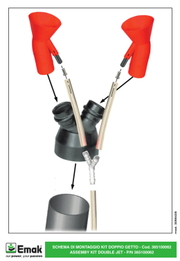
Scaricare