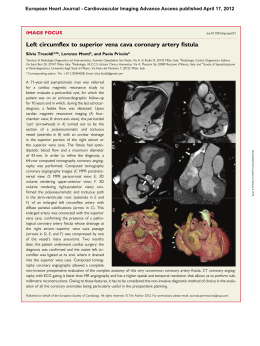
Eur Radiol
DOI 10.1007/s00330-008-1239-8
CARD IAC
Riccardo Marano
Francesco De Cobelli
Irene Floriani
Christoph Becker
Christopher Herzog
Maurizio Centonze
Giovanni Morana
Gian Franco Gualdi
Guido Ligabue
Gianluca Pontone
Carlo Catalano
Dante Chiappino
Massimo Midiri
Giovanni Simonetti
Filippo Marchisio
Lucio Olivetti
Rossella Fattori
Lorenzo Bonomo
Alessandro Del Maschio
NIMISCAD Study Group
Italian multicenter, prospective study
to evaluate the negative predictive value
of 16- and 64-slice MDCT imaging in patients
scheduled for coronary angiography
(NIMISCAD-Non Invasive Multicenter Italian
Study for Coronary Artery Disease)
Received: 14 May 2008
Accepted: 8 October 2008
# European Society of Radiology 2008
G. Morana
Cà Foncello Hospital,
Treviso, Italy
L. Olivetti
Istituti Ospitalieri of Cremona,
Cremona, Italy
G. F. Gualdi
DEA Umberto I Hospital,
La Sapienza University,
Rome, Italy
R. Fattori
S. Orsola University Hospital,
Bologna, Italy
R. Marano (*)
Department of Bioimaging and
Radiological Sciences, Institute of
Radiology, “A. Gemelli” Hospital Catholic University,
L.go A Gemelli 8,
00168 Rome, Italy
e-mail: [email protected]
Tel.: +39-06-30154977
Fax: +39-06-35501928
G. Ligabue
University of Modena
and Reggio Emilia,
Modena, Italy
G. Pontone
Centro Cardiologico Monzino,
Milan, Italy
F. De Cobelli
S. Raffaele Scientific Institute,
Vita-Salute University,
Milan, Italy
C. Catalano
Umberto I Hospital,
La Sapienza University,
Rome, Italy
I. Floriani
Mario Negri Institute,
Milan, Italy
D. Chiappino
G. Pasquinucci Hospital,
Massa, Italy
C. Becker
Ludwig-Maximilians University,
Munich, Germany
M. Midiri
DIBIMEL, University of Palermo,
Palermo, Italy
C. Herzog
J. W. Goethe University,
Frankfurt, Germany
G. Simonetti
Tor Vergata University,
Rome, Italy
M. Centonze
S.Chiara Hospital,
Trento, Italy
F. Marchisio
University of Turin,
Turin, Italy
L. Bonomo
A. Gemelli Hospital,
Catholic University,
Rome, Italy
A. Del Maschio
S. Raffaele Scientific Institute,
Vita-Salute University,
Milan, Italy
Abstract This was a prospective,
multicenter study designed to evaluate
the utility of MDCT in the diagnosis
of coronary artery disease (CAD) in
patients scheduled for elective coronary angiography (CA) using different
MDCT systems from different manufacturers. Twenty national sites prospectively enrolled 367 patients
between July 2004 and June 2006.
Computed tomography (CT) was
performed using a standardized/
optimized scan protocol for each type
of MDCT system (≥16 slices) and
compared with quantitative CA performed within 2 weeks of MDCT. A
total of 284 patients (81%) were
studied by 16-slice MDCT systems,
while 66 patients (19%) by 64-slice
MDCT scanners. The primary analysis
was on-site/off-site evaluation of the
negative predictive value (NPV) on a
per-patient basis. Secondary analyses
included on-site evaluation on a perartery and per-segment basis. On-site
evaluation included 327 patients
(CAD prevalence 58%). NPV, positive
predictive value (PPV), sensitivity,
specificity, and diagnostic accuracy
(DA) were 0.91 (95% CI 0.85–0.95),
0.91 (95% CI 0.86–0.95), 0.94 (95%
CI 0.89–0.97), 0.88 (95% CI 0.81–
0.93), and 0.91 (95% CI 0.88–0.94),
respectively. Off-site analysis included 295 patients (CAD prevalence
56%). NPV, PPV, sensitivity, specificity, and DA were 0.73 (95% CI
Introduction
Coronary angiography (CA) represents the standard of
reference to assess epicardial vessels and establish the
presence of significant coronary artery disease (CAD). CA
is a bi-dimensional diagnostic technique that allows evaluation of the vessel lumen [1] at high spatial (50 lps/cm) and
temporal (<20 ms) resolution. However, CA has its
limitations: it is an invasive technique, involves high costs,
requires hospitalization, and has low patient compliance. The
risks associated with CA are related to its invasiveness and
result in a morbidity rate of 1.5% and a mortality rate of
0.15% [2]. In addition, a considerable proportion of patients
undergoing CA are subsequently found not to have clinically
significant disease [3, 4]. These limitations may justify the
increasing clinical use of alternative non-invasive imaging
techniques for the evaluation and exclusion of clinically
significant CAD.
Many centers have reported their experiences in using
different multidetector computed tomography (MDCT)
technologies, all of which have been characterized by high
diagnostic accuracy in the assessment of patients scheduled
for CA. Whether the reported diagnostic accuracy can be
replicated in clinical practice needs to be established at
different centers with varying expertise, in non-selected
patient populations and with different MDCT systems before
extensive clinical application of MDCT for CAD assessment.
The primary aim of this study was to assess the negative
predective value (NPV) of MDCT in a multicenter study
using different MDCT machines/manufacturers to determine if the technique could replace an invasive procedure
to exclude the presence of significant CAD. Secondary
objectives were to investigate whether MDCT performance
is affected by patient characteristics such as pre-test CAD
risk, body mass index (BMI), and coronary artery calcium
score (CACS). Safety was also investigated.
Materials and methods
This was a national, multicenter, prospective trial sponsored by the Italian Society of Medical Radiology (SIRM).
0.65–0.79), 0.93 (95% CI 0.87–0.97),
0.73 (95% CI 0.65–0.79), 0.93 (95%
CI 0.87–0.97), and 0.82 (95% CI
0.77–0.86), respectively. The results
of this study demonstrate the utility of
MDCT in excluding significant CAD
even when conducted by centers with
varying degrees of expertise and using
different MDCT machines.
Keywords Multidetector CT .
Coronary artery disease . Cardiac CT
Adult patients previously scheduled for an elective CA
evaluation for a diagnostic workup at each of the
participating centers were consecutively evaluated for
inclusion in the study. Exclusion criteria included pregnancy or lactation, contraindication to intravenous administration of iodinated contrast media, heart rate >70 beats
per minute (bpm) despite beta-blocker treatment, absence
of sinus rhythm, New York Heart Association (NYHA)
class III–IV, previous coronary artery bypass graft surgery
(CABG) or stenting, and creatinine value >2 mg/dl. The
Institutional Review Boards of all centers approved the
study protocol, and written informed consent was obtained
from all patients before study inclusion in accordance with
the national legislation and the Declaration of Helsinki.
Patient preparation
Patients with heart rates >70 bpm were given an oral dose
of metoprolol tartrate 45–60 min before MDCT imaging or
an intravenous beta-receptor blocker just before the scan.
Heart rate, electrocardiogram (ECG), and blood pressure
were monitored.
MDCT protocol and image reconstructions
Patients underwent 16- or 64-slice MDCT with a
standardized and optimized protocol for each type of
CT machine. Unenhanced prospective ECG-gated
MDCT was performed for the assessment of calcium
deposits by Agatston Score (AS). A bolus of 100–
120 ml of non-ionic iodinated contrast medium Ultravist® (Iopromide 370; Bayer Schering Pharma, Berlin,
Germany) was intravenously injected, preferably
through the right antecubital vein, at a flow rate of 3–
4 ml/s for the 16-slice MDCT system and 4–6 ml/s for
the 64-slice MDCT system, and followed by a 40–50-ml
saline chaser at the same flow rate.
The main scan parameters have been given for each
scanner type in Table 1, including the number of patients
scanned on each CT system.
Table 1 Main technical and scan parameters of the CT systems and
number of studied patients
Invasive angiographic analysis
CT system
Rot. T
(ms)
TR
(ms)
ST (mm)
Patients
(N=350)
GE Lightspeed16pro
Siemens Sensation16
Philips Brilliance16
Toshiba Aquilon16
GE Volume CT
Siemens Sensation64
Philips Brilliance64
Toshiba Aquilon64
400
375
420
400
350
330
400
400
200
188
210
200
175
165
200
200
16×0.625
16×0.75
16×0.75
16×0.5
64×0.625
32×2×0.6
64×0.625
64×0.5
108
95
41
40
20
29
12
5
CA was performed by standard techniques within 2 weeks
after MDCT procedure, using a transfemoral or transradial
technique. CA was evaluated on-site by a single experienced cardiologist blinded to the MDCT findings. Segmental disease was analyzed in each vessel using the same
15-segment model and form employed for MDCT analysis.
Stenosis severity in each segment was classified according
to the maximal luminal diameter stenosis present in each
segment. Lesions were examined in orthogonal views and
stenosis severity quantitatively determined using an
automated edge detection system (quantitative CA, QCA).
Rot. T = rotation time, TR = temporal resolution, ST = slice
thickness
All images were reconstructed in all phases of the entire
cardiac cycle, by means of retrospective synchronization
with the ECG track, expressed as a percentage of the R-R
interval (relative delay) or as absolute temporal distance
(ms) before the next R wave.
MDCT angiographic analysis
MDCT angiograms were analyzed both on-site and off-site.
The on-site analysis was conducted by a single radiologist
at each center who was aware of the patient’s clinical
condition, but blinded to the results of the invasive CA.
Among the data sets reconstructed at several phases of the
cardiac cycle, the three with the best image quality were
saved on CD and sent for centralized off-site evaluation
that was performed by two independent radiologists with
proven experience in cardiac CT imaging, but who were
not involved in the clinical study and who were blinded to
any patient-related information. Any disagreement between off-site readers was resolved by consensus. An
evaluating form with coronary arteries segmented according to the guidelines of the American Heart Association
(AHA) [5] was used for both analyses. Lesions were
classified by the maximal luminal diameter stenosis by
qualitative analysis of the most severe well-defined lesion
in each segment. Segments were classified as normal
(smooth, parallel, or tapering borders), as non-significant
disease (luminal irregularities or <50% lumen reduction),
as significant stenoses (lumen reduction between 50% and
99%), or occluded. In the case of multiple lesions in a given
segment, the segment was classified by the worst lesion. In
the case of multiple abnormal segments per artery, the
vessel was classified by the worst segment. Patients were
classified as positive for the presence of significant CAD if
there was at least one stenosis >50%.
Data analysis
The assessment of MDCT accuracy was performed
according to a per-patient on-site and off-site evaluation;
an on-site artery- and segment-based approach analysis
was also performed.
The per-patient analysis included all eligible patients,
except those negative at MDCT with at least one of the
following segments not assessable at MDCT: 1, 2, 3, 5, 6,
7, 8, 9, 11, and 13. A patient was classified as true positive
if at least one significant coronary stenosis (>50% or
occlusion) detected by both modalities was present,
regardless of location; a patient was considered as true
negative if no significant lesion was found by both
modalities.
In the per-artery analysis, segments were combined as
right coronary artery (RCA: 1, 2, 3), left main artery (LMA:
5), anterior descending artery (ADA: 6, 7, 8, 9), and
circumflex artery (CXA: 11, 13). Arteries deemed negative
at MDCT, but with at least one non-assessable segment
were excluded. An artery was classified as true positive if
at least one significant lesion was detected by both
investigations, regardless of location and irrespective of
whether or not it was considered for patient inclusion; truenegative arteries appeared normal on all assessable and
negative segments.
The per-segment analysis included only segments that
were assessable on both investigations. True positives were
defined as correct identification by MDCT of significant
coronary artery stenosis and true negatives were defined as
correct identification by MDCT of segments with stenosis
of 50% or less.
Once the concordance of the MDCT with the reference
standard had been assessed, the following statistics were
calculated: sensitivity, specificity, NPV, positive predictive
value (PPV), and diagnostic accuracy (DA).
The primary analysis was the evaluation of the NPVon a
per-patient basis, obtained both in on-site and off-site
evaluation. Expecting a prevalence of CAD ranging from
0.50 to 0.70, and a specificity of 0.95, the NPV was a
function of the sensitivity. Since no precise data were
available for this parameter, the sample size was calculated
in order to obtain a 95% confidence interval for the
estimate of sensitivity of not more than 0.1, with the lower
limit >0.80. The required number of evaluable patients
varied from 254 (prevalence 0.70) to 392 (prevalence
0.50). Anticipating a 10% drop-out rate, it was considered
necessary to enroll between 280 and 431 subjects. After the
enrollment of about 200 patients, an interim analysis was
foreseen in order to obtain a more precise estimate of the
prevalence of the disease in the sample studied. Based on
the analysis results, a recalculation of the necessary number
of subjects was performed.
Secondary analyses included the on-site evaluation on a
per-artery and per-segment basis. Furthermore, the on-site
per-patient analysis was also conducted in subgroups
defined by the National Cholesterol Education Program
(NCEP) coronary heart disease (CHD) risk (low, intermediate, high) [6], by BMI (<25 kg/m2, 25–30 kg/m2, >30 kg/
m2), calcium score (<100, 101–400, 401–1,000, >1,000
AS), and type of MDCT scanner (16- and 64-slice). A χ2
test for trend was used to test for differences in the modality
results in different subgroups of patients. Secondary
analysis also included a safety assessment reporting the
occurrence of adverse events. Results are expressed as
point estimates and their exact 95% confidence intervals
(95% CIs). All p-values are two-tailed. Analyses were
carried out using SAS System Version 8.20 (SAS, Cary, NC).
Results
Between July 2004 and June 2006, a total of 367 patients
scheduled for elective CA for suspected CAD at 20 Italian
centers were screened. Seventeen patients (5%) were
subsequently excluded due to: CA erased (n=1), major
protocol violation (n=9), temporary MDCT equipment
unavailability (n=5), and patient refusal (n=2).The final
study sample included 350 eligible patients.
Academic institutions comprised 60% of the centers:
35% were located in northern Italy, 45% in central Italy,
and 20% in southern Italy and the islands. Both the type
and geographical distribution of the institutions were well
distributed and representative of the national situation.
Figure 1 shows the trial flow diagram and details of reasons
for patient ineligibility. A total of 284 (81%) patients were
scanned by a 16-slice MDCT scanner, while the remaining
66 (19%) by a 64-slice MDCT. The characteristics of the
350 eligible patients are summarized in Table 2.
Per-patient analyses
Results of the per-patient analyses are reported in Table 3.
Out of 295 patients analyzed in the off-site evaluation, 166
had at least one segment with a stenosis >50% by QCA,
which translates into a disease prevalence of 56%. NPV of
MDCT was 0.73 (95% CI 0.65–0.79) and PPV was 0.93
(95% CI 0.87–0.97). Sensitivity was 0.73 (95% CI 0.65–
0.79), specificity was 0.93 (95% CI 0.87–0.97), and DA
was 0.82 (95% CI 0.77–0.86).
The on-site evaluation included 327 patients with a CAD
prevalence of 58%. NPV of MDCT was 0.91 (95% CI 0.85–
0.95), and PPV was 0.91 (95% CI 0.86–0.95). Sensitivity
was 0.94 (95% CI 0.89–0.97), specificity was 0.88 (95% CI
0.81–0.93), and DA was 0.91 (95% CI 0.88–0.94).
Evaluating the CHD risk in our population, the prevalence was 0.38 for the low risk, 0.65 for the intermediate
risk, and 0.77 for the high risk. Altough non-statistically
significant, there was evidence in favor of a trend of
367 patients scheduled for elective
X-ray CA for CAD suspicion
17 patients excluded due to:
- 9 protocol deviations
- 5 MDCT not available
- 1 CA not conducted
- 2 patient refusal
350 eligible patients
OFF-SITE EVALUATION
ON-SITE EVALUATION
Per patient analysis
N=295
Per patient analysis
N=327
Per artery analysis
N=1328
Per segment analysis
N=4628
166 (56%) QCA stenosis >50%
129 (44%) QCA stenosis <50%
189 (58%) QCA stenosis >50%
138 (42%) QCA stenosis <50%
296 (22%) QCA stenosis >50%
1032 (78%) QCA stenosis <50%
530 (11%) QCA stenosis >50%
4098 (89%) QCA stenosis <50%
Fig. 1 Study flow chart
Table 2 Patient characteristics, n (%)
On-site per-artery analysis
Eligible patients
350
Age1 - years
Male
BMI- kg/m2
<25
25–30−
≥30
Total cholesterol1 - mg/dl
HDL1 - mg/dl
SBP - mmHg
DBP - mmHg
HR- beat/min
Current smoking habit
NCEP CHD risk
Low
Intermediate
Peripheral disease
Statin treatment
Diabetes
Beta-blocker treatment
Agatston Calcium Score
0–100
101–400
401–1,000
>1,000
64.0 (19–83)
265 (75.7)
110 (31.4)
193 (55.1)
47 (13.4)
201.3 (95–347)
49 (22–99)
130.0 (95–210)
80.0 (60–100)
64.0 (35–100)
113 (32.3)
124 (35.4)
167 (47.7)
59 (16.9)
140 (40.0)
87 (24.9)
128 (36.6)
168 (48)
90 (25.7)
44 (12.6)
48 (13.8)
1
Median (range)
BMI = Body mass index; SBP = systolic blood pressure; DBP =
diastolic blood pressure; HR = heart rate; NCEP = National
Cholesterol Education Program; CHD = coronary heart disease
decreasing NPV with increasing CHD risk: 0.93 in low risk
and 0.79 in high risk patients (χ2 test for trend=2.11; p=
0.147). No differences in DA were observed.
Assessing the influence of BMI on MDCT performance,
false-positive results were more frequent in obese patients,
specificity fell from 0.92 in normal patients to 0.67 in obese
subjects (χ2 test for trend=6.29; p=0.012). PPV was slightly
negatively affected by BMI: 0.92 in normal and 0.76 in
obese subjects (χ2 test for trend=4.00; p=0.046). Nonsignificant differences resulted for sensitivity, NPV, and DA
because of a high number of correct negatives (14/21).
The analysis of calcium score effect was performed by
collapsing the two higher categories (401–1,000 and
>1,000 AS) into one, because of the low number of patients
in each category. Specificity significantly (p<0.001) decreased when CACS increased: 0.94 (0–100 AS), 0.76 (101–
400 AS), and 0.50 (>400 AS). NPV was negatively affected
by increasing CACS: 0.94 (0–100 AS) vs. 0.86 (101–400
AS) vs. 0.62 (>400 AS). No effect on MDCT performance
was observed for the type of CT machine.
Results of the per-artery analysis are reported in Table 4. Of
the 1,328 arteries analyzed on-site, 296 (22%) had a
stenosis >50% by CA. NPV of MDCT was 0.96 (95% CI
0.93–0.98) for RCA, 0.99 (95% CI 0.97–1.00) for LMA,
0.91 (95% CI 0.86–0.95) for ADA, and 0.94 (95% CI
0.90–0.96) for CXA, respectively, with an overall NPV of
0.96 (95% CI 0.94–0.97).
On-site per-segment analysis
Results of the per-segment analysis are reported in Table 5.
Of 4,628 segments analyzed on-site, 530 (11%) had a
stenosis >50% by CA. The assessable segments were 3,746
(77.59%) by using the 16-slice MDCT system and 882
(78.61%) by using the 64-slice MDCT machine. NPV of
MDCT was 0.96 (95% CI 0.95–0.97), and PPV was 0.72
(95% CI 0.68–0.76). Sensitivity was 0.70 (95% CI 0.66–
0.74), specificity was 0.96 (95% CI 0.96–0.97), and DA
was 0.93 (95% CI 0.93–0.94).
The overall image quality resulted in good (moderate
and/or excellent) in 4,292 (93.95%) segments in the on-site
analysis and in 3,396 (80.17%) segments in the off-site
analysis.
Safety
Six adverse events related to MDCT occurred in 6/350
patients (1.7%): four cases of cutaneous rash, one case of
vomiting, and one case of nausea. All cases were related to
contrast medium injection, all completely recovered, and
none was considered serious.
Discussion
Since the first reports [7, 8], MDCT has been characterized
by continuous and rapid technical evolution, making it
possible to examine the heart and the whole course of
coronary arteries reliably within a single breath-hold,
within a few heart beats, and with sub-millimeter spatial
resolution. Many single-center experiences have been
reported [9–28] using different MDCT technologies,
most of which have been characterized by high values of
DA in the assessment of patients scheduled for CA [9–18;
20–28]. On a per-patient basis, the average weighted
sensitivity reported in these series for the detection of at
least one coronary artery with >50% lumen stenosis is 94%
(range 75% to 100%), whereas the average specificity is
77% (range 49% to 100%) [29–30]. In most of these
studies using 16-slice MDCT, up to 23% of coronary
segments were considered not assessable [19]; similarly,
data analysis from 64-slice MDCT studies could not be
Table 3 Per patient analysis
Evaluation
N
Prevalence
Off-site
Overall
295 0.56
On-site
Overall
327 0.58
By NCEP CHD risk
Low
115 0.38
Intermediate 156 0.65
High
56
0.77
χ2 test for trend
By BMI
102 0.50
<25 kg/m2
25–30
181 0.63
≥30
44
0.52
χ2 test for trend
By calcium score
0–100
151 0.32
101–400
86
0.71
>400
90
0.89
χ2 test for trend
By multidetector
16-slice
264 0.58
64-slice
63
0.57
χ2 test
Sensitivity
Specificity
PPV
NPV
DA
0.73 (0.65–0.79)
0.93 (0.87–0.97)
0.93 (0.87–0.97)
0.73 (0.65–0.79)
0.82 (0.77–0.86)
0.94 (0.89–0.97)
0.88 (0.81–0.93)
0.91 (0.86–0.95)
0.91 (0.85–0.95)
0.91 (0.88–0.94)
0.89 (0.75–0.96)
0.96 (0.90–0.99)
0.93 (0.81–0.99)
χ2 =7.21;p=0.396
0.93 (0.84–0.98)
0.81 (0.69–0.91)
0.85 (0.55–0.98)
χ2 =2.84;p=0.092
0.89 (0.75–0.96)
0.91 (0.84–0.95)
0.95 (0.84–0.99)
χ2 =1.15;p=0.283
0.93 (0.84–0.98)
0.92 (0.80–0.98)
0.79 (0.49–0.95)
χ2 =2.11;p=0.147
0.91 (0.85–0.96)
0.91 (0.85–0.95)
0.91 (0.80–0.97)
χ2 =0.00;p=0.945
0.94 (0.84–0.99)
0.93 (0.87–0.97)
0.96 (0.78–1.00)
χ2 =0.01;p=0.913
0.92 (0.81–0.98)
0.91 (0.81–0.97)
0.67 (0.43–0.85)
χ2 =6.29;p=0.012
0.92 (0.81–0.98)
0.95 (0.89–0.98)
0.76 (0.56–0.90)
χ2 =4.00;p=0.046
0.94 (0.83–0.99)
0.88 (0.78–0.95)
0.93 (0.68–1.00)
χ2 =0.29;p=0.590
0.93 (0.86–0.97)
0.92 (0.87–0.96)
0.82 (0.67–0.92)
χ2 =3.43;p=0.064
0.88 (0.75–0.95)
0.95 (0.86–0.99)
0.96 (0.89–0.99)
χ2 =3.46;p=0.063
0.94 (0.88–0.98)
0.76 (0.55–0.91)
0.50 (0.19–0.81)
χ2 =20.0;p<0.001
0.88 (0.75–0.95)
0.91 (0.81–0.96)
0.94 (0.86–0.98)
χ2 =1.59;p=0.208
0.94 (0.88–0.98)
0.86 (0.65–0.97)
0.62 (0.24–0.92)
χ2 =8.70;p=0.003
0.92 (0.87–0.96)
0.90 (0.81–0.95)
0.91 (0.83–0.96)
χ2 =1.47;p=0.225
0.94 (0.89–0.97)
0.92 (0.78–0.98)
χ2 =2.93;p=0.588
0.86 (0.79–0.92)
0.93 (0.76–0.99)
χ2 =0.74;p=0.388
0.91 (0.85–0.95)
0.94 (0.81–0.99)
χ2 =4.94;p=0.482
0.91 (0.84–0.96)
0.89 (0.72–0.98)
χ2 =1.23;p=0.726
0.91 (0.87–0.94)
0.92 (0.82–0.97)
χ2 =0.08;p=0.772
performed in all coronary segments, with a reported
percentage of non-assessable segments of up to 12% [22,
26–28]. In a recent meta-analysis [31], it was demonstrated
that NPV was sufficiently high with 16-section CT (92%),
making this an excellent tool to rule out CAD, strengthening
our choice to include centers with CT technology ≥16-slice
MDCT. Before extensive clinical application of MDCT for
CAD assessment can be advocated, it is necessary to
establish whether or not the reported DA can be replicated in
clinical practice in different centers with varying expertise
and MDCT manufacturer. In fact, the main limitations of the
current data are the strict eligibility criteria used for patient
selection, the restriction of high image-quality data sets to
selected cohorts or subgroups of patients, and the predominance of a single MDCT manufacturer.
We conducted a prospective national multicenter study,
reflecting the technical aspects of different MDCT systems
(≥16-slice MDCT) available in Italy at the time of the study
to determine whether MDCT can replace the invasive
procedure in patients scheduled for elective CA in order to
Table 4 Per artery analysis by on-site evaluation
Artery
N
Prevalence
Sensitivity
Specificity
PPV
RCA
LMA
ADA
CXA
322
346
334
326
0.28
0.03
0.42
0.17
0.90
0.75
0.88
0.69
0.92
0.99
0.89
0.95
0.82
0.69
0.85
0.75
(0.82–0.95)
(0.43–0.95)
(0.81–0.93)
(0.55–0.81)
(0.88–0.95)
(0.97–1.00)
(0.83–0.93)
(0.92–0.97)
NPV
(0.73–0.89)
(0.39–0.91)
(0.78–0.90)
(0.60–0.86)
0.96
0.99
0.91
0.94
DA
(0.93–0.98)
(0.97–1.00)
(0.86–0.95)
(0.90–0.96)
0.92
0.98
0.88
0.91
(0.88–0.94)
(0.96–0.99)
(0.84–0.92)
(0.87–0.94)
Table 5 Per segment analysis by on-site evaluation
N
Prevalence
Sensitivity
Specificity
PPV
NPV
DA
4,628
0.11
0.70 (0.66–0.74)
0.96 (0.96–0.97)
0.72 (0.68–0.76)
0.96 (0.95–0.97)
0.93 (0.93–0.94)
exclude the presence of significant CAD. Bearing in mind
that relevant coronary segments are essentially those
suitable for a mechanical (PTCA/stenting) or surgical
(CABG) treatment (diameter ≥1.5–2 mm), our study
provides a per-patient on-site analysis of sufficiently high
sensitivity (94%) and NPV (91%) of ECG-gated MDCT
coronary angiography to rule out significant stenosis in
patients scheduled for an elective CA in a large population.
Our study confirms the role of MDCT coronary angiography for reliably excluding significant CAD in patients
with an equivocal clinical presentation, with uninterpretable or equivocal stress-test results, or at low or intermediate risk for CAD. We found that a negative evaluable
MDCT study could have avoided CA in 120/295 (41%)
patients in the off-site evaluation and in 121/327 (37%)
patients in the on-site assessment.
To the best of our knowledge, this is the first study in
which patients were studied in several independent national
centers with different expertise, using different MDCT
machines from different manufacturers, with a standardized and optimized CT protocol. Only one similar multicenter study has been published to date [32]. In that report,
187 patients were studied in 11 participating sites, all using
the same MDCT manufacturer. All MDCT and CA results
were assessed by a single core laboratory in each case. The
per-patient approach was characterized by sensitivity,
specificity, PPV, and NPV of 75%, 77%, 60%, and 87%,
respectively.
The difference between on-site and off-site evaluation in
our study could, in part, be explained by the intrinsic
characteristics of the latter assessment as well as by the fact
that the two off-site readers were completely unaware of
the patient’s clinical information. It could also be because
blinded assessment of each MDCT was performed by using
only three data sets considered to be those with the best
image quality by the on-site reader. These results were
obtained in patients scheduled for elective CA and with a
wide spectrum of clinical settings. The on-site specificity
was somewhat lower in comparison with most of the
published reports [9–28]. This is because there was a
tendency to overestimate the severity of a lesion on CT
(resulting in a number of false-positive outcomes) rather
than underestimating the lesion severity and thereby
“missing” lesions, which may have had serious consequences in a symptomatic patient population.
The clinical worldwide acceptance of MDCT coronary
angiography will probably require further technological
development to ensure optimal test results and a guiding
management of CAD. The extension of MDCT system
usage with higher temporal resolution (<100 ms) may
extend the application of this technology for the assessment
of CAD in a high probability pre-test population with
extensive calcifications and without heart rate control [33–
36]. The employment of hybrid PET/MDCT [37] or
SPECT/MDCT equipment with image fusion [38] may
offer the opportunity for a comprehensive non-invasive
evaluation of the presence of CAD and its consequences,
both in the coronary arteries and myocardium.
The radiation dose can be considered a limitation of this
technique [39]; however, due to the study design, the dose
analysis was not an objective of our study. The most effective
method to reduce radiation exposure is the prospective ECG
tube modulation (ECG pulsing) [40]. For 64-slice MDCT,
this has been reported to be 14.8±1.8 mSv and 9.4±1.0 mSv
without and with ECG-triggering tube output modulation,
respectively, and for 16-slice MDCT, 10.6±1.2 mSv, and
6.4±0.9 mSv without and with ECG triggering tube
output modulation, respectively [41].
Leading single center experience, with limited and
selected patient populations often including patients who
had previously undergone angiography, may be difficult to
translate into general clinical practice. The multicenter
study reported here should reflect the clinical value of
MDCT coronary angiography more accurately, because a
larger number of patients at different national centers has
been evaluated using all the principal MDCT manufacturers, thus reflecting real-world clinical practice. The high
NPV observed suggests that ECG-gated MDCT coronary
angiography could be useful to discriminate those patients
who really need an invasive CA, ruling out clinically
significant CAD in patients amongst whom the prevalence
of disease is low or intermediate [42] and in patients with
an equivocal stress test, as recently recommended also by
the Task Force on the Management of Stable Angina
Pectoris of the European Society of Cardiology [43].
However, to ensure that this non-invasive test is more
accurate, it is important to combine MDCT findings with
accurate and complete patient clinical information.
Funding The study was sponsored by the Italian Society
of Medical Radiology (SIRM), with the support of Bayer
Schering Pharma, Bayer spa, Milan, Italy
Conflict of Interest None declared
Appendix
The following Investigators and Institutions participated in
NIMISCAD (Non Invasive Multicenter Italian Study for
Coronary Artery Disease):
Naples, Italy); Arcadi Nicola, MD (Ospedali Riuniti of
Reggio Calabria, Italy); Profili Manuel, MD (Istituto
Clinico Humanitas, Rozzano, Milan, Italy); Volterrani
Luca, MD (S. Maria alle Scotte Hospital, University of
Siena, Italy).
Radiologists:
Cardiologists:
Marano Riccardo, MD (A. Gemelli Hospital, Catholic
University, Rome, Italy); Liguori Carlo, MD (A. Gemelli
Hospital, Catholic University, Rome, Italy); Bonomo
Lorenzo, MD (A. Gemelli Hospital, Catholic University,
Rome, Italy); De Cobelli Francesco MD (S. Raffaele
Scientific Institute and Vita-Salute University, Milan,
Italy); Esposito Antonio, MD (S. Raffaele Scientific
Institute and Vita-Salute University, Milan, Italy); Del
Maschio Alessandro, MD (S. Raffaele Scientific Institute
and Vita-Salute University, Milan, Italy); Becker
Christoph, MD (Ludwig-Maximilians University, Munich,
Germany); Herzog Christopher, MD (J.W. Goethe University, Frankfurt, Germany); Centonze Maurizio, MD (S.
Chiara Hospital, Trento, Italy); Coser Daniela, MD (S.
Chiara Hospital, Trento, Italy); Morana Giovanni, MD (Cà
Foncello Hospital, Treviso, Italy); Salviato Elisabetta, MD
(Cà Foncello Hospital, Treviso, Italy); Gualdi Gian Franco,
MD (DEA Umberto I Hospital, La Sapienza University,
Rome, Italy); Casciani Emanuele, MD (DEA Umberto I
Hospital, La Sapienza University, Rome, Italy); Ligabue
Guido, MD (University of Modena and Reggio Emilia,
Italy); Fiocchi Federica, MD (University of Modena and
Reggio Emilia, Italy); Pontone Gianluca, MD (Centro
Cardiologico Monzino, Milan, Italy); Andreini Daniele,
MD (Centro Cardiologico Monzino, Milan, Italy); Catalano
Carlo, MD (Umberto I Hospital, La Sapienza University,
Rome, Italy); Carbone Iacopo, MD (Umberto I Hospital, La
Sapienza University, Rome, Italy); Chiappino Dante, MD
(G. Pasquinucci Hospital, Massa, Italy); Midiri Massimo,
MD (DIBIMEL, University of Palermo, Italy); Simonetti
Giovanni, MD (Tor Vergata University, Rome, Italy);
Marchisio Filippo, MD (University of Turin, Italy);
Olivetti Lucio, MD (Istituti Ospitalieri of Cremona,
Italy); Fattori Rossella, MD (S. Orsola University Hospital,
Bologna, Italy); Scardapane Arnaldo, MD (Policlinico of
Bari, Italy); Principi Massimo, MD (S. Maria Hospital,
Terni, Italy); Romano Luigia, MD (A. Cardarelli Hospital,
Trani Carlo, MD (A. Gemelli Hospital, Catholic University, Rome, Italy); Colombo Antonio, MD (S. Raffaele
Scientific Institute and Vita-Salute University, Milan,
Italy); Bonmassari Roberto, MD (S.Chiara Hospital,
Trento, Italy); Chirillo Fabio, MD (Cà Foncello Hospital,
Treviso, Italy); Pastore Raffaele, MD (DEA Umberto I
Hospital, La Sapienza University, Rome, Italy); Modena
Maria Grazia, MD (University of Modena and Reggio
Emilia, Italy); Bartorelli Antonio, MD (Centro Cardiologico Monzino, Milan, Italy); Gaudio Carlo, MD (Umberto
I Hospital, La Sapienza University, Rome, Italy); Vaghetti
Marco, MD (G. Pasquinucci Hospital, Massa, Italy); Novo
Salvatore, MD (DIBIMEL, University of Palermo, Italy);
Romeo Francesco, MD (Tor Vergata University, Rome,
Italy); Sheiban Imad, MD (University of Turin, Italy);
Pirelli Salvatore, MD (Istituti Ospitalieri of Cremona,
Italy); Marzocchi Antonio, MD (S. Orsola University Hospital, Bologna, Italy); Brizio Leonardo Corlianò, MD
(Policlinico of Bari, Italy); Dominici Marcello, MD (S.
Maria Hospital, Terni, Italy); Caruso Aurelio, MD (A.
Cardarelli Hospital, Naples, Italy); Cacciola Maria Teresa,
MD (Ospedali Riuniti of Reggio Calabria, Italy);
Aldrovandi Annachiara, MD (Istituto Clinico Humanitas,
Rozzano, Milan, Italy).
Statistical analysis:
Floriani Irene, PhD (Mario Negri Institute, Milan, Italy);
Tinazzi Angelo, CompSc (SENDO-Tech s.r.l. Milan, Italy).
Industrial support:
Ciceri Marco, MD (Bayer Schering Pharma, Bayer spa,
Milan, Italy); Gatti Simona, MSc (Bayer Schering Pharma,
Bayer spa, Milan, Italy).
References
1. Topol EJ, Nissen SE (1995) Our preoccupation with coronary luminology.
The dissociation between clinical and
angiographic findings in ischemic heart
disease. Circulation 92:2333–2342
2. Davidson CJ, Fishman RF, Bonow RO
(1997) Cardiac catheterization.
In: Braunwald E (ed) Heart
disease: A textbook of
cardiovascular medicine. WB
Saunders, Philadelphia, Pa,
pp 177–203
3. Haberl R, Becker A, Leber A et al (2001)
Correlation of coronary calcification and
angiographically documented stenoses in
patients with suspected coronary artery
disease: results of 1.764 patients. J Am
Coll Cardiol 37:451–457
4. Thom T, Haase N, Rosamond W et al
(2006) American Heart Association
Statistics Committee and Stroke Statistics Subcommittee. AHA Statistical
Update. Heart Disease and Stroke Statistics-2006 Update. A Report From the
American Heart Association Statistics
Committee and Stroke Statistics Subcommittee. Circulation 113(6):e85–151
5. Austen WG, Edwards JE, Frye RL et al
(1975) A reporting system on patients
evaluated for coronary artery disease.
Report of the Ad-Hoc Committee for
Grading of Coronary Artery Disease,
Council on Cardiovascular Surgery.
Circulation 51:5–40
6. Expert Panel on Detection, Evaluation
and Treatment of High Blood Cholesterol in Adults. Executive Summary of
the Third Report of the National Cholesterol Education Program/Adult
treatment Panel III (2001) JAMA
285:2846–2897
7. Achenbach S, Ulzheimer S, Baum U
et al (2000) Noninvasive coronary
angiography by retrospectively ECGgated multislice spiral CT. Circulation
102:2823–2828
8. Nieman K, Oudkerk M, Rensing BJ
et al (2001) Coronary angiography with
multi-slice computed tomography.
Lancet 357:599–603
9. Achenbach S, Giesler T, Ropers D et al
(2001) Detection of coronary artery
stenoses by contrast-enhanced, retrospectively electrocardiographically
gated, multislice spiral computed
tomography. Circulation 103:2535–
2538
10. Vogl TJ, Abolmaali ND, Diebold T et al
(2002) Techniques for the detection of
coronary atherosclerosis: multi-detector
row CT coronary angiography. Radiology 223:212–220
11. Kopp AF, Schroeder S, Kuettner A et al
(2002) Non-invasive coronary angiography with high resolution multidetector-row computed tomography. Results
in 102 patients. Eur Heart J 23:1714–
1725
12. Nieman K, Cademartiri F, Lemos PA
et al (2002) Reliable noninvasive coronary angiography with fast submillimeter multislice spiral computed
tomography. Circulation 106:2051–
2054
13. Ropers D, Baum U, Pohle K et al
(2003) Detection of coronary artery
stenoses with thin-slice multi-detector
row spiral computed tomography and
multiplanar reconstruction. Circulation
107:664–666
14. Hoffmann U, Moselewski F, Cury RC
et al (2004) Predictive value of 16-slice
multidetector spiral computed tomography to detect significant obstructive
coronary artery disease in patients at
high risk for coronary artery disease.
Patient- versus segment-based analysis.
Circulation 110:2638–2643
15. Kuettner A, Kopp AF, Schroeder S et al
(2004) Diagnostic accuracy of multidetector computed tomography coronary angiography in patients with
angiographically proven coronary artery disease. J Am Coll Cardiol
43:831–839
16. Mollet NR, Cademartiri F, Nieman K
et al (2004) Multislice spiral computed
tomography coronary angiography in
patients with stable angina pectoris.
J Am Coll Cardiol 43:2265–2270
17. Kuettner A, Beck T, Drosch T et al
(2005) Diagnostic accuracy of noninvasive coronary imaging using
16-detector slice spiral computed
tomography with 188 ms temporal
resolution. J Am Coll Cardiol 45:123–
127
18. Mollet NR, Cademartiri F, Krestin GP
et al (2005) Improved diagnostic accuracy with 16-row multi-slice computed
tomography coronary angiography.
J Am Coll Cardiol 45:128–132
19. Kaiser C, Bremerich J, Haller S et al
(2005) Limited diagnostic yield of noninvasive coronary angiography by 16slice multi-detector spiral computed
tomography in routine patients referred
for evaluation of coronary artery disease. Eur Heart J 26:1987–1992
20. Hoffman MH, Shi H, Schmitz BL et al
(2005) Non invasive coronary angiography with multislice computed tomography. JAMA 293:2471–2478
21. Leschka S, Alkadhi H, Plass A et al
(2005) Accuracy of MSCT coronary
angiography with 64-slice technology:
first experience. Eur Heart J 26:1482–
1487
22. Raff GL, Gallagher MJ, O’Neill WW
et al (2005) Diagnostic accuracy of noninvasive coronary angiography using
64-slice spiral computed tomography.
J Am Coll Cardiol 46:552–557
23. Mollet NR, Cademartiri F, van
Mieghem CA et al (2005) Highresolution spiral computed tomography
coronary angiography in patients referred for diagnostic conventional coronary angiography. Circulation
112:2318–2323
24. Leber AW, Knez A, von Ziegler F et al
(2005) Quantification of obstructive
and nonobstructive coronary lesions by
64-slice computed tomography. A
comparative study with quantitative
coronary angiography and intravascular
ultrasound. J Am Coll Cardiol 46:147–
154
25. Pugliese F, Mollet NR, Runza G et al
(2006) Diagnostic accuracy of noninvasive 64-slice CT coronary angiography in patients with stable angina
pectoris. Eur Radiol 16:575–582
26. Nikolaou K, Knez A, Rist C et al
(2006) Accuracy of 64-MDCT in the
diagnosis of ischemic heart disease.
Am J Roentgenol 187:111–117
27. Ehara M, Surmely JF, Kawai M et al
(2006) Diagnostic accuracy of 64-slice
computed tomography for detecting
angiographically significant coronary
artery stenosis in an unselected consecutive patient population: Comparison with conventional invasive
angiography. Circ J 70:564–571
28. Ropers D, Rixe J, Anders K et al (2006)
Usefulness of multidetector row spiral
computed tomography with 64-×
0.6-mm collimation and 330-ms rotation for the noninvasive detection of
significant coronary artery stenoses.
Am J Cardiol 97:343–348
29. U.S. Department of Health and Human
Services; Medicare Coverage Database
http://www.cms.hhs.gov/mcd/view
mcac.asp?where_index&mid_24
(5 June 2006)
30. Di Carli MF, Hachamovitch R (2007)
New technology for non-invasive evaluation of coronary artery disease. Circulation 115:1464–1480
31. Hamon M, Morello R, Riddell JW et al
(2007) Coronary arteries: diagnostic
performance of 16- versus 64-section
spiral CT compared with invasive coronary angiography — Meta-Analysis.
Radiology 245:720–731
32. Garcia MJ, Lessick J, Hoffman MH
(2006) Accuracy of 16-row multidetector computed tomography for the
assessment of coronary artery stenosis.
JAMA 296:403–411
33. Flohr TG, McCollough CH, Bruder H
et al (2006) First performance evaluation of a dual-source CT (DSCT)
system. Eur Radiol 16:256–268
34. Achenbach S, Ropers D, Kuettner A
et al (2006) Contrast-enhanced coronary artery visualization by dual-source
computed tomography—Initial experience. Eur J Radiol 57:331–335
35. Johnson TRC, Nikolaou K, Wintersperger BJ et al (2006) Dual-source CT
cardiac imaging: initial experience. Eur
Radiol 16:1409–1415
36. Scheffel H, Alkadhi H, Plass A et al
(2006) Accuracy of dual-source CT
coronary angiography: first experience
in a high pre-test probability population
without heart rate control. Eur Radiol
16:2739–2747
37. Sampson UK, Dorbala S, Limaye A
et al (2007) Diagnostic accuracy of
rubidium-82 myocardial perfusion imaging with hybrid positron emission
tomography/computed tomography in
the detection of coronary artery disease.
J Am Coll Cardiol 49:1052–1058
38. Gaemperli O, Schepis T, Kalff V et al
(2007) Validation of a new cardiac
image fusion software for three-dimensional integration of myocardial
perfusion SPECT and stand-alone 64slice CT angiography. Eur J Nucl Med
Mol Imaging 34:1097–1106
39. Einstein AJ, Henzlova MJ,
Rajagopalan S (2007) Estimating risk
of cancer associated with radiation
exposure from 64-slice computed
tomography coronary angiography.
JAMA 298(3):317–323
40. Flohr T, Kuttner A, Bruder H et al
(2003) Performance evaluation of a
multi-slice CT system with 16-slice
detector and increased gantry rotation
speed for isotropic submillimeter imaging of the heart. Herz 28(1):7–19
41. Hausleiter J, Meyer T, Hadamitzky M
et al (2006) Radiation dose estimates
from cardiac multislice computed
tomography in daily practice: Impact of
different scanning protocols on effective dose estimates. Circulation
113:1305–1310
42. Meijboom WB, van Mieghem CAG,
Mollet NR et al (2007) Sixty-four-slice
computed tomography coronary angiography in patients with high, intermediate, or low pretest probability of
significant coronary artery disease.
J Am Coll Cardiol 50:1469–1475
43. Fox K, Alonso Garcia MA, Ardissino
D et al (2006) Task Force on the
Management of Stable Angina Pectoris
of the European Society of Cardiology;
ESC Committee for Practice Guidelines
(CPG). Eur Heart J 27:1341–1381
Scaricare