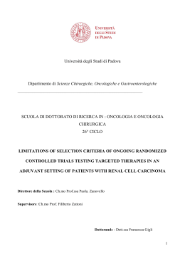
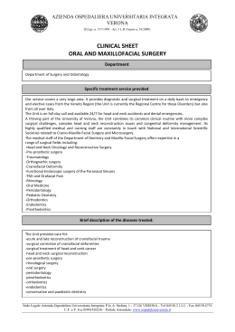
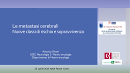
ACTA otorhinolaryngologica italica 2011;31:181-185
Case report
Basaloid squamous cell carcinoma of the larynx:
report of a early laryngeal cancer
Carcinoma squamocellulare basaloide della laringe: case report di uno stadio
precoce
F. Soprani, V. Armaroli, A. Stefanelli1, E. Emiliani1, D. Padovani, D. Casolino
Department of Otorhinolaryngology - Head and Neck Surgery, 1 Department of Radiation Oncology,
S. Maria delle Croci Hospital, Ravenna, Italy
Summary
Basaloid squamous cell carcinoma is a recently recognized, rare and aggressive variant of squamous cell carcinoma with a predilection to
occur in base of the tongue, hypopharynx and larynx (especially the supraglottic tract). It is usually diagnosed in advanced stage, frequently
with distant metastases, requiring aggressive surgical intervention. The prognosis is remarkably poor even after the association of radiotherapy or chemotherapy. Nevertheless recently it has been reported that in the early stage this neoplasm seems to have a behaviour less
aggressive, similar to conventional squamous carcinoma. The therapeutic approach is not clearly defined when the neoplasm is diagnosed
at an early stage. We present a case of early stage of basaloid squamous cell carcinoma of the supraglottic larynx.
Key words: Laryngeal tumours • Supraglottic larynx • Basaloid squamous cell carcinoma • Radiotherapy
Riassunto
Il carcinoma squamoso basaloide è una variante, recentemente codificata, rara ed aggressiva del carcinoma a cellule squamose con localizzazione preferenziale a livello della base lingua, ipofaringe e laringe (in particolar modo la porzione sovraglottica). Nella maggior
parte dei casi è diagnosticato in stadi avanzati, frequentemente con presenza di metastasi a distanza, che richiedono interventi chirurgici
demolitivi. La prognosi è solitamente infausta anche dopo associazione di terapia chirurgica e radioterapica. Recentemente, tuttavia,
secondo vari studi tale neoplasia negli stadi iniziali sembra avere un comportamento meno aggressivo, simile a quello del carcinoma squamocellulare classico. In tali stadi precoci l’approccio terapeutico non è ancora chiaramente definito. Presentiamo la nostra esperienza in
un caso di carcinoma basaloide del laringe ad uno stadio iniziale
Parole chiave: Tumori della laringe • Sopraglottide • Carcinoma squamoso basaloide • Radioterapia
Acta Otorhinolaryngol Ital 2010;31:181-185
Introduction
Basaloid squamous cell carcinoma (BSCC) is a highgrade variant of squamous cell carcinoma (SCC), with a
frequent localization in head and neck. The most frequent
sites of occurrence in the upper aero-digestive tract are
oropharynx (base of tongue), hypopharynx (pyriform sinus) and supraglottic larynx 1-4.
Although BSCC has predilection for cervico-facial region, it can also occur at other sites such as oesophagus,
lung, thymus, anus and cervix uteri 5-9.
Wain et al. recognized the BSCC as a separate variant of
SCC in 1986 and since then only few tens of cases of laryngeal localizations have been reported 10.
It affects mainly men in sixth or seventh decade of life, with
frequent cervical node metastases at presentation 11 12.
The clinical presentation is similar to other laryngeal carcinomas, but especially in supraglottic localizations the
diagnosis is of advanced laryngeal cancer classified by
American Joint Committee 8 13 14.
Metastases in loco-regional nodes are reported in 64% of
patients and disseminated spread (lung, liver, bone, brain
and skin) in 44% of cases 3.
The diagnosis is often delayed so that reasonable treatment options are radical surgery, chemoradiation or radiation alone (5 years survival is reported to be 17.5%) 13 14.
In early BSCC the neoplasm appears to have a behaviour
less aggressive similar to conventional SCC 15 16.
We would like to report a case of T1 stage BSCC of the
supraglottic larynx.
Case report
S.L., a 50 year-old woman, presented to our ENT department complaining a moderate hoarseness. She was a moder181
F. Soprani et al.
Fig. 1. Videolaryngoscopy image showing a polypoid mass of the right false
vocal fold.
Fig. 3. Negative computed tomography (CT) scan of neck.
ate smoker by several years; the medical history was negative. A videofiberlaryngoscopy revealed the presence of a
polypoid mass of the medial margin right (anterior third) of
the right false vocal fold, with an aspect of a papillomatous
like lesion (Fig. 1). The laryngeal mobility was normal as
well as the other portions of the upper aero-digestive tract.
The patient underwent direct microlaryngoscopy under
general anaesthesia with macroscopically wide excision
of the mass with sharp dissection and bleeding control
with monopolar needle dedicated instrument.
Histological examination revealed the presence of a BSCC
of the supraglottic larynx (Fig. 2).
A computed tomography (CT) scan of neck and thorax
was negative (Fig. 3). We have chosen CT rather than
Magnetic Resonance (MRI) considering that the patient
complained claustrophobia.
Also a total-body [18F]-2-fluorodeoxyglucose-PET
(FDG-PET) was negative for distant metastases.
The neoplasm was consequently classified as pT1N0M0
TNM stage.
For the choice of the best therapeutic strategy, considering
on one hand the aggressiveness of this kind of tumor and
on the other hand the very limited extension and the age
of the patient, we have decided, rather than for a surgical
option (partial reconstructive laryngectomy), to execute
an external radical radiotherapy.
After collegiate evaluation, the patient underwent postoperative treatment with external radical radiotherapy.
A CT simulation have been done, after immobilization of
the patient with thermoplastic mask, in supine position.
We have acquired slices of 5 mm thickness from temporomandibular joint to supraclavicular fossa.
The slices have been contoured using a treatment planning sistem Pinnacle 3 (ADAC Philips).
We have contoured a Clinical Target Volume (CTV), considering as CTV1 the larynx and as CTV2 the nodes of
II and III level bilaterally, following the Radiotherapy
Oncology Group (RTOG) consensus guidelines (RTOG
91-11).
A total dose 60 Gy for CTV1 and 44 Gy for CTV2 was
administered in 30 fractions, both at isocenter.
The treatment was not interrupted with good clinical tolerance; at the end of the therapy we reported Grade 1 mucositis of the larynx and a Grade 1 dermatitis of the neck
region (following the RTOG criteria) with a moderate
dysphonia, resolved in few weeks 17.
After a follow up period of 36 months the patient is free
from disease as confirmed by laryngeal endoscopy, CT
scan and FDG-PET.
Fig. 2. Histological examination revealing the typical aspects of a BSCC of
the supraglottic larynx.
182
Basaloid squamous cell carcinoma of the larynx
Discussion
The basaloid subtype of SCC represents less than 1% of
laryngeal carcinomas; to our knowledge since it was recognized in 1986 as a separate entity approximately 170
cases of BSCC were reported in literature and one fourth
of them were located in the larynx, with a predilection
of supraglottic tract; very few cases between these, cases
with exclusive supraglottic involvement diagnosed at early stage are very rare 15 16 18.
BSCC has typical histopathologic features; it is considered an highly malignant laryngeal tumor characterized
by areas of typical in situ and/or invasive SCC associated
with nests of small crowded cells. These cells have typically hyperchromatic nuclei, scant cytoplasm, small cystic
spaces, necrosis, prominent hyalinisation and peripheral
palisading. The overall appearance suggests an attempt of
differentiation toward glandular structures 10 13 19-22.
Many authors agree with Wain et al. that BSCC derives
from totipotential cells located at the base of the ciliated
columnar epithelium or in the proximal salivary gland
ducts, cells that are also precursor of squamous carcinoma, adenocarcinoma, oat cell carcinoma and reverse cell
carcinoma 10 19 23.
Differential diagnosis should mainly include conventional
SCC, small cell neuroendocrine carcinoma, adenoid cystic carcinoma, basal cell adenocarcinoma, adenosquamous
carcinoma, spindle cell squamous carcinoma, mucoepidermoid carcinoma and adenoid squamous cell carcinoma 1 8 13 15 24.
Differential diagnosis is relevant because BSCC differ in
biological behaviour, prognosis and therapeutic management 1 8 11 14 15 24 25.
Even if there is some report that BSCC can be diagnosed
by fine needle aspiration, it is well recognized the importance of obtaining a biopsy specimen as deep and wide as
possible (because basaloid cells could not be included in
the specimen and so it should be difficult to distinguish
especially between adenoid cystic carcinoma and small
cell undifferentiated carcinoma) 3 11 24 25.
Endoscopically BSCC is indistinguishable from the common forms of squamous cancer or, such happened in our
case, even from a papillomatous mass.
The tumor borders are indiscreet because of their infiltrative growth beneath the surface epithelium; this feature
make the neoplasm often deeply invasive and consequently
multifocal or metastatic even at initial presentation 8 13 19 23.
Becker et al. in 1998 observed in cases of BSCC a distinct radiological aspect at Magnetic Resonance Imaging
(MRI) consisting in a typical “lobulated” enhancement
pattern on contrast-enhanced T1-weighted Spin Echo images; the authors did not observed this particular aspect in
the common form of SCC, which typically shows a relatively homogenous enhancement with or without associated area of ulceration or necrosis 23.
Another important instrument that can be helpful in detecting, staging and following BSCC of the head and neck
area is represented by FDG-PET.
Kunkel et al. demonstrated that FDG-PET screening was
able to depict a clinically and radiologically invisible site
of recurrence allowing for salvage surgery and that normalized posttreatment glucose metabolism was predictive for tumor control even in the “high risk population of
BSCC patients” 26.
After the diagnosis and staging the suggested treatment in
literature is a combination of radical surgery followed by
radiation and chemotherapy, especially in presence of metastases, considering that most of reports agree that BSCC
is a biologically high grade malignancy with a tendency
for locally aggressive behaviour and early regional and
distant metastases, with a poor prognosis 1 4 8 10-16 18-20 24 27.
Ferlito, furthermore, found that this neoplasm show a tendency for the development of second primary tumors in
the upper aerodigestive tract 13.
For Erdamar et al. the survival rate of BSCC was poorer
(even if not statistically significant) compared with conventional SCC; they determined moreover that, in comparison with the control group, for patients with supraglottic T2 disease and undergoing partial laryngectomy
exists a statistically significant differences for local recurrences with the consequence that conservative laryngeal surgery should be employed only with great caution.
They concluded that postoperative irradiation should be
encouraged as an appropriate therapy against local aggressivity of tumor; in addition to local control, adjuvant
chemotherapy should also be considered for prevention of
distant metastases 27.
The poor prognosis of BSCC justifies an aggressive treatment: more specific antibasaloid chemotherapeutic drugs
should be sought 18.
Despite these studies referring to BSCC such us a particularly aggressive pathology, some other authors suggest that behaviour of BSCC, especially in early stage, is
similar to conventional squamous cell carcinoma.
A study by Luna et al. revealed that notwithstanding its
aggressiveness, the biologic course of BSCC is similar to
that of conventional SCC when clinical stage, site, and
treatment are matched, emphasizing in particular that survival and incidence of cervical lymph node metastases
were similar to those of conventional SCC 28.
The concept that an early stage of tumor may be regarded
as a different category and treated more conservatively is
also emphasized in three others studies 15 20 29.
For Sheen et al. although BSCC of the upper aerodigestive tract is aggressive, an early glottic type of tumor may
be regarded as a different category and treated more conservatively 29.
In its study Erisen et al. found that their early stage cases
with no evidence of neck nodes metastases (N0) had a good
prognosis. Even if their cases with advanced stage cancer
183
F. Soprani et al.
with pathologically N+ necks had poor prognosis despite
aggressive combined treatment, the authors believe that
early stage BSCC act similarly to their counterparts in conventional SCC, justifying a more conservative treatment.
In the study of Alkan et al., in contrast to the literature, the
two cases of BSCC of the larynx reported were both young
and had no metastatic spread to cervical nodes even with
a presentation in advanced stage (both N0); after a follow
up of at least 24 months, following a surgical and radiant
treatment, the patients are free of disease 20.
Because most cases of BSCC present with advanced stage
tumor it is generally believed that histological features
were reflective of the neoplasm’s aggressive clinical behaviour and poor prognosis.
Very few cases of early stage BSCC are described in
literature and at our knowledge there is not a sufficient
number of case to evaluate the prognosis of this initial
neoplasia; greater number of patients must be studied
comparing BSCC and conventional SCC matching site of
occurrence, stage and age of presentation.
References
Paulino AF, Singh B, Shah JP, et al. Basaloid squamous cell carcinoma of the head and neck. Laryngoscope
2000;110:1479-82.
1
Campman SC, Gandour-Edwards RF, Sykes JM. Basaloid
squamous carcinoma of the head and neck. Report of a case
occurring in the anterior floor of the mouth. Arch Pathol Lab
Med 1994;118:1229-32.
2
3
Raslan WF, Barnes L, Krause JR, et al. Basaloid squamous
cell carcinoma of the head and neck: a clinicopathologic
and flow cytometric study of 10 new cases with review of
the English literature. Am J Otolaryngol 1994;15:20411.
Sheen Ts, Chang Y, Ko J, et al. Basaloid squamous cell
carcinoma of the larynx. Otolaryngol Head Neck Surg
1999;121:647-50.
4
Abe K, Sasano H, Itakura Y, et al. Basaloid-squamous carcinoma of the esophagus. A clinicopathologic, DNA ploidy,
and immunohistochemical study of seven cases. Am J Surg
Pathol 1996;20:453-61.
cell carcinoma of the larynx and hypopharynx. Ann Otol
Rhinol Laryngol 1997;106:1024-35.
Ferrario F, Spriano G, Macchi A, et al. Basaloid-squamous
cell carcinoma of the larynx: a new morphologic entity. Case
report. Acta Otorhinolaryngol Ital 1998;18:338-41.
14
15
Kruslin B, Tomasovic C, Cupic H, et al. Basaloid squamous
cell carcinoma of the larynx: report of two cases. Croat Med
J 1998;39:450-2.
16
Cox JD, Stetz J, Pajak TF. Toxicity criteria of the Radiation
Therapy Oncology Group (RTOG) and the European Organization for Research and Treatment of Cancer (EORTC). Int
J Radiat Oncol Biol Phys 1995;31:1341-6.
17
Bahar G, Feinmesser R, Popovtzer A, et al. Basaloid squamous
carcinoma of the larynx. Am J Otolaryngol 2003;24:204-8.
18
5
Ferry JA, Scully RE. “Adenoid cystic” carcinoma and adenoid basal carcinoma of the uterine cervix. A study of 28
cases. Am J Surg Pathol 1988;12:134-44.
6
Khaldi L, Apostolidis TCh, Pappa DA, et al. Basaloid squamous carcinoma of the larynx. A potential diagnostic pitfall.
Ann Diagn Pathol 2006;10:297-300.
19
Alkan S, Coskun BU, Ugur S, et al. Basaloid squamous cell
carcinoma of the larynx. Auris Nasus Larynx 2006;33:71-4.
20
Unal M, Polat A, Gorur K, et al. Basaloid squamous carcinoma of the larynx and hypopharynx: immunohistopathologic
features of two cases. J Otolaryngol 2005;34:212-5.
21
Moro D, Brichon PY, Brambilla E, et al. Basaloid bronchial
carcinoma. A histologic group with a poor prognosis. Cancer
1994;73:2734-9.
22
Zbaren P, Nuyens M, Stauffer E. Basaloid squamous cell carcinoma of the head and neck. Curr Opin Otolaryngol Head
Neck Surg 2004;12:116-21.
23
7
Salerno G, Di Vizio D, Staibano S, et al. Prognostic value of
p27Kip1 expression in basaloid squamous cell carcinoma of
the larynx. BMC Cancer 2006;6:146.
8
Vincent-Salomon A, de la Rochefordiere A, Salmon R, et al.
Frequent association of human papillomavirus 16 and 18
DNA with anal squamous cell and basaloid carcinoma. Mod
Pathol 1996;9:614-20.
9
10
Wain SL, Kier R, Vollmer RT, et al. Basaloid-squamous carcinoma of the tongue, hypopharynx, and larynx: report of 10
cases. Hum Pathol 1986;17:1158-66.
Barnes L, Ferlito A, Altavilla G, et al. Basaloid squamous
cell carcinoma of the head and neck: clinicopathological
features and differential diagnosis. Ann Otol Rhinol Laryngol 1996;105:75-82.
11
Akyol MU, Seckin S, Akbayrak L, et al. Basaloid squamous
cell carcinoma of the larynx. Eur Arch Otorhinolaryngol
1995;252:485-7.
12
Ferlito A, Altavilla G, Rinaldo A, et al. Basaloid squamous
13
184
Erisen LM, Coskun H, Ozuysal S, et al. Basaloid squamous
cell carcinoma of the larynx: a report of four new cases.
Laryngoscope 2004;114:1179-83.
Becker M, Moulin G, Kurt AM, et al. Atypical squamous cell
carcinoma of the larynx and hypopharynx: radiologic features
and pathologic correlation. Eur Radiol 1998;8:1541-51.
Morice WG, Ferreiro JA. Distinction of basaloid squamous
cell carcinoma from adenoid cystic and small cell undifferentiated carcinoma by immunohistochemistry. Hum Pathol
1998;29:609-12.
24
Sato M, Horiuchi M, Ogata T, et al. Basaloid squamous carcinoma of the larynx: report of a case. Auris Nasus Larynx
1997;24:417-22.
25
Kunkel M, Helisch A, Reichert TE, et al. Surveillance of
basaloid oral squamous cell carcinoma: the value of [18F]
FDG-PET. Oral Oncol 2004;40:56-62.
26
27
Erdamar B, Suoglu Y, Sirin M, et al. Basaloid squamous
cell carcinoma of the supraglottic larynx. Eur Arch Otorhinolaryngol 2000;257:154-7.
Basaloid squamous cell carcinoma of the larynx
Luna MA, el Naggar A, Parichatikanond P, et al. Basaloid
squamous carcinoma of the upper aerodigestive tract. Clinicopathologic and DNA flow cytometric analysis. Cancer
1990;66:537-42.
28
29
Sheen Ts, Chang Y, Ko J, et al. Basaloid squamous cell
carcinoma of the larynx. Otolaryngol Head Neck Surg
1999;121:647-50.
Received: March 18, 2010 - Accepted: August 20, 2010
Address for correspondence: Prof. D. Casolino, Divisione di Otorinolaringoiatria e Chirurgia Testa e Collo, Ospedale S. Maria delle
Croci, viale Randi 5, 48121 Ravenna, Italy. Fax: +39 0544 285818.
E-mail: [email protected]
185
Scaricare