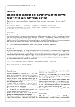
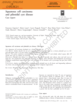
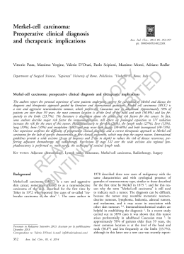
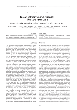
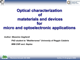
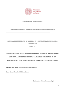
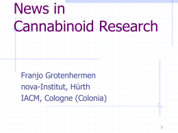
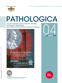
Published in IVIS with the permission of SIVE
Published in IVIS with the permission of SIVE
Close window to return to IVIS
9° CONGRESSO NAZIONALE MULTISALA SIVE
PISA, 1-2 FEBBRAIO 2003
Derek C. Knottenbelt
BVM&S, DVM&S, MRCVS
Philip Leverhulme Hospital
University of Liverpool, UK
Tumori della cute:
il carcinoma squamocellulare
Skin neoplasia: squamous cell carcinoma
Domenica, 2 Febbraio 2003, 11.30
nd
Sunday, February 2 2003, 11.30
SALA PACINOTTI
MEDICINA INTERNA
Chairperson: Francesca Abramo
This manuscript is reproduced in the IVIS website with the permission of SIVE www.sive.it 123
Proceedings of the Annual Meeting of the Italian Association of Equine Veterinarians, Pisa, Italy 2003
Published in IVIS with the permission of SIVE
Close window to return to IVIS
Riassunto
Il carcinoma squamocellulare è al terzo posto in ordine di prevalenza fra i tumori cutanei degli equini (dopo il sarcoide ed il melanoma). Nella maggior parte dei casi, la condizione si riscontra a livello della cute delle palpebre, del muso e del perineo, ma formazioni importanti si
osservano anche a livello del pene dei maschi castrati (di solito risultano colpiti con maggiore
frequenza gli animali più anziani). La razza Clydesdale sembra essere predisposta all’insorgenza della malattia in tutti i siti.
Esiste una stretta relazione fra carcinoma squamocellulare e cute non pigmentata e, più in particolare, nella zone in cui la cute è esposta alla luce solare. Tuttavia, in alcune sedi possono essere coinvolti altri agenti cancerogeni (ad es., lo smegma causa formazioni peniene e clitoridee,
rispettivamente, nei maschi castrati anziani e nelle fattrici). Le neoformazioni perianali e quelle delle labbra vulvari sono relativamente comuni, ma risulta difficile ipotizzare che in questa
sede il problema sia causato dallo smegma o dalla luce solare.
La localizzazione più comune è indubbiamente rappresentata dall’occhio e dalla cute periorbitale. Le palpebre sono spesso colpite nei cavalli con occhi azzurri o colorati o Cremello e la
terza palpebra non pigmentata e la congiuntiva bulbare limbare laterale costituiscono una sede
comune in tutti i tipi di equini. Il carcinoma in situ è un carcinoma squamocellulare ristretto all’epitelio corneale superficiale.
Alterazioni precarcinomatose si possono spesso riconoscere sulla cute del pene e delle palpebre (dove è utile un test di ricerca del sangue occulto nelle lacrime); in questi casi l’adozione
di semplici misure come l’igiene prepuziale o l’applicazione di un paraocchi può prevenire del
tutto la comparsa del tumore.
Si riconoscono due forme principali, una ulcerativa o distruttiva ed una proliferativa superficiale, che esita in una massa tumorale in espansione. La prima può essere molto distruttiva per
i tessuti e sono comuni i sanguinamenti e le infezioni secondarie. Questo tipo è più frequente a
livello di palpebre e muso. Le forme proliferative sono comuni sulla terza palpebra, sul pene e
sulle labbra vulvari e sul clitoride.
La malignità non è una caratteristica comune delle forme cutanee, mentre è molto più probabile in quelle gastriche ed in quelle che si localizzano a livello di bocca, faringe e cavità nasale. I carcinomi squamocellulari del pene nei cavalli castrati più giovani (< 10 anni di età) sono
di gran lunga più frequentemente maligni di quelli che si verificano nei cavalli castrati più anziani nella stessa sede. Sono state descritte forme polmonari secondarie e generalizzate in cui
erano presenti gravi effetti secondari sia locali che sistemici. In un caso relativo ad un pony anziano portato alla visita perché presentava atassia/paraplegia dei quarti posteriori è stata descritta una diffusione metastatica ai corpi vertebrali. La forma gastrica è di solito altamente aggressiva e si ha una disseminazione secondaria direttamente attraverso ed all’interno del peritoneo e del fegato ed ematogena a fegato, polmoni e, da qui, ad altri organi principali.
Le localizzazioni sono rappresentate da:
1. Cute
a. Muso
b. Perineo
c. Pene
d. Vulva
e. Clitoride
2. Palpebre/occhi
a. Congiuntiva
b. Cornea
Proceedings of the Annual Meeting of the Italian Association of Equine Veterinarians, Pisa, Italy 2003
Published in IVIS with the permission of SIVE
Close window to return to IVIS
3. Forme interne
a. Faringee
b. Nasali
c. Orali
d. Gastriche
Il trattamento è sempre difficile, ma almeno la maggior parte delle forme che risultano accessibili ha una natura più benigna e può essere più suscettibile di escissione chirurgica. Il carcinoma squamocellulare è sempre molto sensibile alle radiazioni gamma e beta e, quando risulta fattibile e disponibile, questa terapia può essere utilizzata come protocollo di trattamento efficace. Spesso, tuttavia, la chirurgia rappresenta l’unica opzione e l’escissione meticolosa risulta frequentemente curativa. Recentemente, l’uso di cisplatino e 5-fluorouracile ha offerto una
nuova possibilità ma, anche così, esistono molti casi in cui le difficoltà di accesso o di realizzazione pratica precludono il trattamento.
Le forme palpebrali e peniene/clitoridee e vulvari rappresentano i migliori candidati al trattamento chirurgico. In alcune localizzazioni, è anche attuabile la criochirurgia, ma risulta difficile trattare totalmente l’area coinvolta.
La prognosi per la maggior parte delle forme è buona, ma di solito si consiglia di adottare un
approccio riservato, data la difficoltà di garantire l’ablazione totale del tumore. Le associazioni
di chirurgia e radioterapia o farmaci antimitotici topici di solito rappresentano la migliore soluzione. L’amputazione completa o parziale del pene è un trattamento molto efficace, a condizione che non vi sia alcun coinvolgimento dell’anello prepuziale o dei linfonodi iliaci.
Proceedings of the Annual Meeting of the Italian Association of Equine Veterinarians, Pisa, Italy 2003
Published in IVIS with the permission of SIVE
Close window to return to IVIS
Summary
Squamous cell carcinoma is the third most prevalent skin tumour of horses (behind sarcoid
and melanoma). The condition is most often found in the skin of the eyelids, face and perineum
but important forms occur on the penis of geldings (usually older aged animals are more often
affected). The Clydesdale bred seems to be predisposed to the disease at all sites.
There is a close relationship between SCC and non-pigmented skin and more particularly
where the skin is exposed to sunlight. However in some locations other carcinogens may be involved (e.g. smegma causes penile and clitoral forms in older geldings and mares respectively).
Perianal forms and vulvar labial forms are relatively common but it is hard to visualise smegma or sunshine causing the problem here.
The commonest site is undoubtedly the eye and periorbital skin. The eyelids are often affected in wall eyed or coloured or Cremello horses and non-pigmented third eyelid and lateral limbal bulbar conjunctiva is a common site in all types of horses. Carcinoma in situ is a SCC that
is restricted to the superficial corneal epithelium.
Pre-carcinomatous changes can often be recognised on penile skin and the eyelids (in the latter cases a lacrimal occult blood test is helpful) and in these cases simple measures including
preputial hygiene or blinkers may prevent the tumour appearing at all.
Two major forms are recognised. These include an ulcerative or destructive form and a superficial proliferative form that results in an expanding tumour mass. The former can be very
destructive of tissue and bleeding and secondary infection is common. This type is commonest
on the eyelids and the face. The proliferative forms are common on the third eyelid, the penis
and the vular lips and clitoris.
Malignancy is not common in the cutaneous forms but the gastric form and those forms
that occur in the mouth, pharynx and nasal cavity are much more likely to be malignant.
Penile SCC in younger geldings (<10 years of age) are far more often malignant that
those that occur in older geldings at the same site. Secondary pulmonary and generalised
forms have been described in which there have been serious secondary effects both locally
ands systemically. Metastatic spread to the vertebral bodies have been reported in a case
of an old pony that was presented for hindquarter ataxia/paraplegia. The gastric form is
usually highly aggressive and secondary dissemination occurs directly across and into the
peritoneum and liver and haematogenously to the liver, lungs and thence to other major organs.
Locations include:
1. Skin
a. Face / Muzzle
b. Perineum
c. Penis
d. Vulva
e. Clitoris
2. Eyelids / Eyes
a. Conjunctiva
b. Cornea
3. Internal forms
a. Pharyngeal
b. Nasal
c. Oral
d. Gastric
Proceedings of the Annual Meeting of the Italian Association of Equine Veterinarians, Pisa, Italy 2003
Published in IVIS with the permission of SIVE
Close window to return to IVIS
Treatment is always difficult but at least most forms that are accessible have a more benign
nature and may be amenable to surgical excision. SCC is always very sensitive to gamma and
beta radiations and so where these are feasible and available this is an effective treatment protocol. Often however, surgery is the only option and then meticulous excision is often curative.
Recently the use of cisplatin and 5-fluorouracil have provided a new possibility but again there
are many cases in which access or practicality preclude treatment.
Eyelid forms and penile / clitoral and vulvar forms are the best candidates for surgical
treatment. Cryosurgery is also feasible in some locations but it is difficult to treat the total area
involved.
The prognosis for most forms is good but a guarded approach is usually advised because of
the difficulty of ensuring total ablation of the tumour. Combinations of surgery and radiation
or topical antimitotic drugs usually provide the best outlook. Complete or partial amputation
of the penis is a very effective treatment provided that there is no involvement of the preputial
ring or the iliac lymph nodes.
Proceedings of the Annual Meeting of the Italian Association of Equine Veterinarians, Pisa, Italy 2003
Published in IVIS with the permission of SIVE
Primary squamous cell carcinoma is a common tumour of horses that is restricted to
squamous epithelium. Within the broad pathological group there is wide variation in the
prevalence of each type. The histological
characteristics are typical and easily recognised and the extent of malignancy is likewise
easily recognised by the extent of differentiation and the character / normality (or otherwise) of the keratin production. The less aggressive the tumour, the greater the normality
and the greater the extent of production of keratin. Highly malignant forms produce virtually no keratin and have a particularly aggressive nature with rapid metastatic spread via
blood and lymphatics to either remote or organs or to the local draining lymph node.
Solar radiation, geographical location, and
individual susceptibility (which may relate to
breed, age and lack of adnexal pigment) are
probably the most important risk factors associated with SCC. Predispositions to squamous
cell carcinoma occur within breeds (the
Clydesdale Horse is probably predisposed)
and within colour variations.
There is little doubt amongst clinicians that
depigmented skin exposed to carcinogens
such as smegma (in the case of penile and clitoral forms) and sunlight (in cutaneous and
Close window to return to IVIS
ocular forms) are implicated but there are no
scientific studies that confirm this widely held
belief. Certainly most clinicians will recognise that certain animals including coloured
and cream ponies are predisposed. Furthermore smegma is a likely carcinogen in geldings because breeding stallions appear to be
much less likely to succumb to the penile
forms than aged geldings.
Sites:
Integumentary
1. Facio-nasal
2. Palpebral
3. Conjunctival
4. Corneal
5. (Perineal)
Alimentary Tract
1. Oro-palatine
2. Oro-Pharyngeal
3. Gastric
4. Anal
Respiratory Tract
1. Nasal
2. Paranasal sinuses
3. Nasopharynx
Reproductive tract
1. Penile
2. Vulval
3. Clitoral
Perianal squamous cell carcinoma. Note the non-pigmented skin.
Forms: From a purely clinical perspective
three major forms of the disease are recognised.
1. Proliferative forms in which the tumour
develops in a proliferative fashion. A
common site for this type is the penile
form in older geldings and the carcinoma in situ on the cornea.
2. Ulcerative / Destructive forms that result
in tissue loss and erosions and ulcerations.
These are commonly found for example
on the non-pigmented eyelids and third
eyelids. It is also the predominate form in
perianal or perineal forms of the disease
(as opposed to the vulvar form in mares)
Proceedings of the Annual Meeting of the Italian Association of Equine Veterinarians, Pisa, Italy 2003
Published in IVIS with the permission of SIVE
3. Combined ulcerative / Destructive forms
are actually quite common and are typified by the vulvar or clitoral forms.
Younger geldings are liable to this form
and where it occurs in horses under 10
years of age there is a very high degree
of malignancy associated with it.
OCULAR AND PERIOCULAR
SQUAMOUS CELL CARCINOMA
Squamous cell carcinoma (SCC) occurs in
the eyelids, the conjunctiva (particularly of
the third eyelid and the lateral limbus of the
eye) and on the surface of the cornea (carcinoma in situ). Palpebral SCC (pSCC) is often
extensive, highly destructive, and invasive.
However, a milder proliferative, slower growing form is sometimes encountered. Non-pigmented eyelids are much more liable to develop the destructive and ulcerative form and certain breeds seem more liable to its development. Although for the most part SCC of the
eyelids and orbital structures are very seldom
malignant they can cause extensive destruction or infiltration and so treatment becomes
progressively more problematical.
The least common ocular forms include a
lateral limbal form that is invariably proliferative (at least until the condition is advanced)
and a carcinoma in situ where the corneal epithelium is involved. The latter are sometimes
difficult to diagnose. Usually cases are pre-
Early ulcerative (white arrow) and proliferative palpebral carcinoma (open arrow).
Close window to return to IVIS
sented for investigation of ocular discharges
(usually with a characteristic purulent appearance).
Clinical diagnosis of the condition is usually
simple when the changes are advanced but in the
early stages lacrimation is a feature and testing
the tears with a urine blood dipstick usually reveals blood. Sometimes the lacrimation is plainly haemorrhagic. Pathological diagnosis of all
the forms is easily achieved by biopsy but
some sites are not amenable to excision of large
pieces of tissue so pathologists need to be informed of the nature of the lesion and the location of the biopsy within it.
Severe ulcerative (destructive) carcinoma of the lower lid
and caruncle. The tumour also involved the third eyelid
and was treated successfully with gamma radiation.
A typical proliferative carcinoma on the nictitans. This
was treated successfully by surgical excision of the affected area leaving a distorted but functional nictitans.
Proceedings of the Annual Meeting of the Italian Association of Equine Veterinarians, Pisa, Italy 2003
Published in IVIS with the permission of SIVE
A severe highly destructive carcinoma on the muzzle. This
is only treatable by radiation and even then there is likely to be gross deformity. (Slide courtesy of E.R Singer)
CUTANEOUS SQUAMOUS CELL
CARCINOMA
Close window to return to IVIS
Pre-carcinomatous changes on the penile skin of an aged
gelding (left) and a single smaller, haemorrhagic carcinoma on the penile skin of a younger horse. Both were
successfully treated. Hygiene forms a major part of the
management of these forms.
Cutaneous forms of squamous cell carcinoma
occur in the facial region, the ears and the perineal skin (see Figure above) . The large majority of these are ulcerative / destructive forms and
the site and extent of tissue loss at the time of
initial presentation dictate the prognosis.
PENILE SQUAMOUS CELL
CARCINOMA
A pre-cancerous change in the skin of the
penis and the glans may be detectable – usually such cases are presented for investigation of a preputial discharge [often containing some (stale) blood]. Penile squamous
cell carcinoma is recognised in two major
forms that have different implications for
prognosis and treatment.
The common form occurs in old geldings
– stallions are very rarely affected and this
suggests that smegma may be a significant
carcinogen. The lesions may be proliferative
or ulcerative / destructive or there may be a
combination of the two. The glans is most
often affected while the preputial reflection
and the skin of the shaft of the penis may
sometimes be involved. Lesions may be
multiple and may be ulcerated or proliferative.
Malignant squamous cell carcinoma of the glans in a 9 year
old gelding. There was iliac
lymph node involvement and
the shaft of the penis felt
“wooden”. Treatment was not
attempted.
In younger horses (< 10 – 14 years of age)
there is a much higher malignancy rate and
these cases are often beyond treatment on
presentation. The lesions are often highly malignant in appearance and often the glans is
the primary lesion.
The more malignant forms and those that
have a very chronic course may sometimes
“seed” into the skin at the preputial ring or
preputial orifice. These lesions are also very
aggressive and they may occur independently
of the penile forms. The prognosis for these
cases is extremely poor and en bloc resection
of the penis and the prepuce with perineal ure-
Proceedings of the Annual Meeting of the Italian Association of Equine Veterinarians, Pisa, Italy 2003
Published in IVIS with the permission of SIVE
Close window to return to IVIS
throstomy is the only therapeutic course of action to take. The site may become strictured
by a combination of fibrous tissue scarring
and tumour expansion and so the first signs of
this complication may be difficulty with urination.
erative (at least to some degree) facial distortions and swellings may be detected. These
forms often have an aggressive destructive
nature and so it is possible to have a large
proliferative lesion with extensive surrounding destruction.
VULVAR / CLITORAL SQUAMOUS
CELL CARCINOMA
GASTRIC CARCINOMA
Vulvar or clitoral forms occur almost exclusively in older mares. It is suggested that
the carcinogenic properties of smegma may
be involved in the development of the clitoral
form and at this site carcinomas can develop
on pigmented skin. The vulvar forms are usually proliferative when they develop within
the vestibule and ulcerative when they involve
the lips of the vulva. The clitoral form is invariably proliferative. Treatment is usually a
combination of radiation (where feasible and
available) and surgical excision. Topical 5fluorouracil (5% cream) has been used successfully but it is very slow and can be an uncomfortable and therefore difficult treatment
to complete. Intralesional cisplatin has also recently gained some favour but the logistics of
this material are difficult and a stable emulsion must be used. The aqueous solutions or
other solutions are not likely to be effective
and in any case require to be repeated frequently over some weeks or months.
MUCOSAL SQUAMOUS CELL
CARCINOMA
Squamous cell carcinoma can occur in any
squamous epithelium. The commonest sites
for these forms are in the oral and nasal cavities (including the paranasal sinuses) and the
in the pharynx. The main presenting signs include chronic serosanguinous / fetid nasal
discharge or oral bleeding with the possibility of secondary signs including nasal obstruction in the nasal forms and dysphagia in the
oral forms. However, in spite of an aggressive tumour the clinical signs may not be obvious. Because these forms are usually prolif-
Gastric squamous cell carcinoma by definition can only occur in the oesophageal (squamous) portion of the stomach. The suggested
carcinogens include recurrent gastric infestation with Habronema and Draschia spp.
worms or Gastrophilus spp (Bots) but there
are many horses with high infestations of all
these that never develop any pathology let
alone carcinoma. There is no evidence in
horses of a bacterial carcinogen. Usually the
lesions are a combination of proliferation and
destruction and this is probably the most aggressive for of the condition in horses. There
is a high rate of direct spread to the liver and
secondary metastatic spread to the lungs. The
initial signs are not usually obvious and so on
presentation these cases are usually clinically
ill with weight loss, recurrent anterior abdominal pain and inappetance. They may be marginally anaemic but usually recurrent colic,
weight loss and a harsh dry coat are the main
signs. Because these signs are often attributed
to other conditions and because a long endoscope is required for diagnosis cases are often
presented late in the course. It is sometimes
possible to obtain a tentative diagnosis from
gastric washings (containing both blood and
abnormal squamous cells) or from peritoneal
fluid (containing blood, protein and abnormal
squamous cells that should definitively never
appear in peritoneal fluid) usually in the latter
cases there are reactive mesothelial cells also
and so the diagnosis may be confused with
mesothelioma or simply a reactive peritoneal
cavity.
Management:
Established SCC is usually locally aggressive and the prognosis is poor if periocular
and/or orbital tissues have been invaded. Squa-
Proceedings of the Annual Meeting of the Italian Association of Equine Veterinarians, Pisa, Italy 2003
Published in IVIS with the permission of SIVE
mous cell carcinoma of the upper and lower
eyelids can be treated by surgical excision/debulking combined with cryotherapy (usually a
double freeze-thaw cycle with liquid nitrogen)
or brachytherapy (usually via a strontium-90
applicator).
Note:
Laser therapy has been used to treat a
variety of equine eyelid tumours, but
that it is too early to assess the overall
long-term success rate.
Squamous cell carcinoma (SCC) occurs in
the eyelids, the conjunctiva (particularly of
the third eyelid and the lateral limbus of the
eye) and on the surface of the cornea (carcinoma in situ). Palpebral SCC (pSCC) is often extensive, highly destructive, and invasive. However, a milder proliferative, slower growing form is sometimes encountered.
Non-pigmented eyelids are much more liable to develop the destructive and ulcerative form and certain breeds seem more liable to its development. Although for the
most part SCC of the eyelids and orbital
structures are very seldom malignant they
can cause extensive destruction or infiltration and so treatment becomes progressively
more problematical.
Interestingly a particularly invasive form of
SCC occurs in the nasal chambers, the
paranasal sinuses and the hard palate. The former are characterised by a combined form of
proliferation and expanding destruction. This
is a common cause of exophthalmos in aged
horses and of oral and dental difficulties in all
ages of horse.
The management of squamous cell carcinoma relies upon surgical removal or cryonecrosis, where these are feasible or to the application of various antimitotic and cytotoxic
chemicals. Radiation is the gold standard of
therapy in suitable sites where surgical removal cannot be contemplated or where the
tumour margins cannot be defined sufficiently
to allow total removal. There are no immunological methods for the management of this
tumour type.
Close window to return to IVIS
a) Benign neglect is ill advised for this
condition. Those tumours that are small and
benign may however be observed carefully at
regular intervals. Extension into the orbit from
the paranasal sinuses or nasal chambers is
usually a bad prognostic sign and surgery becomes increasingly more difficult.
b) Surgical excision: This is a realistic
proposition for nictitans and conjunctival
forms and for most cases of early penile carcinoma; indeed it is the treatment of choice for
all defined types of carcinoma at all sites but
the criteria for selection of this method are
strict. Surgical removal of corneal carcinoma
in situ is extremely difficult requiring microsurgical techniques involving a superficial
keratectomy of the affected area and a margin
of 1 – 2 mm. Eyelid tumours are seldom
amenable to this option. But very small, localised proliferative lesions can be excised if
the margin of the lid can be preserved or can
be reconstructed satisfactorily to preclude the
complications arising from poor eyelid function. Surgical ablation should not be contemplated if the tumour has already invaded the
local lymph node or there are difficulties with
margin definition. Surgical amputation of the
penis in suitable cases is curative and recurrences are rare provided that the surgery is
properly performed and that all the tumour tissue is removed. Attempts at en bloc resection
for cases of aggressive carcinoma carry a correspondingly poor prognosis because the surgery is a temporary salvage procedure in most
cases. Some sites of course totally preclude
surgical removal. Clitoral and some vulvar
forms can be surgically removed with a reasonable prognosis.
c) Cryosurgery: Cryonecrosis is a fast but
rarely effective method of treatment for eyelid
lesions and small lesions elsewhere. Nasal lesions and some oral lesions may be managed in
this way but the results are dependent upon the
surgical efficiency of total tumour ablation.
Thermocouple assistance is obligatory. Lesions
that are ulcerated and which bleed significantly
can be managed by careful cryosurgery. Nictitans, limbal SCC, and carcinoma in situ have all
Proceedings of the Annual Meeting of the Italian Association of Equine Veterinarians, Pisa, Italy 2003
Published in IVIS with the permission of SIVE
been treated by cryosurgery with variable successes. The secondary effects of the freeze may
be very serious and so its use should be strictly
restricted to small eyelid lesions and to limbal
superficial lesions. There are areas of course
where this is totally impossible.
d) 5-Fluoro-uracil: The value of this chemical in ointment formulation has been described
for oral SCC (Patterson, 1997) but there are no
reports of its use in the eyelids or for ocular lesions. It would seem that this is an area that
could have some prospect. A 5% solution of 5F-U has been used by the author to treat a small
limbal carcinoma that regrew at the site of a
previous surgical excision but the true nature of
the lesion (carcinoma V granuloma) was never
established pathologically. Topical 5-FU requires persistent application over some weeks
or even longer and highly invasive or aggressive lesions do not seem to respond effectively
in the time available. The commercial ointment
is 5% but it may be that this is plainly too weak
to control a large or difficult lesion.
e) Cisplatin: Intralesional infiltration of an
emulsion of cisplatin in almond (or sesame)
oil or a stable emulsion containing a higher
concentration of cisplatin can be used. It is effective in some cases although there are few
reports of efficacy compared to other standard
methods. The method is difficult logistically
because the material needs to be injected intralesionally in a stable form and 1 mg cisplatin needs to be delivered to 1 ml of tumour
and so the volume and concentration of the
chemical are critical. A stable emulsion is difficult to obtain in some cases because of the
lack of water solubility of the chemical and
the need to use an aqueous solution.
f) Immunomodulation using BCG: The author has had little success with this approach,
in stark contrast to its value in the treatment of
bovine ocular and periocular SCC. The reasons for this are not clear. In any case it is not
of any value in sites away from the eye.
g) Radiation: Radiation treatment is probably the best available option for all forms of
Close window to return to IVIS
SCC in the horse but in the periorbital region
there are logistic complications that significantly reduce its make its value much less.
SCC is very susceptible to both gamma and
beta radiation. Gamma radiation is used ex192
tensively (via linear iridium sources) in Liverpool for the treatment of palpebral forms of
SCC. Beta radiation delivered via a Strontium
90
plaque has also had considerable benefit on
limbal. Nictitans and carcinoma in situ lesions. The cosmetic effects of the treatment
are remarkably good with a relatively rapid
resolution and minimal secondary complications even in severely destructive eyelid cases.
The relatively poor penetration of beta radiation (limited to 1 – 2 mm deep) means that
surgical treatment may need to be applied before application of radiation. As is always the
case every piece of tissue removed from a
surgical site should be submitted for
histopathology so that rational treatment/dose
calculations can be made. Where ever possible this option should be considered to be the
definitive method and is a useful fall back
method when others are either not feasible or
fail to resolve the problem.
Prognosis:
The prognosis for squamous cell carcinoma
is dependent on:
1. The site of the lesion and its suitability
for any effective therapy
2. The malignancy of the tumour itself
3. The duration of the lesion and failed attempts to treat it
4. The extent of secondary consequences
arising from the tumour.
For the most part SCC in the horse has less
malignancy than in most other species but
gastric carcinoma, oropharyngeal carcinoma
and penile carcinoma in younger geldings are
usually (but not invariably!) far more malignant than the others. The tumour may metastasise by haematogenous or lymphatic extension but at some sites (especially the stomach
forms) spread may be by contiguous expansion into adjacent organs. Thus the gastric
form may extend into the liver and cause severe liver damage.
Proceedings of the Annual Meeting of the Italian Association of Equine Veterinarians, Pisa, Italy 2003
Published in IVIS with the permission of SIVE
Conjunctival SCC of the nictitans tested by urine dipstick
showing heavy blood contamination.
Secondary squamous cell carcinoma lesions
have been described in the lumbar vertebral
body in an aged pony with senile carcinoma
(Harrison et al., 1994) and in the lungs from
most forms where these have become highly
aggressive.
Close window to return to IVIS
Typically of any neoplastic disease the prognosis is heavily dependent on the time interval
between onset and diagnosis. An early diagnosis is critical but the clinical signs are usually
not distinctive and could easily be missed. For
example the earliest signs of SCC of the conjunctiva or cornea is haemorrhagic lacrimation.
This rather non-specific sign can be detected by
the simple expedient of inserting a urine dipstick into the conjunctival sac and reading the
“blood” square. However, there are other causes of this also! There is certainly no justification for routine gastroscopy or peritoneal tap on
the off chance that the horse may have a gastric
carcinoma!
Tumour markers would seem to be the future hope for early diagnosis but as yet no specific (or non-specific) marker has yet been
identified for horses. Until we have methods
for early diagnosis and better treatments (with
radiation teletherapy being the best overall
option) the SCC is likely to provide both a diagnostic challenge and a therapeutic problem.
References
Patterson SD (2000) Treatment of SCC with
topical 5 fluouracil.
Harrison LD, May *** (199*) Penile carcinoma
with secondary metastasis to the lumbar
vertebrae. Vet Rec.
Proceedings of the Annual Meeting of the Italian Association of Equine Veterinarians, Pisa, Italy 2003
Scaricare