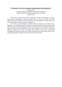
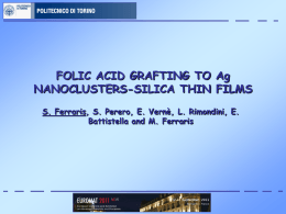
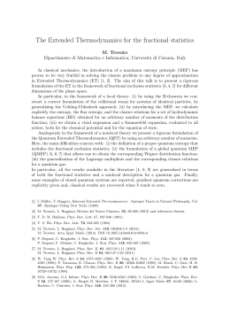
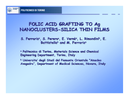
Photoluminescence of radiation-induced color centers in lithium fluoride thin films for
advanced diagnostics of proton beams
M. Piccinini, F. Ambrosini, A. Ampollini, L. Picardi, C. Ronsivalle, F. Bonfigli, S. Libera, E. Nichelatti, M. A.
Vincenti, and R. M. Montereali
Citation: Applied Physics Letters 106, 261108 (2015); doi: 10.1063/1.4923403
View online: http://dx.doi.org/10.1063/1.4923403
View Table of Contents: http://scitation.aip.org/content/aip/journal/apl/106/26?ver=pdfcov
Published by the AIP Publishing
Articles you may be interested in
Optical spectroscopy and microscopy of radiation-induced light-emitting point defects in lithium fluoride crystals
and films
Low Temp. Phys. 38, 779 (2012); 10.1063/1.4740241
Two-beam interferometric encoding of photoluminescent gratings in LiF crystals by high-brightness tabletop soft
x-ray laser
Appl. Phys. Lett. 85, 4163 (2004); 10.1063/1.1812841
Femtosecond-laser-encoded distributed-feedback color center laser in lithium fluoride single crystals
Appl. Phys. Lett. 84, 311 (2004); 10.1063/1.1640784
Spectroscopic analysis and persistent photon-gated spectral hole burning in LiF:F 2 − color center crystal
Appl. Phys. Lett. 79, 2318 (2001); 10.1063/1.1406563
Photoluminescence and the thermal stability of color centers in γ-irradiated LiF and LiF(Mg)
J. Appl. Phys. 82, 3722 (1997); 10.1063/1.365734
This article is copyrighted as indicated in the article. Reuse of AIP content is subject to the terms at: http://scitation.aip.org/termsconditions. Downloaded to IP:
192.107.52.30 On: Tue, 30 Jun 2015 14:04:49
APPLIED PHYSICS LETTERS 106, 261108 (2015)
Photoluminescence of radiation-induced color centers in lithium fluoride
thin films for advanced diagnostics of proton beams
M. Piccinini,1,a) F. Ambrosini,2 A. Ampollini,1 L. Picardi,1 C. Ronsivalle,1 F. Bonfigli,1
S. Libera,1 E. Nichelatti,3 M. A. Vincenti,1 and R. M. Montereali1
1
ENEA, C.R. Frascati, UTAPRAD, Technical Unit for Development and Applications of Radiations,
Via E. Fermi 45, 00044 Frascati (Rome), Italy
2
University Sapienza-Roma I, Piazzale Aldo Moro 5, 00185 Rome, Italy
3
ENEA, C.R. Casaccia, UTTMAT, Technical Unit for Materials Technologies, Via Anguillarese 301,
00123 S. Maria di Galeria (Rome), Italy
(Received 18 March 2015; accepted 22 June 2015; published online 30 June 2015)
Systematic irradiation of thermally evaporated 0.8 lm thick polycrystalline lithium fluoride films on
glass was performed by proton beams of 3 and 7 MeV energies, produced by a linear accelerator, in
a fluence range from 1011 to 1015 protons/cm2. The visible photoluminescence spectra of radiationinduced F2 and F3þ laser active color centers, which possess almost overlapping absorption bands at
about 450 nm, were measured under laser pumping at 458 nm. On the basis of simulations of the linear energy transfer with proton penetration depth in LiF, it was possible to obtain the behavior of the
measured integrated photoluminescence intensity of proton irradiated LiF films as a function of
the deposited dose. The photoluminescence signal is linearly dependent on the deposited dose in the
interval from 103 to about 106 Gy, independently from the used proton energies. This behavior is
very encouraging for the development of advanced solid state radiation detectors based on optically
C 2015
transparent LiF thin films for proton beam diagnostics and two-dimensional dose mapping. V
AIP Publishing LLC. [http://dx.doi.org/10.1063/1.4923403]
Various kinds of ionizing radiations generate stable primary and aggregate defects, also known as color centers
(CCs), in lithium fluoride (LiF) crystals1 and thin films.2
CCs in LiF are well known for applications in light-emitting
miniaturized devices3–6 and tunable solid-state lasers,7 but
they were proposed also for the development of novel highspatial resolution solid-state soft X-ray imaging detectors.8,9
Such applications exploit the broad photoluminescence (PL)
bands of CCs in LiF, stable at room temperature (RT), which
efficiently emit in the visible and near-infrared spectral
ranges. Among them, the laser-active F3þ and F2 CCs (two
electrons bound to three and two anion vacancies, respectively) possess almost overlapped absorption bands, at about
450 nm (M band),10 so that, under light excitation in this
spectral range, they simultaneously emit green (F3þ) and red
(F2) luminescence, peaking at 541 and 678 nm, respectively.10,11 In the last years, their optical emission properties
under light excitation were investigated in pure and doped
LiF materials in different forms12–16 for application in radiation dosimetry.
Very recently, we have started the investigation of the
optical absorption and emission properties of CCs induced in
LiF crystals and thin films by low-energy protons.17,18 In this
letter, we present experimental results about the PL response
of F3þ and F2 point defects in optically transparent thermally
evaporated LiF thin films, irradiated by 3 and 7 MeV protons
in a large interval of irradiation dose.
The exposed samples were polycrystalline LiF films,19
nominally about 1 lm thick, grown by thermal evaporation
on 1 mm thick glass substrates kept at a constant temperature
a)
Author to whom correspondence should be addressed. Electronic mail:
[email protected].
0003-6951/2015/106(26)/261108/4/$30.00
of 300 C during the deposition process, performed in a vacuum chamber at a pressure below 1 mPa, at the Solid State
Laser Laboratory in ENEA C.R Frascati. The starting material consisted of LiF microcrystalline powder (Merck
Suprapur, 99.99% pure), heated at about 800 C in a watercooled tantalum crucible. The evaporation rate, monitored in
situ by an INFICON quartz oscillator, was automatically
controlled at a fixed value of 1 nm/s during the growth.
Several substrates were mounted on a rotating sample holder,
in order to achieve a better thickness uniformity.20
With the aim of determining the optical properties of the
deposited LiF thin films, one of them, grown on a silica substrate, was carefully analyzed to estimate its thickness and
the spectral dispersion of the optical constants of it within a
suitable wavelength range (190 nm k 1200 nm). The
thickness of the film was first evaluated by measuring with a
mechanical profilometer Tencor P-10 the step height due to
the coating border at 9 evenly distributed positions and then
averaging the data. The resulting mean thickness was
ð0:8060:04Þ lm. As far as the optical constants of the LiF
layer are concerned, they were estimated by best fitting the
parameters of a thin-film theoretical model21 to the measured
absolute specular reflectance and direct transmittance spectra, which were taken with a Perkin-Elmer Lambda 900
spectrophotometer. In this case, a one-term Sellmeier and a
Gaussian function in the photon-energy domain were used to
represent the refractive index and extinction coefficient of
the film, respectively. The best-fitting dispersion of the refractive index is shown in Figure 1, where it is also compared to tabulated data for bulk LiF22 modified, by using
Maxwell Garnett’s equation, for a suitable packing density
of 90.5% (i.e., 9.5% of voids in the film). The main film parameters resulting from the analysis were film thickness of
106, 261108-1
C 2015 AIP Publishing LLC
V
This article is copyrighted as indicated in the article. Reuse of AIP content is subject to the terms at: http://scitation.aip.org/termsconditions. Downloaded to IP:
192.107.52.30 On: Tue, 30 Jun 2015 14:04:49
261108-2
Piccinini et al.
FIG. 1. Dispersion curve (Sellmeier model) of the thermally evaporated LiF
film refractive index as resulting from the best fit of reflectance and transmittance experimental spectra. The corresponding dispersion of LiF, obtained
by applying to bulk refractive index data a correction for a packing density
of 90.5%, is also shown.
ð85461Þ nm, linear inhomogeneity along the film growth
axis21 of ð0:560:3Þ%, deviation from parallelism of the
film faces21 amounting to ð2:060:1Þ%, and surface r.m.s.
roughness of ð18:360:2Þ nm. The extinction coefficient was
estimated to be less then 103 in the visible range, a value
which still ensures a very good degree of transparency for a
film which is less than 1 lm thick.
Proton beams of 3 and 7 MeV energy were produced by
a linear accelerator (PL7 model by ACCSYS-HITACHI)
working as the injector of the prototype of a protontherapy
linac under development at ENEA C.R. Frascati.23 A 50 lm
thick kapton window was placed at the output of the machine
beamline. The LiF samples were irradiated at RT in air at a
distance of 10 mm from this exit window and were attached
on an aluminum mask with a 3 mm pin-hole, in order to irradiate them on circular spots with the most uniform transversal intensity distribution of the proton beam. The beam
current was 1 lA in 60 ls-long pulses at a repetition rate of
50 Hz. The irradiation fluence covered the range from 1011
to 1015 protons/cm2 by varying the total number of pulses
delivered to different LiF films.
The PL spectra were measured at RT in the wavelength
range between 480 nm and 800 nm by pumping in a
continuous-wave regime with the 457.9 nm line of an argon
laser, which allows to simultaneously excite the green and red
emissions of F3þ and F2 CCs in the irradiated LiF films.2 The
PL signal was spectrally filtered by a monochromator and
acquired by means of a photomultiplier with lock-in technique. The PL spectra were corrected for the instrumental calibration. The laser power was 25 mW with a power density
on the sample of 8.9 102 W/cm2, which caused no detectable photobleaching effects during the acquisition of spectra.
All the PL measurements were performed 24 h after irradiation, which assured aggregate CCs stabilization.24 Samples
were kept in darkness, while measurements repeated within
some months after irradiation showed no significant change in
the PL response, confirming the CCs stability. By comparison,
bare glass substrates, irradiated in similar conditions, showed
no detectable PL under 457.9 nm laser excitation, although
some darkening was observed in white light. Measurements
Appl. Phys. Lett. 106, 261108 (2015)
of as grown unirradiated LiF films were also performed, but
they showed no detectable PL.
PL spectra of two samples irradiated with 3 and 7 MeV
protons at the same fluence of 1.5 1012 protons/cm2 are
shown in Figure 2. They consist of two broad emission bands
peaked at about 540 nm and 680 nm, ascribed to F3þ and F2
centers, respectively.11,12 The PL spectral features (peak
positions and half-widths) remained unchanged in the investigated fluence range (not shown). Figure 3 shows the PL signal integrated in the whole examined spectral range as a
function of the fluence of 3 and 7 MeV protons. It increases
with fluence and shows a linear behavior up to 8 1013 protons/cm2, while at higher fluences saturation effects take
place and a plateau appears. At all the investigated fluence
values, the integrated PL intensity is always higher in the
LiF films irradiated at 3 MeV than at 7 MeV.
Such a behavior can be explained by simulations performed using SRIM software.25 The inset of Figure 3 reports
the linear energy transfer (LET) calculated by SRIM as a
function of the proton implantation depth in the whole LiF
film-based detector for 3 and 7 MeV proton beams in the experimental conditions described above. LET is continuously
increasing with depth, reaching the maximum value (Bragg
peak) almost at the end of the implantation path. In LiF films,
due to their limited thickness (0.8 lm), LET can be considered as constant and only a very small fraction of the total
proton energy is lost in them, the rest being deposited in the
glass substrate. As shown in the inset of Figure 3, the LET is
lower for 7 MeV protons (12 keV/lm) than for 3 MeV protons
(28.4 keV/lm), which is consistent with the lower PL intensity of 7 MeV proton irradiated LiF films in Figures 2 and 3.
The LET calculated by SRIM software allows one estimating the total dose (D) deposited in the LiF films by using
the following formula:
D¼
LU
;
q
where L is LET, U is the proton fluence, and q is the material
density. The film density can be estimated in a simplified
way by considering the film as an aggregate of material
“grains” separated by air interstices. The fraction of the total
FIG. 2. Photoluminescence spectra of 0.8 lm thick LiF films irradiated by 3
and 7 MeV proton beams at a fluence of 1.5 1012 protons/cm2, measured
at RT under laser pumping at 457.9 nm.
This article is copyrighted as indicated in the article. Reuse of AIP content is subject to the terms at: http://scitation.aip.org/termsconditions. Downloaded to IP:
192.107.52.30 On: Tue, 30 Jun 2015 14:04:49
261108-3
Piccinini et al.
FIG. 3. Integrated visible photoluminescence signal as a function of the 3
and 7 MeV proton fluence in colored 0.8 lm thick LiF films grown on a glass
substrate. (Inset) SRIM simulation of the linear energy transfer as a function
of the proton implantation depth in the LiF film-based detector.
volume occupied by the LiF material is assumed to be the
packing density value derived from the refractive index spectrometric characterization (see Figure 2).
Figure 4 shows the PL signal integrated in the whole
spectral emission range as a function of the estimated dose
deposited by both 3 and 7 MeV proton beams in the used LiF
films. It increases with dose and shows a linear behavior from
the lowest dose value of 103 Gy up to 4 105 Gy. Moreover,
the PL response of the LiF films is independent on the proton
beam energies in the investigated dose interval.
An important characteristic of the solid state LiF film
radiation imaging detectors based on PL by CCs is the possibility of storing information about the transverse proton
beam intensity. The inset of Figure 4 shows the image of a
3 MeV proton beam stored in a LiF film detector, read by a
conventional fluorescence microscope (Nikon Eclipse 80-i)
equipped with a color CCD camera. By a careful calibration
of the visible PL signal as a function of the irradiation dose
(see Figure 4), one can obtain a transverse two-dimensional
dose mapping of proton beams.
In conclusion, the integrated and spectral PL intensities
of 3 and 7 MeV proton-induced F2 and F3þ electronic defects
were carefully measured in optically transparent 0.8 lm thick
FIG. 4. Integrated visible photoluminescence signal as a function of the 3
and 7 MeV proton dose in colored 0.8 lm thick LiF films grown on a glass
substrate. (Inset) Photoluminescence image of a 3 MeV proton beam stored
by color centers in the LiF film-based detector.
Appl. Phys. Lett. 106, 261108 (2015)
LiF films, thermally evaporated on glass substrates, in a
wide interval of irradiation dose. A linear PL response was
obtained and the PL intensity values are independent of the
beam energy in a large dose range.
The high emission efficiency of the F2 and F3þ centers
and the good optical quality of the thermally evaporated LiF
films allow to record the transversal proton beam intensity
profile by directly acquiring the PL image of the irradiated
spots on LiF films using a conventional fluorescence microscope as reading instrument.
The presented results are very encouraging for the use
of LiF thin films as high spatial resolution solid state proton
imaging detectors and dosimeters at high doses. Systematic
experimental investigations will be essential to investigate
the PL response dependence on both the dose-rate and at
higher proton energies, as well as the effect of temperature
on the PL measurements around RT. The possibility of reusing the LiF films after CCs bleaching, by thermal annealing
at 400 C for 30 min, was recently reported for X-ray irradiated LiF films by a Japanese group26 and experiments are
planned also on proton irradiated LiF films grown in different conditions.
Research carried out within the TOP-IMPLART
(Oncological Therapy with Protons—Intensity Modulated
Proton Linear Accelerator for RadioTherapy) Project,
funded by Finanziaria Laziale di Sviluppo (Lazio Region
Financial Agency), Italy.
1
W. B. Fowler, Physics of Color Centers (Academic Press, New York and
London, 1968).
R. M. Montereali, “Point defects in thin insulating films of lithium fluoride
for optical microsystems,” in Handbook of Thin Film Materials,
Ferroelectric and Dielectric Thin Films, edited by H. S. Nalwa (Academic
Press, 2002), Chap. 7, Vol. 3, pp. 399–431.
3
V. V. Ter-Mikirtychev and T. T. Tsuboi, Prog. Quantum Electron. 20, 219
(1996).
4
R. M. Montereali, A. Mancini, G. C. Righini, and S. Pelli, Opt. Commun.
153, 223 (1998).
5
A. Belarouci, F. Menchini, H. Rigneault, B. Jacquier, R. M. Montereali, F.
Somma, and P. Moretti, Opt. Commun. 189, 281 (2001).
6
K. Kawamura, M. Hirano, T. Kurobori, D. Takamizu, and H. Hosono,
Appl. Phys. Lett. 84, 311 (2004).
7
T. T. Basiev, S. B. Mirov, and V. V. Osiko, IEEE J. Quantum Electron.
24, 1052 (1988).
8
G. Baldacchini, F. Bonfigli, A. Faenov, F. Flora, R. M. Montereali, A.
Pace, T. Pikuz, and L. Reale, J. Nanosci. Nanotechnol. 3(6), 483 (2003).
9
R. M. Montereali, F. Bonfigli, M. A. Vincenti, and E. Nichelatti, Il Nuovo
Cimento C 36(2), 35 (2013).
10
J. Nahum and D. A. Wiegand, Phys. Rev. 154(3), 817 (1967).
11
G. Baldacchini, E. De Nicola, R. M. Montereali, A. Scacco, and V.
Kalinov, J. Phys. Chem. Solids 61, 21 (2000).
12
W. L. McLaughlin, J. M. Puhl, A. Kovacs, M. Baranyai, I. Slezsak, M. C.
Saylor, S. A. Saylor, S. D. Miller, and M. Murphy, Radiat. Phys. Chem.
55, 767 (1999).
13
S. D. Miller and G. W. R. Endres, Radiat. Prot. Dosim. 33, 59 (1990).
14
L. Oster, Y. S. Horowitz, and L. Podpalov, Radiat. Prot. Dosim. 128(3),
261 (2008).
15
B. Marczewska, P. Bilski, E. Mandowska, and A. Mandowski, Cent. Eur.
J. Phys. 10(4), 1009 (2012).
16
A. Piaskowska, B. Marczewska, P. Bilski, A. Mandowski, and E.
Mandowsai, Radiat. Meas. 56, 209 (2013).
17
M. Piccinini, F. Ambrosini, A. Ampollini, M. Carpanese, L. Picardi, C.
Ronsivalle, F. Bonfigli, S. Libera, M. A. Vincenti, and R. M. Montereali,
Nucl. Instrum. Methods Phys. Res. B 326, 72 (2014).
18
M. Piccinini, F. Ambrosini, A. Ampollini, M. Carpanese, L. Picardi, C.
Ronsivalle, F. Bonfigli, S. Libera, M. A. Vincenti, and R. M. Montereali,
J. Lumin. 156, 170 (2014).
2
This article is copyrighted as indicated in the article. Reuse of AIP content is subject to the terms at: http://scitation.aip.org/termsconditions. Downloaded to IP:
192.107.52.30 On: Tue, 30 Jun 2015 14:04:49
261108-4
19
Piccinini et al.
R. M. Montereali, G. Baldacchini, S. Martelli, and L. C. Scavarda do
Carmo, Thin Solid Films 196, 75 (1991).
20
M. A. Vincenti, F. Bonfigli, R. M. Montereali, A. Rufoloni, F. Basoli, E.
Di Bartolomeo, S. Licoccia, and E. Nichelatti, ENEA Technical Report
No. RT/2011/19/ENEA, 2011, ISSN/0393-3016.
21
M. Montecchi, R. M. Montereali, and E. Nichelatti, Thin Solid Films 396,
264 (2001); 402, 311 (2002).
22
E. D. Palik, Handbook of Optical Constants of Solids (Academic Press,
London, 1985), Vol. 1, pp. 675–693.
Appl. Phys. Lett. 106, 261108 (2015)
23
C. Ronsivalle, M. Carpanese, C. Marino, G. Messina, L. Picardi, S. Sandri,
E. Basile, B. Caccia, D. M. Castelluccio, E. Cisbani, S. Frullani, F. Ghio, V.
Macellari, M. Benassi, M. D’Andrea, and L. Strigari, Eur. Phys. J. 126, 68
(2011).
24
G. Baldacchini, S. Bigotta, and R. M. Montereali, J. Lumin. 94–95, 299 (2001).
25
J. F. Ziegler, M. D. Ziegler, and J. P. Biersac, Nucl. Instrum. Methods
Phys. Res. B 268, 1818 (2010).
26
T. Kurobori and A. Matoba, Jpn. J. Appl. Phys., Part 1 53, 02BD14
(2014).
This article is copyrighted as indicated in the article. Reuse of AIP content is subject to the terms at: http://scitation.aip.org/termsconditions. Downloaded to IP:
192.107.52.30 On: Tue, 30 Jun 2015 14:04:49
Scaricare