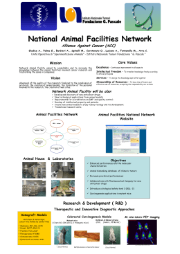
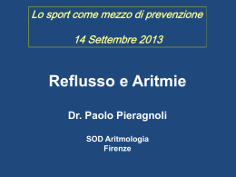
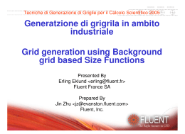
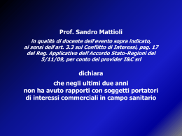
Deep seroma after incisional hernia repair
Case reports and review of the literature
Ann. Ital. Chir.
Published online (EP) 12 May 2015
pii: S2239253X15022938
www.annitalchir.com
PR RE
IN AD
TI -O
N
G NL
PR Y
O CO
H P
IB Y
IT
ED
Giuseppe Salamone, Leo Licari, Antonino Agrusa, Giorgio Romano, Gianfranco Cocorullo,
Gaspare Gulotta.
Department of Surgical, Oncological and Oral Sciences University of Palermo.
General Surgery and Emergency Policlinico Universitario “P. Giaccone”, Palermo, Italy
Deep seroma after incisional hernia repair. Case report and review of the literature
AIM: Wound-related complications are common after incisional hernia repair with mesh; seroma formation is the most
frequent problem. The formation of a deep seroma has been rarely reported in the literature.
MATERIAL OF STUDY: In one year, September 2012-2013, 136 patients underwent surgery for incisional hernia repair,
both elective and urgent. Results: The following complications were observed: one dislocation of polypropylene prosthesis,
a massive relapsed seroma and two deep seromas described in this article. A 63- years-old female underwent open incisional hernia repair with an intraperitoneal PTFE patch. She developed recurrent seroma under the mesh drained percutaneously, and finally the prosthesis was removed. A 72- years-old male underwent open incisional hernia repair with
an intraperitoneal PTFE patch. After several months the patient had seroma infection. The prosthesis was then removed.
CONCLUSIONS: Seroma is a wellknown complication of postoperative ventral hernia repair, especially where prosthetic mesh
is used. The formation of a deep seroma is rare. Only few works mention this complication in literature. In the development of these chronic seromas a role may be played by a long-term inflammatory reaction, more pronounced with
polypropylene and polyester meshes than with ePTFE. A conservative follow up of the seromas is recommended because
drainage can introduce infection. In cases where the seroma causes discomfort or is infected then drainage is necessary.
From experience at our institution we suggest that patients with the deep subtype of mesh-associated seromas may require
closer clinical follow up. When possible, we recommend attempting the drainage of the liquid, eventually followed by
microbiological examination.
KEY
WORDS:
Deepseroma, Incisional hernia, Intraperitoneal mesh
Introduction
Wound-related complications are common after incisional hernia repair with mesh; among them the seroma
formation is the most frequent problem. When mesh is
Pervenuto in Redazione Aprile 2014. Accettato per la pubblicazione
Marzo 2015.
Correspondence to: Giuseppe Salamone MD, Department of Surgicl
Oncological and Oral Sciences, University of Palermo,General Surgery
and Emergency, Policlinico Universitario “P. Giaccone”, Via Liborio
Giuffrè 5, 90127 Palermo, Italy (e-mail: [email protected])
used for repair of larger and more complex incisional
hernias, the risk of seroma formation increases and
appears in 30-50% of cases 1. The formation of a deep
seroma has been rarely reported in the literature. Scott
et al. reported two cases2, Heniford et al reported a frequency of 0,36% (3/819 patients) of long-term fluid collection develop under the mesh3. Tsereteli et al. reported 7 cases (of 442 patients in 5 years) with persistent
seroma posterior to the mesh4. The different biomaterials used to construct commonly used prosthetic mesh
influence the permeability to fluid and affect seroma formation rates. Previously reported rates of seroma occurrence with different types of mesh range from 4% to
8% with polypropylene grafts and 5% to 15% with polytetrafluorethylene (PTFE) grafts 5,6.
Published online (EP) 12 May 2015 - Ann. Ital. Chir.
1
G. Salamone, et al.
CASE HISTORY 1
A 63-years-old female with a medical history of diabetes
mellitus type II, ulcerative colitis and hypertension,
underwent at the age of 60 open right nephrectomy for
a voluminous cyst. In October 2012 she underwent open
incisional hernia repair. The hernia was repaired with an
intraperitoneal PTFE patch. Five months after surgery,
she developed a seroma, without infection, and abdominal pain, localized in the left side of the abdomen. An
abdominal CT revealed a large, well-defined intraabdominal fluid collection (23x11x25 cm) under the mesh
(Figs. 1, 2, 3). A percutaneous closed suction aspiration
PR RE
IN AD
TI -O
N
G NL
PR Y
O CO
H P
IB Y
IT
ED
Susmallian et al. evaluated the true incidence of seroma formation after laparoscopic repair of incisional
hernia with PTFE patch; in 20 patients seroma was
diagnosed clinically in only 35% of cases, while ultrasound examination revealed the presence of seroma in
100% of patients7. The extent of seroma formation is
also known to vary between subjects despite the use
of identical mesh materials and surgical technique. In
some patients only a small amount of fluid is found
using ultrasonography, others suffer from a large volume of seroma fluid that has to be aspirated or treated during operation8. In most instances, these seromas
resolve either spontaneously or with the insertion of
drains or serial percutaneous aspirations. The best way
to recognize and differentiate postoperative fluid collection from hernia recurrence seems to be a computed tomography (CT) scan of the abdomen and its wall
9. CT is a useful imaging tool in patients with repair
of incisional ventral hernia, showing the correct site of
the mesh, subclinical fluid collections in the abdominal wall and recurrent hernia 10.
Material and Methods
In one year, from September 2012 to September 2013,
136 patients underwent surgery for incisional hernia
repair, of which 106 midline (78%) and 30 lateral
(22%), both elective and urgent. 19% of those with
median line were treated with direct suture, 22% treated with polypropylene sublay and for the remaining
57% the intraperitoneal mesh was used (40% polyester
with a three dimensional weave material, 10% PTFE,
7% other dual-meshes). Finally in 2 patients a biological mesh and syntheticbioabsorbable mesh (for tissue reinforcement) was used.
10% of those with lateral surgical treatment direct
suture was used (size less than 3 cm), 36.7% were
treated with polypropylene sublay and for the remaining 53.3% an intraperitoneal mesh was used (20%
polyester with a three dimensional weave material,
23.3% PTFE, 10% other dual-meshes).
Intraperitoneal meshes were fixed along the circumference, with cautious hemostasis, resection of the hernia
sac, placement of suction drain in the subcutaneous
tissue (removed on day 3 post-operative) and application of pressure dressing(removed in 7th post-operative day). The following complications were observed:
in a patient with liver disease HBV related and postoperative paralytic ileus; the dislocation of the prosthesis (polypropylene) and subsequent incisional hernia recurrence; in a patient with chronic heart failure
a massive seroma relapsed, twice drained percutaneously (biological prosthesis); two deep seromas, the
only complications that required removal of the prosthesis, described in more detail in the sections below.
2
Ann. Ital. Chir. - Published online (EP) 12 May 2015
Fig. 1: Case 1: intra-abdominal fluid collection under the mesh.
Fig 2: Case 1 3D. Reconstruction with OsiriX Imaging Software
Lateral view.
PR RE
IN AD
TI -O
N
G NL
PR Y
O CO
H P
IB Y
IT
ED
Deep seroma after incisional hernia repair. Case reports and review of the literature
Fig. 5: Case 1: serous material under the mesh.
ma cavity consisted of thick granulation tissue that separated the mesh and underlying fluid from the abdominal contents. This made it possible to repair the abdominal wall without the use of a mesh, in doubt of infection. The microbiological examination came back negaFig. 3: Case 1 3D. Reconstruction with OsiriX Imaging Software tive, highlighting the absence of infection. The patient
Front view.
had no postoperative complications and complete remission of the painful symptoms.
CASE HISTORY 2
A 72-years-old male with a medical history of hypertension, chronic obstructive pulmonary disease, underwent at the age of 66 abdominal aortic aneurysm. At
the age of 70 underwent open incisional hernia repair
Fig. 4: Case 1: percoutaneous drainage with instillation of fibrin glue.
was subsequently performed. After four months the seroma recurred (20 x 10 x 24) and it was treated with percutaneous drainage (Fig. 4) with instillation of fibrin glue
as a sclerosant. Despite these attempts, 3 months later,
a large seroma recurred. The patient was then hospitalized; abdominal CT was performed revealing the seroma under the mesh (18 x 8 x 24 cm). The patient
underwent surgery, the prosthesis was removed and 2 L
of serous material was aspirated (Fig. 5), partly taken for
microbiological examination. The posterior wall of sero- Fig. 6: Case 2: intra-abdominal fluid collection under the mesh.
Published online (EP) 12 May 2015 - Ann. Ital. Chir.
3
G. Salamone, et al.
PR RE
IN AD
TI -O
N
G NL
PR Y
O CO
H P
IB Y
IT
ED
used. An exact etiology for seroma formation is not clear,
but an immediate local inflammatory response to the
presence of mesh is thought to be a culprit, such as an
abnormal disposition of reticulin fibres 11. Commonly
applied mesh materials are made from expanded PTFE
or polypropylene. Both materials may cause a variety of
tissue reactions leading to postoperative cytokine release,
fever or a persistent formation of seroma 12. The extent
of seroma formation varies between subjects despite the
use of identical mesh materials and surgical technique.
Although in some patients only a small amount of fluid
is found using ultrasonography, others suffer from a large
volume of seroma fluid that has to be aspirated or treated during operation. The latter occurs during the immediate postoperative period and usually resolves with or
without drainage within 4-6 weeks. A conservative follow
up of the seromas is recommended because drainage can
introduce infection. In cases where the seroma causes discomfort or is infected then drainage is necessary13.
The formation of a deep seroma is rare. In literature,
only few works mention this complication. Scott P.D.
reports two cases, Heniford observed 3 cases out of 850
patients (0,3%)3.The largest number of cases reported
was by Tsereteli Z with, a group of 6 patients out of
442 (1,3%), this study proposes a treatment algorithm
for seroma deep persistent, which takes into account,
among the various consequential options, drainage of the
fluid collection and the removal of the mesh.
We report two cases out of 136 patients (1.4%). The
development of these chronic seromas may involve other mechanisms other than the ones described in the previous studies because long-term inflammatory reaction is
more pronounced with polypropylene and polyester
meshes than with ePTFE14. Our patients, as also
described by Tsereteli, have developed an extensive peel
or rind that separated the mesh and underlying fluid
from the abdominal contents, which allowed, once
removed the mesh, to repair the wall only with suture.
Cauterization of the hernia sac and the application of a
central full-thickness suture to reduce the dead space
between the hernia sac and the patch significantly
decrease the incidence of seroma, hematoma, and infection.
From experience at our institution we suggest that
patients with the deep subtype of mesh-associated seromas may require closer clinical follow up. When possible, we recommend attempting the drainage of the liquid, eventually followed by microbiological examination.
Finally, as also suggested by Tsereteli’s algorithm, we recommend the removal the prosthetic mesh after the first
seroma occurrence.
Fig. 7: Case 2: the removal of infected mesh.
Fig. 8: Case 2: use of synthetic bioabsorbable mesh onlay.
with an intraperitoneal PTFE patch, followed by cauterization and partial excision of the hernia sac.
After two months the partial wound dehiscence was treated surgically. After 2 years recurrent incisional hernia and
deep seroma was observed (17x14x6 cm) (Fig. 6), without symptoms. After 6 months, the patient developed
cutaneous fistula with consequent infection of seroma.
The prosthesis was then removed, and the recurrent incisional hernia repaired with the components separation
technique and a synthetic bioabsorbable mesh onlay
(Figs. 7, 8).
Results and Conclusions
Seroma is a well-known complication of postoperative
ventral hernia repair, especially where prosthetic mesh is
4
Ann. Ital. Chir. - Published online (EP) 12 May 2015
Riassunto
La formazione di sieromi a seguito di correzione chirurgica di laparoceli con mesh è una evenienza frequen-
Deep seroma after incisional hernia repair. Case reports and review of the literature
te oramai non più considerata una vera e propria complicanza. Il “Deep Seroma” rappresenta invece un raro
riscontro, come peraltro confermato dai rari casi riportati in letteratura, che talvolta può raggiungere grandi
dimensioni e, se complicati da infezione, possono avere
una percentuale elevata di mortalità. In un anno, nel
nostro istituto 136 pazienti sono stati sottoposti ad intervento chirurgico di plastica della parete addominale per
laparocele con posizionamento di protesi: tra le complicanze osservate desideriamo segnalare due rari casi di
deep seroma. In entrambi i casi la protesi utilizzata è
stata in PTFE al di sotto della quale, sul versante viscerale, si sono formati due voluminosi deep seromas.
L’esordio dei due casi è stato differente: sintomatico il
primo senza sequele infettive, mentre il secondo, asintomatico ma con l’evidenza di fistola cutanea e successiva
infezione del sieroma. Importante la diagnosi precoce
che deriva da un stretto follow-up dei pazienti operati,
che può essere agevolmente sospettata con una ETG e
perfezionata con TC addome ed ricostruzione 3D della
lesione che eseguito in uno dei due casi ha permesso
una valutazione anatomica più precisa.
4. Tsereteli Z, Ramshaw B, Ramaswamy A: Chronic posterior seroma with neoperitoneum following laparoscopic ventral hernia repair:
Treatment algorithm. Hernia, 2008; 12(4):363-66.
References
11. Salamone G, Licari L, Atzeni J, Tutino R, Gulotta G: Histologic
considerations about a rare case of recurrent incisional hernia on
McBurney incision. Ann Ital Chir, 2014; 85 (ePub).
5. Turkcapar AG, Yerdel MA, Aydinuraz K, Bayar S, Kuterdem
E: Repair of midline incisional hernias using polypropylene grafts. Surg
Today, 1998; 8(1):59-63.
6. Chrysos E, Athanasakis E, Saridaki Z, et al.: Surgical repair of
incisional ventral hernias: Tensionfree technique using prosthetic materials (expanded polytetrafluoroethylene Gore-Tex Dual Mesh). Am Surg,
2000; 66(7):679-82.
7. Susmallian S, Gewurtz G, Ezri T, Charuzi I: Seroma after laparoscopic repair of hernia with PTFE patch: Is it really a complication?
Hernia, 2001; 5(3):139-41.
PR RE
IN AD
TI -O
N
G NL
PR Y
O CO
H P
IB Y
IT
ED
8. Schachtrupp A, Klinge U, Junge K, et al.: Individual inflammatory response of human blood monocytes to mesh biomaterials. Br J
Surg, 2003; 90(1):114-20.
1. Berger D, Bientzle M, Muller A: Postoperative complications after
laparoscopic incisional hernia repair. Incidence Treatment. Surg
Endosc, 2002; 16 (12):1720-723.
2. Scott PD, Harold KL, Craft RO, Roberts CC: Postoperative
seroma deep to mesh after laparoscopic ventral hernia repair: Computed
tomography appearance and implications for treatment. Rad Case Rep
2008
3. Heniford BT, Park A, Ramshaw BJ, Voeller G: Laparoscopic
repair of ventral hernias. Nine years’ experience with 850 consecutive
hernias. Ann Surg, 2003; 238(3):391-99.
9. Dukhno O, Pinsk I, Hertzano Y, Levy I, Ovnat A: An unusual presentation of a huge seroma following ventral hernia repair. Surg
Pract, 2005.
10. Gossios K, Zikou A, Vazakas P, Passas G, Glantzouni A,
Glantzounis G, Kontogiannis D, Tsimoyiannis E: Value of CT after
laparoscopic repair of postsurgical ventral hernia. Abdom Imaging,
2003; 28(1):99-102.
12. Schachtrupp A, Klinge U, Junge K, et al.: Individual inflammatory response of human blood monocytes to mesh biomaterials. BJS,
2003; 90(1):114-20.
13. Gutierrez DIP, Vargas RJ, Dieguez Garcia JA: The value of CT
diagnosis of hernia recurrence after prosthetic repair of ventral incisional hernias. Eur Radiol, 2001; 11(7):1161-164.
14. Klinge U, Klosterhalfen B, Muller M, Schumpelick V: Foreign
body reaction to meshes used for the repair of abdominal wall hernias.
Eur J Surg, 1999; 165(7):665-73.
Published online (EP) 12 May 2015 - Ann. Ital. Chir.
5
Scaricare