!
UNIVERSITA’ DEGLI STUDI DI SASSARI
SCUOLA DI DOTTORATO IN
SCIENZE BIOMOLECOLARI E BIOTECNOLOGICHE
Indirizzo: Biochimica, Biochimica clinica e Biologia Molecolare
XXII CICLO
Coordinatore Prof. Bruno Masala
A novel Role of Cdk9/CyclinT2 complexes in skeletal
muscle and Rhabdomyosarcoma cells
Il coordinatore
Dottorando
Prof. Bruno Masala
Irene Marchesi
Tutor
Prof. Luigi Marco Bagella
Anno Accadenico 2008-2009
CONTENTS
CONTENTS
1. Abstract ............................................................................................................. 1
2. Introduction........................................................................................................ 2
2.1 Differentiation of skeletal muscle................................................................ 2
2.2 Interplay between proliferation, differentiation and rhabdomyosarcoma ... 4
2.3 Cdk9, Cyclin T and role in muscle differentiation ...................................... 6
2.4 EZH2 and role in the regulation of muscle differentiation.......................... 9
3. Aim of the project .............................................................................................. 14
4. Materials and methods ....................................................................................... 15
4.1 Cloning and Sequencing.............................................................................. 15
4.2 Plasmids....................................................................................................... 15
4.3 Lentivirus production and infection ............................................................ 16
4.4 Cell culture and differentiation.................................................................... 17
4.5 Immunoblotting ........................................................................................... 17
4.6 Total RNA extraction, cDNA synthesis, Real Time PCR........................... 18
4.7 Transient transfections and luciferase assay................................................ 19
4.8 Nuclear extraction and Co-immunoprecipitation ........................................ 19
4.9 !"#$%""&'()*(+),-$&.&/*0&'()'.)1234.-"&'()#$'0%&("5)3$*("6*0&'()*(+)
7&(+&(8)&()9&0$' :::::::::::::::::::::::::::::::::::::::::::::::::::::::::::::::::::::::::::::::::::::::::::::::::::::::::::::::;<
5. Results................................................................................................................ 21
5.1 Isolation and characterization of the murine Cyclin T2 cDNA................... 21
5.2 During the myogenesis, mRNA and protein levels of CycT2b are
significantly higher respect to CycT2a........................................................ 24
5.3 Cyclins T2 activate the muscle specific genes promoters; CycT2b have
predominat role in the latest stages of the myogenesis ............................... 26
5.4 RD cells show mRNA and protein levels of muscle specific genes lower
and EZH2 levels significantly higher respect to C2C12 ............................. 27
5.5 EZH2 inhibit the promoters of several muscle specific gene...................... 28
5.6 EZH2 binds to Cdk9 and Cyclins T2 both in vitro and in vivo ................... 29
6.
Discussion ......................................................................................................... 31
7.
References......................................................................................................... 34
)
Irene Marchesi, A novel role of Cyclin T2 complexes in skeletal muscle and Rhabdomyosarcoma
cells, Scuola di Dottorato in Scienze Biomolecolari e Biotecnologiche - indirizzo Biochimica,
Biochimica Clinica e Biologia Molecolare, Universita’ degli Studi di Sassari
1. ABSTRACT
Cyclin dependent kinase 9 (Cdk9) is a member of the cyclin dependent kinase family. This
protein is a seorine-threonine kinase, involved in many cellular processes. The regulatory units of
Cdk9 are the T family Cyclins (T1, T2) and Cyclin K1. Cyclin T2 has two forms termed CycT2a
and CycT2b that arise by an alternative splicing of the primary transcript. Human Cyclins T2a
and T2b share the first 642 amino acids but have different carboxyl termini. Previous studies
underscored a crucial role for Cdk9 in association of Cyclin T2 during skeletal myogenesis.
Upon induction of muscle differentiation, MyoD recruits Cdk9/CycT2 on muscle-specific gene
promoter sequences. This complex is able to phosphorylate the C-terminal domain of RNA
polymerase II, enhancing Myod function and promoting myogenic differentiation.
Rhabdomyosarcoma (RMS), one of the most common childhood solid tumor, arises from muscle
precursor cells and fails to complete both the differentiation program both the irreversibly cell
cycle exit, resulting in uncontrolled proliferation and incomplete myogenesis. In RMS, Cdk9
fails to phosphorylate MyoD and the ability of MyoD to arrest cell proliferation and to activate
the myogenic program is repressed. The result of this study confirmed the involvement of Cdk9/
CyclinT2 complexes during the myogenesis. Both isoforms of Cyclin T2 are able to activate the
myogenic program at different stages of differentiation but CycT2b have a predominant role in
particular during the latest stages. Moreover we demonstred that EZH2 is probably responsible to
inhibition of Cdk9 in RMS cells and her overexpression contribuite to inhibition of muscle
differentiation program.
Irene Marchesi, A novel role of Cyclin T2 complexes in skeletal muscle and Rhabdomyosarcoma
cells, Scuola di Dottorato in Scienze Biomolecolari e Biotecnologiche - indirizzo Biochimica,
Biochimica Clinica e Biologia Molecolare, Universita’ degli Studi di Sassari
2. INTRODUCTION
2.1 Differentiation of skeletal muscle
The identity, proliferation and terminal differentiation of skeletal muscle cells is controlled by
combinatorial activities of several transcription factors (Sartorelli and Caretti, 2005).
In particular, an important “modulatory” role in the development of skeletal muscle tissue is
performed by a family of transcription factors, which have in common a basic helix-loop-helix
DNA binding domain, called myogenic bHLH family. This transcription factors family includes
MyoD (reviewed by Weintraub et al., 1991), myogenin (Braun et al., 1989a; Edmondson and
Olson, 1989; Wright et al., 1989), Myf-5 (Braun et al., 1989b), and MRF4, or Myf-6/herculin,
factors. (Rhodes and Konieczny, 1989; Braun et al., 1990; Miner and Wold, 1990; Rudnicki et
al., 1993).
During the skeletal muscle differentiation process, this family of muscle-restricted bHLH
proteins activate the differentiation program by binding to sequence-specific DNA elements, E
box sites (CANNTG), located in enhancer and promoter sequences of muscle specific genes
(Lassar et al., 1989), and by inducing the transcription of regulatory and structural muscle
specific genes (Lassar and Munsterberg, 1994; Molkentin and Olson, 1996; Yun and Wold, 1996;
Arnold and Winter, 1998). Notably, efficient MyoD DNA-protein-binding is achieved by
heterodimerization with other non-myogenic bHLH proteins, which include the products of the
E2A gene (E12, E47) and HEB, also referred as E proteins (Murre et al., 1989; Lassar et al.,
1991; Puri and Sartorelli, 2000).
The cardinal role of MyoD in skeletal myogenesis is evinced by the amount of its target genes. In
fact, MyoD regulates more than 300 genes that could be grouped into at least eight categories:
1. adhesion/matrix
2. cell cycle/DNA replication
3. grow factors/ligand
4.
metabolism
5. nuclear regulatory factors
Irene Marchesi, A novel role of Cyclin T2 complexes in skeletal muscle and Rhabdomyosarcoma
cells, Scuola di Dottorato in Scienze Biomolecolari e Biotecnologiche - indirizzo Biochimica,
Biochimica Clinica e Biologia Molecolare, Universita’ degli Studi di Sassari
6. proteolysis/apoptosis/chaperone
7. receptors/signaling
8. structural/cytoskeletal
(Bergstrom et al., 2002; Giacinti et al., 2006).
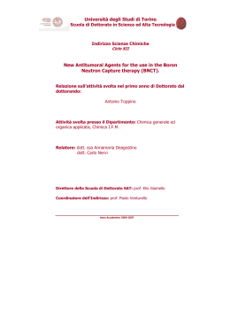
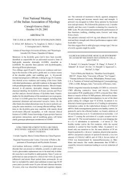
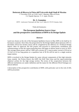
Each MyoD monomer forms two α helics interrupted by a short stretch of aminoacids modeled
as a loop. The first α helix (H1) includes the basic and the helix1 domains; the second helix (H2)
starts immediately after the loop and ends at aminoacid 166. The basic domain fits in the major
groove of the DNA, establishes most of the DNA-protein interactions and is involved in the
activation of transcription (Davis and Weintraub, 1992; Puri and Sartorelli, 2000). Instead, the
two α helics organize the dimerization interface for the formation of the heterodimers (Figure 1).
A
B
C
Figure 1: A) Domains organization of Myod. B) bHLH domain C) Schematic representation of
MyoD-DNA binding.
Irene Marchesi, A novel role of Cyclin T2 complexes in skeletal muscle and Rhabdomyosarcoma
cells, Scuola di Dottorato in Scienze Biomolecolari e Biotecnologiche - indirizzo Biochimica,
Biochimica Clinica e Biologia Molecolare, Universita’ degli Studi di Sassari
Interestingly, the bHLH proteins MyoD, its interacting partners E12, E47 and c-myc recognize
similar, yet distinct, E-boxes. In fact, in vitro experiments have established that MyoD prefers
the CAGCTC sequence, whereas E47 selects the CACCTG, and c-Myc the CACGTG motif,
respectively (Blackwell et al., 1990; Blackwell and Weintraub, 1990; Blackwell et al., 1993; Puri
and Sartorelli, 2000).
Full activation of muscle gene expression by MRFs is also dependent by their association with
members of the MEF2 transcription factors family. In fact, it has been reported that MEF2
factors cannot activate muscle genes on their own, but they potentiate the activity of MRFs.
(Sartorelli and Caretti 2005; Lluìs et al., 2006).
Other than bHLH non-myogenic factors, MyoD can recruit transcriptional co-activators p300
and PCAF, two histone acetyltransferases (HATs) that promote muscle gene transcription by
inducing acetylation of both chromatin and sequence-specific transcription factors, as for MyoD
(Eckner et al., 1994; Eckner et al., 1996; Yuan et al., 1996; Sartorelli et al., 1997; Puri et al.,
1997a,c; Puri et al., 1997c; Struhl, 1998; Giordano and Avantaggiati, 1999; Sartorelli et al.,
1999; Puri and Sartorelli, 2000; Iezzi et al., 2002). Significantly, p300/CBP directly interacts
with MyoD and conveys transcriptional competency by contacting proteins present in the TFIID
complex and the basal transcription machinery (Eckner et al., 1996; Yuan et al., 1996; Sartorelli
et al., 1997; Puri et al., 1997c; Giacinti et al., 2006).
2.2 Interplay between proliferation, differentiation and rhabdomyosarcoma
A clear and effective statement asserts that cell proliferation and differentiation are mutually
exclusive events. In muscle cells, as in other cell types, the decision to divide or differentiate is
determined by a balance of opposing cellular signals (Olson, 1992).
Several HLH proteins have been characterized as inhibitors of myogenesis which exert their
activity by distinct mechanisms:
1. Inhibition of myogenic bHLH proteins by direct protein-protein interaction: Id (Benezra et al.,
1990), Twist (Hamamori et al., 1997), I-mfa (Chen et al., 1996), Cdk4-CyclinD1 (Zhang et al.,
1999a)
Irene Marchesi, A novel role of Cyclin T2 complexes in skeletal muscle and Rhabdomyosarcoma
cells, Scuola di Dottorato in Scienze Biomolecolari e Biotecnologiche - indirizzo Biochimica,
Biochimica Clinica e Biologia Molecolare, Universita’ degli Studi di Sassari
2. Displacement of E12/47: Id- E12/47 complex (Jen et al., 1992)
3. Competition for DNA binding sites: MyoR (Lu et al., 1999), Mist (Lemercier et al., 1998) and
ZEB (Postigo and Dean, 1997; Postigo et al., 1999)
4. Cytoplasmic retention of myogenic bHLH proteins: I-mfa (Chen et al., 1996)
Numerous proteins either facilitate or are required in order to enhance myogenic bHLH
transcriptional activity. Some of these are transcriptional activators themselves, whereas others
do not directly interact with sequence-specific DNA targets (coactivators).
For instance, as alredy explained, MyoD forms a multiprotein complex with its heterodimeric
partners, E12 or E47, and with acetyltransferases p300 and PCAF, whose histone acetylation
alters nucleosomal conformation and increase accessibility of transcription factors to DNA.
Furthermore, PCAF-dependent MyoD acetylation stabilizes MyoD binding to DNA, presumably
by inducing conformational changes in MyoD protein structure. Moreover, additional
acetylations of the basal transcription machinery are also involved in the activation of
transcription (Puri and Sartorelli, 2000).
It is essential to stress that other proteins influence the myogenic program through several
mechanisms. For instance, thyroid hormone (TH) and retinoic acid receptors (RAR and RXR)
are functionally related to the activation of muscle-specific promoters (Carnac et al., 1992;
Albagli-Curiel et al., 1993; Downes et al., 1993; Halevy and Lerman, 1993; Downes et al., 1994;
Alric et al., 1998). Moreover, pRb is able to significantly upregulate MyoD activity and is
involved in the hexpression of late muscle differentiation markers, MHC and MCK (Gu et al.,
1993).
Another crucial mechanism in the activation of myogenic program is established by the multiprotein chromatin-remodeling complex SWI/SNF, recruited on myogenic loci by a p38dependent mechanism. In fact, upon MKK6-dependent enzymatic activation, p38 localizes on
the chromatin of muscle-gene regulatory elements promoting the recruitment of SWI/ SNF
(Simone et al., 2004b). Thus, cofactor binding sites might help to expose a crucial E box or
substitute for an E box facilitating the formation of stable and functional MyoD
transcriptioncomplexes (Lluìs et al., 2006).
Irene Marchesi, A novel role of Cyclin T2 complexes in skeletal muscle and Rhabdomyosarcoma
cells, Scuola di Dottorato in Scienze Biomolecolari e Biotecnologiche - indirizzo Biochimica,
Biochimica Clinica e Biologia Molecolare, Universita’ degli Studi di Sassari
Significantly, Rhabdomyosarcoma (RMS), one of the most common childhood solid tumor,
arises from muscle precursor cells and fails to complete both the differentiation program and the
irreversibly cell cycle exit, resulting in uncontrolled proliferation and incomplete myogenesis
(Merlino and Helman, 1999). Surprisingly, RMS cells express MyoD and myogenin to varying
degrees, but they show only limited expression of genes associated with terminal differentiation
(Hiti et al., 1989; Dias et al., 1991). Interestingly, the ability of MyoD to arrest cell proliferation
and to activate the myogenic program is repressed in RMS, hypothesizing a MyoD activitydependent inhibition of myogenesis (Tapscott et al., 1993; Otten et al., 1997).
2.3 Cdk9, Cyclin T and role in muscle differentiation.
Cyclin-dependent kinase 9 (cdk9), previously named PITALRE (Grana et al., 1994), is a cdk2related serine/threonine kinase, widely expressed in human and murine tissues with high protein
levels in terminally differentiated cells (De Luca et al., 1997a; Bagella et al., 1998, 2000;
Simone et al., 2002).
Cdk9 regulation and activity strictly differs from other CDKs. Cdk9 activity is not cell cycledependent and it does not appear to be required in cell cycle progression (MacLachlan et al.,
1995; De Falco and Giordano, 1998). In addition, unlike the other cdks, Cdk9 fails to
phosphorylate histone H1. In fact, it is involved in the promotion of transcription elongation via
phosphorylation of the carboxyl-terminal domain (CTD) of RNA polymerase II, converting the
inactive unphosphorylated, pre-initiation complex into the phosphorylated and active form
(Grana et al., 1994; Dahmus, 1996; Marshall et al., 1996; Zhu et al., 1997; Wei et al., 1998
Simone et al., 2002; Soutoglou and Talianidis, 2002). The essential residues in transcription
elongation are serine 2 and 5 (Zhou et al., 2001; Sano et al., 2002; Soutoglou and Talianidis,
2002).
CDK9 activity is regulated by cyclins T (T1, T2) and cyclin K (Fu et al., 1999).
Cyclin T1 and T2 share a highly conserved amino terminal motif (cyclin box region, 81%
identity in human T-cyclins), a putative coiled-coil motif, a His-rich motif ( responsible of the
protein-protein interactions with the CTD of RNA polymerase II) and a carboxy- terminal PEST
sequence (less conserved than cyclin box region, 46% identity in human T-cyclins) (Peng et al.,
Irene Marchesi, A novel role of Cyclin T2 complexes in skeletal muscle and Rhabdomyosarcoma
cells, Scuola di Dottorato in Scienze Biomolecolari e Biotecnologiche - indirizzo Biochimica,
Biochimica Clinica e Biologia Molecolare, Universita’ degli Studi di Sassari
1998b; De Luca et al., 2003). The ‘‘cyclin homology box,’’ formed by 290 amino acids, is the
most conserved region among different members of the cyclin-family and serves to bind CDK9.
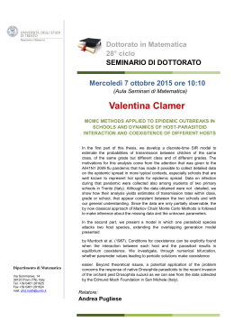
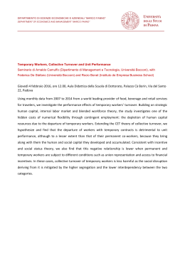
Cyclin T2 has two isoforms, T2a and T2b, that likely arise by an alternative splicing of the
primary transcript, which share the first 642 amino acids but have different carboxyl termini (De
Luca et al., 2003) (Figure 2). Interestingly, CycT2 bears a leucine-rich stretch next to its cyclin
box capable to bind to CTD of RNA polymerase II, thus providing an extra domain capable of
targeting RNAPII (Peng et al., 1998; Kurosu et al., 2004).
Figure 2: Schematic representation of mRNA and protein structure of the two isoforms of cyclin
T2.
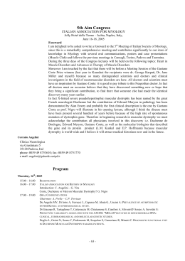
In muscle differentiation, Cdk9 is one of the co-activators of MyoD necessary for the completion
of the myogenic program (Simone and Giordano, 2001, 2007; Simone et al., 2002). Indeed, it
has been demonstrated that Cdk9 directly interacts with MyoD in vitro (Simone et al., 2002).
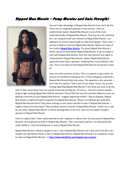
Moreover, recently it has been shown that Cdk9, in muscle cells, takes part of a multimeric
complex containing MyoD, cyclin T2, p300, PCAF and Brg1 (Giacinti et al., 2006). This
complex binds to muscle-specific gene promoter regions and promote gene expression by
Irene Marchesi, A novel role of Cyclin T2 complexes in skeletal muscle and Rhabdomyosarcoma
cells, Scuola di Dottorato in Scienze Biomolecolari e Biotecnologiche - indirizzo Biochimica,
Biochimica Clinica e Biologia Molecolare, Universita’ degli Studi di Sassari
inducing chromatin remodeling, through acetylation of specific lysine residues of histones H3
and H4 and phosphorylation of RNA polymerase II CTD through Cdk9 (Simone et al., 2004;
Giacinti et al., 2006; Simone and Giordano, 2007; Giacinti et al., 2008) (Figure 3).
Figure 3: Schematic representation of the transcriptional complexes regulating gene
expression in differentiation muscle cells.
Significantly, cyclin T1 was not detected on the same regions, suggesting cyclin T2-depedent
Cdk9 activation (Giacinti et al., 2006).
Surprisingly, Rhabdomyosarcoma cells showed upregulation of both Cdk9 and Cyclin T2 and a
strongest Cdk9-cyclinT2 interaction when compared to myoblasts, although Cdk9 fails to
phosphorylate MyoD. It is important to stress that no mutations were detected in the coding
sequences of Cdk9 and Cyclin T2 genes and no significant values muscle-specific gene
expression was detected in presence of overexpressed MyoD in RD cells (Simone and Giordano
2007).
Irene Marchesi, A novel role of Cyclin T2 complexes in skeletal muscle and Rhabdomyosarcoma
cells, Scuola di Dottorato in Scienze Biomolecolari e Biotecnologiche - indirizzo Biochimica,
Biochimica Clinica e Biologia Molecolare, Universita’ degli Studi di Sassari
2.4 EZH2 and role in the regulation of muscle differentiation
Polycomb repressive complex 2 (PRC2) is a histone methyltransferase which is responsible of
the tri-methylation of lysine-27 of histone H3 (H3-K27). (Cao et al., 2002; Czermin et al., 2002;
Kuzmichev et al., 2002; Muller et al., 2002).
PRC2 was initially purified and characterized from human cells and Drosophila embryos (Muller
et al., 2002; Kuzmichev, et al., 2002; R. Cao et al., 2002; Czermin, et al., 2002); it contains a
conserved catalytic subunit, termed EZH2 in human, that include the signature SET domain,
which provides the methyltransferase active site (Rea et al., 2000). The SET domains have an
unusual “thread-the-needle” structure, called pseudoknot (Cheng and Zhang, 2007; Dillon et al.,
2005 for reviews), formed by juxtaposition of two conserved peptide motifs with one peptide
inserted through the loop created by the other. These structures show that the substrate lysine and
methyl donor cofactor bind opposite sides of the SET domain with their binding pockets
connected by an interior channel that aligns the reactive groups for methyl transfer. To attain
robust histone methyltransferase activity, EZH2 must be complexed with at least two of its non
catalytic partners, EED/ESC and SUZ12 (Cao and Zhang 2004; Pasini et al., 2004; Ketel et al.,
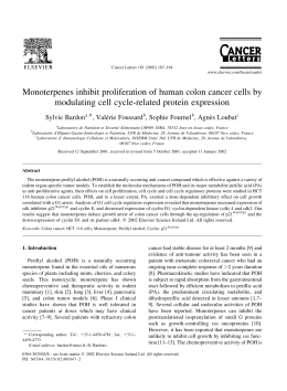
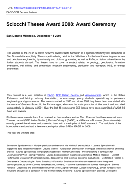
2005; Nekrasov et al., 2005; Montgomery et al., 2005). Both the C-terminal SET domain and the
adjacent cysteine-rich (CXC) domain are required for histone methyltransferase activity (Muller
et al., 2002; Kuzmichev, et al., 2002; R. Cao et al., 2002; Cao and Zhang 2004). Instead, Nterminal domains provide binding sites for assembly with the required partner subunits (Figure
4).
Figure 4: Composition of PRC2 and domain organization of EZH2. (A) The four core
subunits of human PRC2 are EZH2, EED, SUZ12 and RbAp48 (B) Five functional domains
in EZH2.
Irene Marchesi, A novel role of Cyclin T2 complexes in skeletal muscle and Rhabdomyosarcoma
cells, Scuola di Dottorato in Scienze Biomolecolari e Biotecnologiche - indirizzo Biochimica,
Biochimica Clinica e Biologia Molecolare, Universita’ degli Studi di Sassari
PRC2 enzyme function can also be influenced by another associated component, called PHF1
(PCL in flies). Although PHF1 is not a core subunit of PRC2, its association with the complex
can stimulate PRC2 enzyme activity and/or influence its recruitment to target genes in vivo
(Nekrasov et al., 2007; Savla et al., 2008; Sarma et al., 2008; Cao et al., 2008).
Polycomb (PcG)-mediated modifications of histones is an essential mechanism that ensures the
establishment and maintenance of gene expression pattern during mammalian development.
(Sparmann and van Lohuizen 2006). Gene expression silencing by PcG proteins, characterized
by the trimethylation of lysine 27 on histone 3 (H3K27me3) (Czermin et al., 2002; Muller et al.,
2002), is required at specific stages of development for the timely expression of genes involved
in stem cell fate and lineage commitment upon differentiation (Boyer et al., 2006; Lee et al.,
2006; Bracken et al., 2006; Pietersen and van Lohuizen, 2008) and for mammalian Xinactivation and imprinting (Plath et al., 2003; Umlauf et al., 2004).
Polycomb silencing and DNA methylation have often been considered biochemically
independent gene silencing systems. However, recent studies show that EZH2 and DNA
methyltransferases (DNMTs) are physically and functionally linked and that EZH2 acts upstream
of DNMTs to methylate and silence target chromatin (Virė et al., 2006). The mechanism is not
yet clear, but an hypothesis is that target genes are initially silenced through histone H3-K27
methylation by PRC2. PRC2 recruits DNA methyltransferases (DNMTs) which methylate CpG
DNA of target genes, leading to a more permanently or deeply silenced chromatin state (Ohm et
al., 2007; Schlesinger et al., 2007; Widschwendter et al., 2007).
Moreover, in human cells, PRC2 can physically associate with HDAC1 and HDAC2. HDACs
are not core subunits of PRC2 but transient interactions likely still provide functional synergy
between these silencing enzymes in vivo. The precise mechanism of this synergy at target gene
chromatin are not yet clear. HDACs may deacetylate H3-K27 to make the ε-amino group
available for methylation by PRC2. Alternatively, HDACs may deacetylate other histone lysines,
such as H3-K9, H3-K14 or H4-K8, in order to adjust the local histone code for silencing (van
Der Vlag and Otte, 1999; Muller et al., 2002; Kuzmichev et al., 2002; Cao et al., 2002; Czermin
et al., 2002; Varambally et al., 2002; Cao and Zhang, 2004) (Figure 5).
Irene Marchesi, A novel role of Cyclin T2 complexes in skeletal muscle and Rhabdomyosarcoma
cells, Scuola di Dottorato in Scienze Biomolecolari e Biotecnologiche - indirizzo Biochimica,
Biochimica Clinica e Biologia Molecolare, Universita’ degli Studi di Sassari
Figure 5: Model for collaboration of epigenetic silencing enzymes. Target genes are initially
silenced through histone H3-K27 methylation by PRC2. If the lysines are acetylated, may first
require deacetylation by a histone deacetylase (HDAC). PRC2 may also recruit DNA
methyltransferases (DNMTs) which methylate CpG DNA of target genes, leading to a more
permanently or deeply silenced chromatin state
Polycomb-mediated gene silencing and DNA methylation underlie many epigenetic processes
important in normal development as well as in cancer.
Typically, EZH2 is down-regulated in adult differentiated tissues (Varambally et al., 2002;
Bracken et al., 2003; Kleer et al., 2003). Moreover, in contrast to widespread EZH2 roles in
early mouse development (O’Carroll et al., 2001; Erhardt et al., 2003), post-embryonic EZH2
expression is limited (Hobert et al., 1996; Laible et al., 1997). Furthermore, even when detected
Irene Marchesi, A novel role of Cyclin T2 complexes in skeletal muscle and Rhabdomyosarcoma
cells, Scuola di Dottorato in Scienze Biomolecolari e Biotecnologiche - indirizzo Biochimica,
Biochimica Clinica e Biologia Molecolare, Universita’ degli Studi di Sassari
in adult tissues, EZH2 is concentrated in undifferentiated progenitor cell populations, such as
hematopoietic cells of the pro-B lymphocyte lineage (Su et al., 2003).
Interestingly, EZH2 is overexpressed in a variety of different tumors. EZH2 levels are
abnormally elevated in cancer tissues versus corresponding normal tissues, with highest
expression correlating with advanced stages of disease and poor prognosis (for review Simon
and Lange 2008).
Significantly, numerous MyoD-target sequences in muscle-restricted gene promoter regions,
silent in embryonic staminal cells, are occupied by PcG proteins marked by H3K27me3 (Lee et
al., 2006). Moreover, in skeletal muscle cells (SMC), PcG proteins and H3K27me3 are no longer
present at MyoD target-sequences, allowing for their transcriptional activation. Importantly,
although MyoD is expressed in undifferentiated SMC, PcG proteins continue to bind certain
MyoD target genes, which continue to be marked by H3K27me3 and silenced. After additional
molecular signals that promote the complete myogenic program, initiate PcG binding and
H3K27me3 are lost at MyoD target loci, resulting in appropriate muscle gene expression and
SMC differentiation (Caretti et al., 2004; Juan et al., 2009) (Figure 6).
Furthermore, recent studies showed that levels of Ezh2 transcript and protein in RMS compared
to normal myoblasts are consistently higher (Ciarapica et al., 2009).
These data suggest that EZH2 plays a key role in skeletal-muscle differentiation, specifically in
the maintenance of the undifferentiated state of muscle cell precursors. We hyphotesize that
EZH2 overexpression may participate in Rhabdomyosarcoma formation and progression.
Irene Marchesi, A novel role of Cyclin T2 complexes in skeletal muscle and Rhabdomyosarcoma
cells, Scuola di Dottorato in Scienze Biomolecolari e Biotecnologiche - indirizzo Biochimica,
Biochimica Clinica e Biologia Molecolare, Universita’ degli Studi di Sassari
Figure 6: Two-step activation model of muscle gene expression. Regulatory regions of certain
muscle-specific genes are occupied by a protein complex containing the DNA-binding protein
YY1, the methyltransferase Ezh2, and the deacetylase HDAC1. Deacetylation of lysine residues
by HDAC1 and trimethylation of H3-K27 by Ezh2 actively prevent transcription (repressed
state). At the triggering of transcriptional activation, YY1 is displaced from the chromatin, Ezh2
and HDAC1 are replaced by SRF. H3-K27 becomes hypomethylated, and MyoD and HATs are
recruited to the regulatory regions and allowing initiation of transcription (activated state).
Irene Marchesi, A novel role of Cyclin T2 complexes in skeletal muscle and Rhabdomyosarcoma
cells, Scuola di Dottorato in Scienze Biomolecolari e Biotecnologiche - indirizzo Biochimica,
Biochimica Clinica e Biologia Molecolare, Universita’ degli Studi di Sassari
3. AIM OF THE PROJECT
Skeletal muscle differentiation is influenced by multiple pathways which regulate the activity of
myogenic regulatory factors (MRFs) and the MEF2 family members, in positive or negative
ways.
Elucidating the mechanisms governing muscle-specific transcription will provide important
insight to better understand the embryonic development of muscle at the molecular level and will
have important implications in setting out new therapeutic strategy.
Rhabdomyosarcoma is a highly malignant pediatric tumor that derive from mesenchymal cells
already committed to become skeletal muscle cells. In this tumor, the activity of MRFs are
compromised. Furthermore, recent studies showed that levels of EZH2, a protein involved in the
regulation of muscle differentiation process, in rhabdomyosarcoma are consistently higher
compared to normal myoblasts.
The purpose of this study is the characterization of the two murine Cyclin T2 isoforms, CycT2a
and CycT2b and the evaluation of their role in muscle differentiation program. Moreover the
project focus on the modulation by PRC2 of specific skeletal muscle differentiation-related gene
promoters and its involvement in rhabdomyosarcoma formation. In particular the study focus on
the possible interaction between Cdk9/Cyclin T2 complexes and EZH2.
This study will help to clarify the function of these proteins and the molecular regulation during
the myogenic program tumor formation.
Irene Marchesi, A novel role of Cyclin T2 complexes in skeletal muscle and Rhabdomyosarcoma
cells, Scuola di Dottorato in Scienze Biomolecolari e Biotecnologiche - indirizzo Biochimica,
Biochimica Clinica e Biologia Molecolare, Universita’ degli Studi di Sassari
4. MATERIALS AND METHODS
4.1 Cloning and Sequencing
Rapid amplication of cDNA ends (RACE) was employed to generate complete cDNA sequence
encoding the Cyclin T2 isoforms. Mouse skeletal muscle Marathon-ready cDNAs (Clontech)
were used as templates in RACE polymerase chain reaction (PCR) to obtain the 5‘and 3’-end
cDNA fragment according with the manufacture’s protocol
The PCR for 5’-end was carried out using Adaptor primer 1, included in the kit, as sense primer
and CycT2rev1 (GCTTGCAAATGGTCCAATTGGG) as antisense primer.
The PCR for 3’-ends was carried out using CycT2afor and CycT2bfor
(CCACGGTGCTCAGGAGTCCT; CAGCGGATGGAATGCCTCCC respectively) as sense
primers and Adaptor primer 1 as antisense primer.
PCR products were cloned into pGEM-T Easy Vector System II (Promega) and the sequenced
using T7 and SP6 primers. The sequences are analyzed on the 3730 DNA Analyzer from Applied
Biosystems.
The sequence was used to design the primers for full lenght amplification:
CycT2for1: GGATCCATGGCGTCGGGCCGTGGA
CycT2arev: GGATCCCTGGAGTCAGGACCGTGGGGCTCC
CycT2brev: GGATCCTTACATATTCATTCCTTG
4.2 Plasmids
The plasmids Myogenin-luciferase Myh-luciferase promoter were constructed by PCR. The
genomic DNA was extract by DNeasy blood and tissue kit (Qiagen) following the manufacture’s
protocol. The PCR were performed with specific primers:
Myogenin promoter for CAAACGCTAGCCAGCTCTCACGGCTGCTATGA
Myogenin promoter rev GGGAGATCTGGTAGAAATAGGGGGATGTCTC
Myh promoter for CTCCCGGGCTGTATTTCCTCATCTGTGAGGA
Myh4 promoter rev: CTACAAGCTTAGACCAGTTGCTCCTATGCCC
Irene Marchesi, A novel role of Cyclin T2 complexes in skeletal muscle and Rhabdomyosarcoma
cells, Scuola di Dottorato in Scienze Biomolecolari e Biotecnologiche - indirizzo Biochimica,
Biochimica Clinica e Biologia Molecolare, Universita’ degli Studi di Sassari
The amplifed products were cloned in the NheI-BglII site and XmaCI-HindIII of the pGL3 basic
vector (Promega) for Myogenin promoter and Myh promoter respectively.
The plasmids pcDNA3-CycT2a and pcDNA3-CycT2b were constructed by PCR using pGEM-T
Easy-CycT2a and pGEM-T Easy-CycT2b. The PCR were performed with the primers:
mBamHICycT2for: GGATCCACCATGGCGTCGG
mBamHICycT2arev: GGATCCCTGGAGTCAGGACC
mBamHICycT2brev: GGATCCTTACATATTCATTCTTG
The amplifed products were cloned in the BamHI site of the pcDNA3 vector (Invitrogen)
The correct sequences of all these constructs were confirmed by sequencing using the 3730 DNA
Analyzer from Applied Biosystems. The constructs pcDNA3-cdk9wt expressing full-length and
Gst-cdk9 have been previously described (De Falco et al., 2000). The constructs expressing,
MyoD and EZH2 have been described previously. (Simone et al., 2002; Tonini et al., 2004
respectively).
4.3 Lentivirus production and infection
293T packaging cell were seed at 1.5x10⁵ cells/ml (6 ml per plate) in low-antibiotic growth
media (DMEM + 10% FBS) in 6 cm tissue culture plates.
After 24 hours, the packaging cells were transfected with 3 lentivirus plasmids. 1µg of HairpinpLKO.1 vector (shRNA-EZH2 from Sigma), 0.9µg of packaging plasmid psPAX2, 0.1µg of
envelope plasmid pMD2G were diluted in OPTI-MEM to total volume of 250µl. 24µl of FuGene
HD were added to plasmids mix and incubated 20 minutes at room temperature. The transfection
mix was transfered to the the packaging cells. The cells were incubated at 37 °C, 5% CO2. 18
hours post-transfection the medium was replaced with fresh high-serum medium. After 24 hours
the virus in the medium were harvested and the replaced with high-serum media. 24 hours after
the first harvest, the virus were harvested and the packaging cells were discarded. The media
containing virus were filtered with 0.45µm filter. The eluate was transferred to a sterile
polypropylene storage tube.
Irene Marchesi, A novel role of Cyclin T2 complexes in skeletal muscle and Rhabdomyosarcoma
cells, Scuola di Dottorato in Scienze Biomolecolari e Biotecnologiche - indirizzo Biochimica,
Biochimica Clinica e Biologia Molecolare, Universita’ degli Studi di Sassari
RD cells were infected with 1ml (1 moi) of virus solution. After 18-24h of incubation the media
were replaced with growth media. After 48h the media were replaced with growh media added
with puromicine (final concentration 2µg/ml).
4.4 Cell culture and differentiation
Myoblasts (C2C12) were grown in DMEM supplemented with 20% FBS 1% L-glutamine and
antibiotics (Growth Medium, GM). Rhabdomyosarcoma cells RD were grown in DMEM
supplemented with 10% FBS and antibiotics. The differentiation was induced by serum
withdrawal in the presence of 2% horse serum (Differentiation Medium, DM). All cell lines were
obtained from ATCC. Pellets were collected every 24h for 144h or 96h.
4.5 Immunoblotting
Cell were lysated in lysis buffer (20 mM Tris HCl pH 8; 137 mM NaCl; 10% glycerol 1%
Nonidet P-40; 2 mM EDTA; Protease Inhibitor Cocktails)
The protein concentration was determined by Bradford assay (Biorad, CA), following the
manufacturer’s instructions and by using BSA as a standard.
The protein extract (50µg) was resolved in 8% SDS/PAA gel and transferred to a nitrocellulose
membrane at 4°C and at 100V for 1h.The blots were blocked with TBS-T containing 5% non-fat
dry milk.
The protein levels were detected with the followed antibody anti-Cdk9 (Rockland), AntiCyclinT2, anti-MyoD anti-Myogenin, anti-MYH (SANTA CRUZ) anti EZH2 (Invitrogen). Equal
loading was controlled with anti-Gapdh and anti-Hsp70 (SANTA CRUZ).
Antibody were used in TBS-T containing 3% non-fat dry milk. Anti-mouse, rabbit (1:10000),
goat (1:2500) peroxidase conjugated (Pierce) and ECL detection system (PerkinElmer) were
used for detection.
Irene Marchesi, A novel role of Cyclin T2 complexes in skeletal muscle and Rhabdomyosarcoma
cells, Scuola di Dottorato in Scienze Biomolecolari e Biotecnologiche - indirizzo Biochimica,
Biochimica Clinica e Biologia Molecolare, Universita’ degli Studi di Sassari
4.6 Total RNA extraction, cDNA synthesis and Real Time PCR
Total RNA was extracted from 5–10 x 106 cells using High Pure RNA Isolation Kit (Roche). 1µg
of RNA was used for cDNA production with random primers, using High Capacity cDNA
Reverse Transcription Kit (Applied Biosystems), following the manufacture’s protocol. 20µl of
reaction was diluited in 200µl of sterile water.
Real Time was performed using 4,5µl of cDNA and 250 nM primers diluited in FastStart
Universal SYBR Green Master (ROX), (Roche), to a final volume of 10µl. Amplification
conditions for all amplicons were 95°C for 10 minutes, followed by 40 cycles of 95°C for 15s,
60°C for 30s, 72°C for 30s.
Accumulation of fluorescent products was monitored using an Applied Biosystem 7300 system.
Each data point was obtained from at least three independent experiments. Transcripts for
glyceraldehyde-3-phosphate dehydrogenase were used as a reference. To ensure specific PCR
amplification, every real time PCR run was followed by a dissociation phase analysis
(denaturation curve) and by gel electrophoresis. ΔΔCT method was used to calculate relative
changes in gene expression; primer efficiency was calculated for every target using five x 10fold serial dilutions of PCR products. Specific primer sequences are reported in the table below
For
Rev
mGAPDH
AGAAGGTGGTGAAGCAGGCATC
CGAAGGTGGAAGAGTGGGAGTTG
mCycT2a
CGTCTCCTCCGCCTCCAGTG
AGATGTCCGTAGCCCACCTGC
mCycT2b
AGCGAAGCCTCCCACAACC
GTCCGTAGCCCACCTGGTATG
mMyoD
GATGGCATGATGGATTACAGCG
GGAGATGCGCTCCACTATGCT
mMyogenin
CAATGCACTGGAGTTCGGTCC
AGTGATGGCTTTTGACACCAAC
mMYH
CAGAGCTTATTGAGATGCTTCTG
ATCACAGCGCCTGTGAGCTTG
hGAPDH
GAAGGTGAAGGTCGGAGT
CATGGGTGGAATCATATTGGA
hCycT2a
CAGGACTCCTCAGAACAGTGG
TGTCCGTAGCCCACCTGAACT
hCycT2b
CAACCACCACTCCAAAATGAGC
GAGGAGGGGGTAAGGGATGG
hMyoD
GACGGCATGATGGACTACAGC
GGAGATGCGCTCCACGATGC
hMyogenin
CCTGCTCAGCTCCCTCAACC
AGGGTCAGCCGTGAGCAGATG
Irene Marchesi, A novel role of Cyclin T2 complexes in skeletal muscle and Rhabdomyosarcoma
cells, Scuola di Dottorato in Scienze Biomolecolari e Biotecnologiche - indirizzo Biochimica,
Biochimica Clinica e Biologia Molecolare, Universita’ degli Studi di Sassari
4.7 Transient transfections and luciferase assay
Transient transfections were performed using FuGene HD (Roche applied). 2µg of total DNA
diluted in 100µl Opti-MEM (CellGro) was incubated with 8µl of FUGeneHD for 20 minutes to
let the transfection complex form. The transfection complex was added in a ratio 1:16 to the
volume of the incubation medium of the cells (6µl of transfection complex was added to 100µl of
complete medium in 96 wells plate).
Dual luciferase reporter assay (Promega) was used to measure the firefly luciferase and renilla
luciferase activity within the transfected cells. Each experiment was conducted as suggested by
the manifacturer. Luciferase assay was conducted on C2C12 myoblast transfected with the
Myogenin-luc reporter, Myh-luc reporter, RL-TK renilla and expression vectors for CycT2a,
CycT2b, Cdk9 and MyoD. The transfected cells were cultured in DM for 24h and 48h.
Luciferase activity was normalized to TK-directed Renilla expression, in a ratio 1:20 respect to
firefly luciferase vector, to control for variability in transfection efficiencies. The assay were
performed with Sirius Luminometer (Berthold detection systems).
The results are expressed in arbitrary units relative to the activity of the basic luciferase vector
(pGL3-myogenin/Myh promoter).
4.8 Nuclear extraction and Co-immunoprecipitation
For nuclei isolation cells were lysated with NP40 buffer (10mM Tris-HCl; 10mM NaCl; 3mM
MgCl2; 30mM Sucrose; 0.5% NP40; Protease Inhibitor Cocktails) for 30 minutes and
centrifuged for 10 minutes at 3000 g. The pellet with the nuclei was washed with NP40 buffer
and resuspended in non denturing lysis buffer (20 mM Tris HCl pH 8; 137 mM NaCl; 10%
glycerol 1% Nonidet P-40; 2 mM EDTA; Protease Inhibitor Cocktails). Lysates were then
sonicated at amplitude 30% in twice cycles of 30 sec, spaced out 15 sec, using a Fisher Model
550 Sonic Dismembrator (Fisher, Pittsburgh, PA).
2mg of nuclear extract was precipitated with 2µg of anti-Cdk9 antibody (Rockland) or normal
rabbit IgG. The immunoprecipitates were purified by the addition of protein A-agarose (Roche)
following the manufacturer’s instructions. Immunoblot were performed with anti-EZH2
(Invitrogen).
Irene Marchesi, A novel role of Cyclin T2 complexes in skeletal muscle and Rhabdomyosarcoma
cells, Scuola di Dottorato in Scienze Biomolecolari e Biotecnologiche - indirizzo Biochimica,
Biochimica Clinica e Biologia Molecolare, Universita’ degli Studi di Sassari
4.9 Espression and Purification of GST-fusion proteins, Translation and Binding in vitro
GST-Cdk9 and GST were generated by growing 400ml of recombinant E. coli BL21 culture at
37°C to an A600 of 0.4. Cultures were induced for 4 hours with 1 mM IPTG. After induction,
cultures were pelleted, resuspended in NETN buffer (20 mM Tris HCl Ph 8; 100 mM NaCl; 1
mM EDTA; 0.5% NP-40) containing 1mM PMSF and 1mM DTT and sonicated at amplitude
30% in six cycles of 1 minute, spaced out 30 seconds, using a Fisher Model 550 Sonic
Dismembrator (Fisher, Pittsburgh, PA). The bacterial lysates were cleared of cellular debris by
centrifugation and incubated with gluthatione agarose beads over night at 4°C. The complexes
beads-GST and beads GST-Cdk9 were washed twice in NETN buffer 0.2M once with RIPA
buffer (50 mM Tris HCl pH 8; 150 mM NaCl; 1% NP-40; 0.5% Sodium deoxycholate) and once
with NETN buffer 1mM PMSF and 1mM DTT. The complexes were resuspended in 150 µl of
NETN buffer.
The TNT coupled reticulocyte kit was used for in vitro translation (Promega, WI, USA),
according the manufacturer's instructions. All the samples were labeled using 35S- Methionine
10 µl of labeled samples were incubated with 10 µg of GST-CDK9 or GST as a negative control,
in 150 µl of Buffer A (20 mM Tris HCl pH 8; 150 mM KCl; 5 mM MgCl2; 0.2 mM EDTA; 10%
Glycerol; 0.1% NP-40) containing DTT 1mM and PMSF 1mM. The samples then were washed
in buffer A ,c and were resolved on 8% SDS-PAGE. The gels then were fixed with a fixing
solution (50% methanol 10% glacial acetic acid) in slowly agitation for 30 minutes. Finally the
gels were dried for 30 minutes at 80°C and subjected to autoradiography.
Irene Marchesi, A novel role of Cyclin T2 complexes in skeletal muscle and Rhabdomyosarcoma
cells, Scuola di Dottorato in Scienze Biomolecolari e Biotecnologiche - indirizzo Biochimica,
Biochimica Clinica e Biologia Molecolare, Universita’ degli Studi di Sassari
5. RESULTS
5.1 Isolation and characterization of the murine Cyclin T2 cDNA
Murine cDNA of Cyclin T2 isoforms were cloned into pGEM-T Easy Vector System II and
sequenced. The murine CycT2a and CycT2b, show 86% and 87% of similarity respect the
human counteparts. Instead, the comparison between the amino acidic sequences show 85% and
86% of similarity (Figure 7). The predicted molecular weights of CycT2a and CycT2b are 73
kDa and 80 kDa respectively.
A
Figure 7: Cloning and sequencing of murine Cyclin T2 cDNA. (A) Similarity between human
and murine isoforms. (B) Amino acid sequence of the murine Cyclin T2. The difference between
the mouse and human protein sequence is denoted by a bar between the two.
Irene Marchesi, A novel role of Cyclin T2 complexes in skeletal muscle and Rhabdomyosarcoma
cells, Scuola di Dottorato in Scienze Biomolecolari e Biotecnologiche - indirizzo Biochimica,
Biochimica Clinica e Biologia Molecolare, Universita’ degli Studi di Sassari
A
Cyclin T2a
Human
Mouse
Human
Mouse
Human
Mouse
Human
Mouse
Human
Mouse
Human
Mouse
Human
Mouse
Human
Mouse
Human
Mouse
Human
Mouse
Human
Mouse
1
MASGRGASSRWFFTREQLENTPSRRCGVEADKELSCRQQAANLIQEMGQRLNVSQLTINTAIVY
!
!
!
MASGRGASSRWFFTREQLENTPSRRCGVEADEELSHRQQAANLIQDMGQRLNVSQLTINTAIVY
65
MHRFYMHHSFTKFNKNIISSTALFLAAKVEEQARKLEHVIKVAHACLHPLEPLLDTKCDAYLQQ
!
!
MHRFYMHHSFTKFNRNIISPTALFLAAKVEEQARKLEHVIKVAHACLHPLEPLLDTKCDAYLQQ
129
TQELVILETIMLQTLGFEITIEHPHTDVVKCTQLVRASKDLAQTSYFMATNSLHLTTFCLQYKP
!
TQELVLLETIMLQTLGFEITIEHPHTDVVKCTQLVRASKDLAQTSYFMATNSLHLTTFCLQYKP
193
TVIACVCIHLACKWSNWEIPVSTDGKHWWEYVDPTVTLELLDELTHEFLQILEKTPNRLKKIRN
!
!
TVIACVCIHLACKWSNWEIPVSTDGKHWWEYVDPTVTLELLDELTHEFLQILEKTPSRLKRIRN
257
WRANQAARKPKVDGQVSETPLLGSSLVQNSILVDSVTGVPTNPSFQKPSTSAFPAPVPLNSGNI
!!! !
!
!
!
!!
WRA--MAKKPKVDGQVSETPLLGSSLVQNSILVDSVTGVPANPSFQKPSTSTFPAPIPLNSGST
321
SVQDSHTSDNLSMLATGMPSTSYGLSSHQEWPQHQDSARTEQLYSQKQETSLSGSQYNINFQQG
!!
! !
!
!
!!! !
!!
! !
SVQDSRASDNLSVLAAGMPSTSYSLSSHQEWPQHPDSARTDPVYTQKQEATLSGSQY-ISFQQG
385
PSISLHSGLHHRPDKISDHSSVKQEYTHKAGSSKHHGPISTTPGIIPQKMSLDKYREKRKLETL
!!
!
!
!!
!!
PSMALHSGLHHRPDKVADHSSAKQEYTHKAGSSKHHGPIPATPGMLPQKMSLDKYREKRKLETL
449
DLDVRDHYIAAQVEQQHKQGQSQAASSSSVTSPIKMKIPIANT---EKYMADKKEKSGSLKLRI
! !
! !!
! !! !!!!
! !! !!!! !! !
!
DVDTRDHYLAAHAEQQHKHGPAQAVTGTSVTSPIKMKLPLTNSDRPEKHVAEKKERSGSLKLRI
513
PIPPTDKSASKEELKMKIKVSSSERHSSSDEGSGKSKHSSPHISRDHKEKHKEHPSSRHHTSSH
! !!
!
!!
!
PIPPPDKGPSKEELKMKIKVASSERHSSSDEGSGKSKHSSPHISRDHKEKHKEHPANRHH-SSH
577
KHSHSHSGSSSGGSKHSADGIPPTVLRSPVGLSSDGISSSSSSSRKRLHVNDASHNHHSKMSKS
!!!!!!!!!
!
!
!!! ! ! !! ! !!!
K----YLHMHSGGSKHTADGMPPTVLRSPVGLGPEGVSSASS-ARKKLHSSEASHNHHSKMSKS
641
SKSSGGLRTSQHPRETGQEASGDQRSStop
!
! !!
SKSAGGLRTSQHPRETGQETSGAPRSStop
Irene Marchesi, A novel role of Cyclin T2 complexes in skeletal muscle and Rhabdomyosarcoma
cells, Scuola di Dottorato in Scienze Biomolecolari e Biotecnologiche - indirizzo Biochimica,
Biochimica Clinica e Biologia Molecolare, Universita’ degli Studi di Sassari
Cyclin T2b
Human
Mouse
Human
Mouse
Human
Mouse
Human
Mouse
Human
Mouse
Human
Mouse
Human
Mouse
Human
Mouse
Human
Mouse
Human
Mouse
Human
Mouse
Human
Mouse
1
MASGRGASSRWFFTREQLENTPSRRCGVEADKELSCRQQAANLIQEMGQRLNVSQLTINTAIVY
!
!
!
MASGRGASSRWFFTREQLENTPSRRCGVEADEELSHRQQAANLIQDMGQRLNVSQLTINTAIVY
65
MHRFYMHHSFTKFNKNIISSTALFLAAKVEEQARKLEHVIKVAHACLHPLEPLLDTKCDAYLQQ
!
!
MHRFYMHHSFTKFNRNIISPTALFLAAKVEEQARKLEHVIKVAHACLHPLEPLLDTKCDAYLQQ
129
TQELVILETIMLQTLGFEITIEHPHTDVVKCTQLVRASKDLAQTSYFMATNSLHLTTFCLQYKP
!
TQELVLLETIMLQTLGFEITIEHPHTDVVKCTQLVRASKDLAQTSYFMATNSLHLTTFCLQYKP
193
TVIACVCIHLACKWSNWEIPVSTDGKHWWEYVDPTVTLELLDELTHEFLQILEKTPNRLKKIRN
!
!
TVIACVCIHLACKWSNWEIPVSTDGKHWWEYVDPTVTLELLDELTHEFLQILEKTPSRLKRIRN
257
WRANQAARKPKVDGQVSETPLLGSSLVQNSILVDSVTGVPTNPSFQKPSTSAFPAPVPLNSGNI
!!! !
!
!
!
!!
WRA--MAKKPKVDGQVSETPLLGSSLVQNSILVDSVTGVPANPSFQKPSTSTFPAPIPLNSGST
321
SVQDSHTSDNLSMLATGMPSTSYGLSSHQEWPQHQDSARTEQLYSQKQETSLSGSQYNINFQQG
!!
! !
!
!
!!! !
!!
! !
SVQDSRASDNLSVLAAGMPSTSYSLSSHQEWPQHPDSARTDPVYTQKQEATLSGSQY-ISFQQG
385
PSISLHSGLHHRPDKISDHSSVKQEYTHKAGSSKHHGPISTTPGIIPQKMSLDKYREKRKLETL
!!
!
!
!!
!!
PSMALHSGLHHRPDKVADHSSAKQEYTHKAGSSKHHGPIPATPGMLPQKMSLDKYREKRKLETL
449
DLDVRDHYIAAQVEQQHKQGQSQAASSSSVTSPIKMKIPIANT---EKYMADKKEKSGSLKLRI
! !
! !!
! !! !!!!
! !! !!!! !! !
!
DVDTRDHYLAAHAEQQHKHGPAQAVTGTSVTSPIKMKLPLTNSDRPEKHVAEKKERSGSLKLRI
513
PIPPTDKSASKEELKMKIKVSSSERHSSSDEGSGKSKHSSPHISRDHKEKHKEHPSSRHHTSSH
! !!
!
!!
!
PIPPPDKGPSKEELKMKIKVASSERHSSSDEGSGKSKHSSPHISRDHKEKHKEHPANRHH-SSH
577
KHSHSHSGSSSGGSKHSADGIPPTVLRSPVGLSSDGISSSSSSSRKRLHVNDASHNHHSKMSKS
!!!!!!!!!
!
!
!!! ! ! !! ! !!!
K----YLHMHSGGSKHTADGMPPTVLRSPVGLGPEGVSSASS-ARKKLHSSEASHNHHSKMSKS
641
SKSSGSSSSSSSSVKQYISSHNSVFNHPLPPPPPVTYQVGYGHLSTLVKLDKKPVETNGPDANH
!
!
!
!
!! !
SKSAGSSSSSSS-VKQYLSSHSSVFNHPLPPPPPVTYQVGYGHLSTLVKLDKKPVEPHGPEANH
705
EYSTSSQHMDYKDTFDMLDSLLSAQGMNMStop
EYSTSSQHMDYKDTFDMLDSLLSAQGMNMStop
Irene Marchesi, A novel role of Cyclin T2 complexes in skeletal muscle and Rhabdomyosarcoma
cells, Scuola di Dottorato in Scienze Biomolecolari e Biotecnologiche - indirizzo Biochimica,
Biochimica Clinica e Biologia Molecolare, Universita’ degli Studi di Sassari
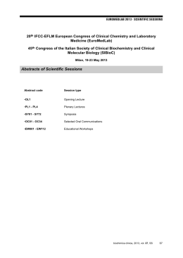
5.2 During the myogenesis, mRNA and protein levels of CycT2b are significantly higher
respect to CycT2a levels
The muscle differentiation process was induced in murine myoblasts (C2C12) through serumdeprivation triggering in presence of horse serum, for 144 hours (Figure 8). The CycT2a and
CycT2b transcripts and proteins levels were analysed by Real-Time PCR and immunoblotting at
several stages of differentiation (every 24 hours).
Immunoblot show that protein levels of CycT2b, in comparison to CycT2a are markedly higher
in all the stages of differentiation. The levels of Cyclin T2 increase during the myogenic
program, (figure 9B). This results were confirmed by Real-Time PCR analysis that show that the
levels of CycT2b cDNA are more or less 3.5 times higher respect the CycT2a cDNA levels
(Figure 9A).
The myogenesis was verified by the evaluation of cells phenotype (figure 8) and the
detemination of mRNA and protein espression of MyoD, Myogenin and MYH (Figure 9).
Figure 8: C2C12 differentiation time course. Myoblasts (C2C12) were grown to 90%
confluence in complete medium (GM, 20% FBS) and induced to differentiate by serum
withdrawal in the presence of 2% horse serum (DM) for 144h. Pellets were collected every 24h.
Irene Marchesi, A novel role of Cyclin T2 complexes in skeletal muscle and Rhabdomyosarcoma
cells, Scuola di Dottorato in Scienze Biomolecolari e Biotecnologiche - indirizzo Biochimica,
Biochimica Clinica e Biologia Molecolare, Universita’ degli Studi di Sassari
B
Figure 9: Espression of Cyclin T2 in C2C12 during the muscle differentiation. (A) Real-time
quantitative PCR (qPCR) was used to determine relative mRNA expression levels upon
induction of myogenesis. qPCR was performed using cDNA from myoblasts at several stage of
differentiation (24h-144h). To distinguish the two Cyclin T2 isoforms, specific primers were
designed in the splicing site region. The myogenesis was verified by detemination of mRNA
espression of MyoD, Myogenin and MYH. The reported data were normalized to
glyceraldehyde-3-phosphate dehydrogenase (GAPDH) levels. (B) Immunoblot analysis The
protein levels were detected with the followed antybody Anti-Cyclin T2, anti-Cdk9, anti-MyoD
anti-Myogenin, anti-MYH. Equal loading was controlled with anti-Gapdh and anti-Hsp70.
Irene Marchesi, A novel role of Cyclin T2 complexes in skeletal muscle and Rhabdomyosarcoma
cells, Scuola di Dottorato in Scienze Biomolecolari e Biotecnologiche - indirizzo Biochimica,
Biochimica Clinica e Biologia Molecolare, Universita’ degli Studi di Sassari
5.3 Cyclins T2 activate the muscle specific genes promoters; CycT2b have a predominat
role in the latest stages of the myogenesis
To establish a functional difference between CycT2a and CycT2b, C2C12 myoblast were
transiently transfected with Myogenin promoter luciferase reporter (Myogenin-luc) and Myh
promoter luciferase reporter (Myh-luc), and expression vectors for CycT2a, CycT2b, Cdk9,
MyoD. The myoblasts were cultured in DM for 24h and 48h.
Transfection of either individual or pairwise combinations of Cdk9, CycT2a and CycT2b
expression vectors had no effect on Myogenin-luc and Myh-luc in the absence of MyoD.
Conversely, MyoD-dependent transactivation of promoters was increased, by coexpression of
both complexes Cdk9/CyclinT2 (Figure 10). At 24h, CycT2a and CycT2b had the same effect on
the myogenin-luc (Figure 10A); at 48h the effect of CycT2b in comparison to the effect of
CycT2a was stronger (Figure 10C). On the Myh-luc the effect of CycT2b was stronger at both
24h and 48h (Figure10 B,D). This suggest a predominant role of Cyct2b in latest stages of
differentiation process.
A
B
C
D
Figure 10: Regulation of Myogenin promoters by Cyclin T2. Luciferase assay was conducted
on C2C12 myoblast transfected with the Myogenin-luc (A, C), Myh-Luc (B, D), RL-TK renilla
and expression vectors for CycT2a, CycT2b, Cdk9 and MyoD. The transfected cells were
cultured in DM for 24h and 48h. Luciferase activity was normalized to TK-directed Renilla
expression. The results are expressed in arbitrary units relative to the activity of the basic
luciferase vector (pGL3-myogenin promoter, pGL3 Myh promoter).
Irene Marchesi, A novel role of Cyclin T2 complexes in skeletal muscle and Rhabdomyosarcoma
cells, Scuola di Dottorato in Scienze Biomolecolari e Biotecnologiche - indirizzo Biochimica,
Biochimica Clinica e Biologia Molecolare, Universita’ degli Studi di Sassari
5.4 RD cells show mRNA and protein levels of muscle specific genes lower and EZH2 levels
significantly higher respect to C2C12
To better understand the skeletal muscle differentiation process and the rhabdomyosarcoma
formation, RD cells were grow 90% confluence in complete medium and induced to differentiate
by serum withdrawal in the presence of 2% horse serum (DM) for 96h. Pellet were collected
every 24h.
After 96h unlike C2C12, RD cells failed to complete the differentiation program and they
continued to proliferate (Figure 11A). Immunoblot showed that protein levels of all markers of
myogenesis (MyoD, Myogenin and Myh) are markedly lower respect C2C12 differentiation
program. Interestingly, the levels of EZH2, a protein frequently overexpressed in several tumors,
were considerably higher (Figure 11B).
A
B
C2C12
RD
Figure11: C2C12 and RD differentiation time course. (A) Comparison between C2C12 and
RD phenotype after 96h in DM. (B) Immunoblot analysis. The protein levels were detected
with the followed antybody anti-Cyclin T2, anti-Cdk9, anti-EZH2 anti-MyoD anti-Myogenin,
anti-MYH. Equal loading was controlled with anti-Hsp70.
Irene Marchesi, A novel role of Cyclin T2 complexes in skeletal muscle and Rhabdomyosarcoma
cells, Scuola di Dottorato in Scienze Biomolecolari e Biotecnologiche - indirizzo Biochimica,
Biochimica Clinica e Biologia Molecolare, Universita’ degli Studi di Sassari
5.5 EZH2 inhibit the promoters of several muscle specific gene
In order to understand the role of PCR2 in the rhabdomyosarcoma formation process a stable
EZH2 knockdown RD cell line was generated using a vector-based shRNA. EZH2 knockdown
cells did not show changes in cellular morphology.
The cells were induced to differentiate by serum withdrawal in the presence of 2% horse serum
for 96h. Pellet were collected every 24h.
Interestingly, analysis of mRNA and protein expression showed that EZH2 knockdown resulted
in a significant increase on the MyoD, Myogenin and MYH levels (Figure 12). The partial
reactivation of muscle specific genes, suggests a role of EZH2 in the inhibition of muscle
differentiation program.
A
B
Figure 12: Comparison between mRNA and protein espression in RD and RD EZH2
knockdown . (A) Real-time quantitative PCR with specific primers for MyoD and Myogenin.
(B) Immunoblot analysis. The protein levels were detected with the followed antybody: antiMyoD, anti-Myogenin, anti-MYH and anti-EZH2. Equal loading was controlled with antiHsp70.
Irene Marchesi, A novel role of Cyclin T2 complexes in skeletal muscle and Rhabdomyosarcoma
cells, Scuola di Dottorato in Scienze Biomolecolari e Biotecnologiche - indirizzo Biochimica,
Biochimica Clinica e Biologia Molecolare, Universita’ degli Studi di Sassari
5.6 EZH2 binds to Cdk9 and Cyclins T2 both in vitro and in vivo
To investigate whether EZH2 was able to interact with Cdk9 and with in vitro, Gst pull-down
experiments were performed using Gst-Cdk9 and GST-EZH2 purified from BL21 E. Coli cells
and four in vitro translated product [35S]-EZH2 full-length, [35S]-Cdk9, [35S]-CycT2a and [35S]CycT2b. As shown in figure 13A, Gst-Cdk9 was able to pull down full-length EZH2, while this
was not the case for the Gst fragment alone. In addition, Gst-EZH2 was able to pull down both
Cyclin T2 isoforms, showing a direct interaction between EZH2 and Cyclins T2 (Figure 13B,C).
Moreover in order to test if EZH2 and Cdk9 were able to interact in Rhabdomyosarcoma cells,
RD cells lysates were subjected to Co-immunoprecipitation with anti-Cdk9 and
immunoprecipitation were then probed with anti-EZH2 (Figure 14). Normal Rabbit IgGs have
been used as negative control.
A
B
Figure 13: Binding in vitro assay: EZH2 binds to Cdk9, CycT2a and CycT2b in vitro. (A)
Physical interaction between Gst-Cdk9 and in vitro translated EZH2. (B) Physical interaction
between Gst-EZH2 and in vitro translated CycT2a and CycT2b.
Irene Marchesi, A novel role of Cyclin T2 complexes in skeletal muscle and Rhabdomyosarcoma
cells, Scuola di Dottorato in Scienze Biomolecolari e Biotecnologiche - indirizzo Biochimica,
Biochimica Clinica e Biologia Molecolare, Universita’ degli Studi di Sassari
Figure 14: Co-immunoprecipitation. RD cells lysates were co-immunoprecipitated with antiCdk9 and Normal Rabbit IgG as negative control. Immunoblotting was performed with antiEZH2.
Irene Marchesi, A novel role of Cyclin T2 complexes in skeletal muscle and Rhabdomyosarcoma
cells, Scuola di Dottorato in Scienze Biomolecolari e Biotecnologiche - indirizzo Biochimica,
Biochimica Clinica e Biologia Molecolare, Universita’ degli Studi di Sassari
6. DISCUSSION
During cell division, cyclins play an essential role being subjected to cyclical expression and
ubiquitin-dependent degradation, and acting as regulatory subunits of complexes with the cyclindependent kinases (CDKs) (Sherr, 1996; Grana and Reddy, 1995).
Some CDKs/cyclin, as CDK7/cyclin H, CDK8/ cyclin C, and CDK9/cyclin T, seem to direct
their activity in a cell cycle independent manner and appear to be involved in other processes as
signal transduction, apoptosis, differentiation and transcription during the initiation or the
elongation steps (Dynlacht, 1997; De Luca et al., 2003).
A Previous study showed that Cdk9, in association with Cyclin T2, plays an important role in the
activation of the myogenic program (Simone et al., 2002b). Upon induction of muscle
differentiation, MyoD recruits Cdk9/CycT2 on muscle-specific gene promoter sequences. This
complex is able to phosphorylate the C-terminal domain (CTD) of RNA polymerase II,
enhancing MyoD function and promoting myogenic differentiation (Giacinti et al., 2006). The
transcriptional activity of MyoD is deficient in rhabdomyosarcoma cells and in these cells, Cdk9
fails to phosphorylate MyoD (Simone and Giordano, 2007).
EZH2 is the catalytic subunit of PRC2 and is responsible of the lysine-27 tri-methylation of
histone H3.
Gene expression silencing by PcG proteins, is required at specific stages of development and is
down-regulated in adult differentiated tissues (Varambally et al., 2002; Bracken et al., 2003;
Kleer et al., 2003). PcG is able to regulate embryonic development of muscle inhibiting the
homeobox gene expression (Caretti et al., 2004). Interestingly, EZH2 is overexpressed in a
variety of different tumors as in the rhabdomyosarcoma.
This work focuses on the evaluation of role of Cdk9/Cyclin T2 complexes during the skeletal
muscle differentiation and in rhadbomyosarcoma cells.
In order to understand the role of Cyclin T2 isoforms in muscle differentiation, myogenesis was
induced in murine myoblast and mRNA and protein levels were analyzed at several stage of
differentiation process. In addition, luciferase assay performed allowed to identify functional
differences between two complexes.
Irene Marchesi, A novel role of Cyclin T2 complexes in skeletal muscle and Rhabdomyosarcoma
cells, Scuola di Dottorato in Scienze Biomolecolari e Biotecnologiche - indirizzo Biochimica,
Biochimica Clinica e Biologia Molecolare, Universita’ degli Studi di Sassari
The results demonstrated that CycT2b levels, both mRNA and protein expressions, in
comparison to CycT2a are markedly higher in all the stages of differentiation. These results
assumed a major role for CycT2b in muscle differentiation process. Moreover the luciferase
assay showed that both cyclins T2 isoforms was able to increased MyoD-dependent
transactivation of Myogenin and Myh promoters. During the first stages of differentiation both
Cyclins T2 activate the muscle differentiation program but during the latest stages the activity of
CycT2b is stronger.
The higher espression and the differences of functional activity show by CycT2b suggest a
predominant role of this protein in the differentiation process, in particular during the latest
stages.
Rhabdomyosarcoma (RMS) is one of the most common childhood solid tumor that arises from
muscle precursor cells. In RMS cells, the ability of MyoD to arrest cell proliferation and to
activate the myogenic program is repressed and the myoblasts fail to complete the differentiation
program (Tapscott et al. 1993; Otten et al. 1997; Merlino and Helman, 1999). Moreover, Cdk9
fails to phosphorylate MyoD but no mutations were detected in the coding sequences of Cdk9
and Cyclin T2 (Simone and Giordano 2007). This allow to hypothesize that the MyoD inhibition
arise from inhibition of Cdk9 activity.
The comparison between normal myoblast (C2C12) and RMS cells (RD) proteins levels
confirmes that the expression of muscle specific genes is inhibited in RD cells whereas Cdk9 is
strongly expressed. Interestingly, EZH2 a protein involved in the regulation of differentiation
process of several tissue as in skeletal muscle differentiation, is overexpressed. EZH2 is the
catalytic subunit of Polycomb repressive complex 2 (PRC2), which is histone methyltransferase
that targets lysine-27 of histone H3 responsible of silencing ot target genes. Typically, EZH2 is
down-regulated in adult differentiated tissues but over-expressed in a wide variety of cancerous
tissue types (for review Simon and Lange 2008). Previous study showed that PRC2 plays a key
role in the maintenance of the undifferentiated state of muscle cell precursors (Caretti et al.,
2004 ).
In order to understand if the overexpression of EZH2 was the cause of rabdomyosarcoma
formation, a stable EZH2 knockdown RD cell line was generated and induced to differentiate.
Irene Marchesi, A novel role of Cyclin T2 complexes in skeletal muscle and Rhabdomyosarcoma
cells, Scuola di Dottorato in Scienze Biomolecolari e Biotecnologiche - indirizzo Biochimica,
Biochimica Clinica e Biologia Molecolare, Universita’ degli Studi di Sassari
Interestingly, EZH2 knockdown allow a significant increase on the MyoD, Myogenin and MYH
levels. The partial reactivation of muscle specific genes confirm the hypothesis that EZH2
overexpression contribuite to inhibition of muscle differentiation program.
To investigate whether the inhibition of Cdk9 in Rabdomyosarcoma cells and overexpression of
EZH2 was interrelated, we assumed an interaction between Cdk9 and EZH2. We confirmed the
interaction between the two proteins by alternative methods, co-immunoprecipitation and in vitro
pull-down assays. Moreover we demonstrate that EZH2 is able to interact with both CycT2a and
CycT2b.
These studies highlight a critical role for Cdk9/CycT2b complex in controlling skeletal muscle
growth and differentiation. Our observation point toward the cooperation with MRFs such as
MyoD and Myogenin suggesting that CycT2b has a crucial role in maintaining cells at the
differentiation terminal stage. In addition, the physical interaction between Cdk9/Cyclin T2
complex and EZH2 emphasizes its commitment to the skeletal muscle lineage. It is reasonable to
assume that during myogenesis Cdk9/Cyclin T2 complex might be implicated in the
downregulation of EZH2, a step that is functionally required for the process of myotube
formation.
The understanding of the functional properties of the Cdk9/Cyclin T2 complexes and the
interaction with specific associated partners will help us to clarify the complex mechanisms that
regulate the myogenic program. Moreover, the characterization of the binding between Cdk9 and
myogenic regulatory factors like EZH2 may contribuite to explain the most important pathways
involved in muscle program regulation and rhabdomyosarcoma formation.
Further studies are on going in our laboratory to define more accurately the molecular
mechanisms underlying the myogenic function of Cdk9/CyclinT2 complexes with EZH2, and to
verify its biological relevance in rhabomyosarcoma formation.
.
Irene Marchesi, A novel role of Cyclin T2 complexes in skeletal muscle and Rhabdomyosarcoma
cells, Scuola di Dottorato in Scienze Biomolecolari e Biotecnologiche - indirizzo Biochimica,
Biochimica Clinica e Biologia Molecolare, Universita’ degli Studi di Sassari
7. REFERENCES
Albagli-Curiel O, Carnac G, Vandromme M, Vincent S, Crépieux P, Bonnieu A. 1993a. Seruminduced inhibition of myogenesis is differentially relieved by retinoic acid and
triiodothyronine in C2 murine muscle cells. Differentiation; research in biological
diversity 52(8387038):201-210.
Albagli-Curiel O, Carnac G, Vandromme M, Vincent S, Crépieux P, Bonnieu A. 1993b. Seruminduced inhibition of myogenesis is differentially relieved by retinoic acid and
triiodothyronine in C2 murine muscle cells. Differentiation; research in biological
diversity 52(8387038):201-210.
Alric S, Froeschlé A, Piquemal D, Carnac G, Bonnieu A. 1998. Functional specificity of the two
retinoic acid receptor RAR and RXR families in myogenesis. Oncogene 16(9464546):
273-282.
Arnold HH, Winter B. 1998. Muscle differentiation: more complexity to the network of
myogenic regulators. Current opinion in genetics & development 8(9794824):539-544.
Bagella L, MacLachlan TK, Buono RJ, Pisano MM, Giordano A, De Luca A. 1998. Cloning of
murine CDK9/PITALRE and its tissue-specific expression in development. Journal of
cellular physiology 177(9766517):206-213.
Bagella L, Stiegler P, De Luca A, Siracusa LD, Giordano A. 2000. Genomic organization,
promoter analysis, and chromosomal mapping of the mouse gene encoding Cdk9. Journal
of cellular biochemistry 78(10797576):170-178.
Bergstrom DA, Penn BH, Strand A, Perry RL, Rudnicki MA, Tapscott SJ. 2002. Promoterspecific regulation of MyoD binding and signal transduction cooperate to pattern gene
expression. Molecular cell 9(11931766):587-600.
Blackwell TK, Huang J, Ma A, Kretzner L, Alt FW, Eisenman RN, Weintraub H. 1993. Binding
of myc proteins to canonical and noncanonical DNA sequences. Molecular and Cellular
Biology 13(8395000):5216-5224.
Blackwell TK, Weintraub H. 1990. Differences and similarities in DNA-binding preferences of
MyoD and E2A protein complexes revealed by binding site selection. Science 250
(2174572):1104-1110.
Boyer LA, Plath K, Zeitlinger J, Brambrink T, Medeiros LA, Lee TI, Levine SS, Wernig M,
Tajonar A, Ray MK, Bell GW, Otte AP, Vidal M, Gifford DK, Young RA, Jaenisch R.
2006. Polycomb complexes repress developmental regulators in murine embryonic stem
cells. Nature 441(16625203):349-353.
Bracken AP, Dietrich N, Pasini D, Hansen KH, Helin K. 2006. Genome-wide mapping of
Polycomb target genes unravels their roles in cell fate transitions. Genes & Development
20(16618801):1123-1136.
Bracken AP, Pasini D, Capra M, Prosperini E, Colli E, Helin K. 2003. EZH2 is downstream of
the pRB-E2F pathway, essential for proliferation and amplified in cancer. The EMBO
journal 22(14532106):5323-5335.
Irene Marchesi, A novel role of Cyclin T2 complexes in skeletal muscle and Rhabdomyosarcoma
cells, Scuola di Dottorato in Scienze Biomolecolari e Biotecnologiche - indirizzo Biochimica,
Biochimica Clinica e Biologia Molecolare, Universita’ degli Studi di Sassari
Braun T, Bober E, Buschhausen-Denker G, Kohtz S, Grzeschik KH, Arnold HH, Kotz S. 1989a.
Differential expression of myogenic determination genes in muscle cells: possible
autoactivation by the Myf gene products. The EMBO journal 8(2583111):3617-3625.
Braun T, Bober E, Buschhausen-Denker G, Kohtz S, Grzeschik KH, Arnold HH, Kotz S. 1989b.
Differential expression of myogenic determination genes in muscle cells: possible
autoactivation by the Myf gene products. The EMBO journal 8(2583111):3617-3625.
Braun T, Bober E, Winter B, Rosenthal N, Arnold HH. 1990. Myf-6, a new member of the
human gene family of myogenic determination factors: evidence for a gene cluster on
chromosome 12. The EMBO journal 9(2311584):821-831.
Braun T, Buschhausen-Denker G, Bober E, Tannich E, Arnold HH. 1989c. A novel human
muscle factor related to but distinct from MyoD1 induces myogenic conversion in 10T1/2
fibroblasts. The EMBO journal 8(2721498):701-709.
Cao Q, Yu J, Dhanasekaran SM, Kim JH, Mani RS, Tomlins SA, Mehra R, Laxman B, Cao X,
Kleer CG, Varambally S, Chinnaiyan AM. 2008. Repression of E-cadherin by the
polycomb group protein EZH2 in cancer. Oncogene 27(18806826):7274-7284.
Cao R, Wang L, Wang H, Xia L, Erdjument-Bromage H, Tempst P, Jones RS, Zhang Y. 2002.
Role of histone H3 lysine 27 methylation in Polycomb-group silencing. Science 298
(12351676):1039-1043.
Cao R, Zhang Y. 2004. SUZ12 is required for both the histone methyltransferase activity and the
silencing function of the EED-EZH2 complex. Molecular cell 15(15225548):57-67.
Caretti G, Di Padova M, Micales B, Lyons GE, Sartorelli V. 2004. The Polycomb Ezh2
methyltransferase regulates muscle gene expression and skeletal muscle differentiation.
Genes & Development 18(15520282):2627-2638.
Caretti G, Schiltz RL, Dilworth FJ, Di Padova M, Zhao P, Ogryzko V, Fuller-Pace FV, Hoffman
EP, Tapscott SJ, Sartorelli V. 2006. The RNA helicases p68/p72 and the noncoding RNA
SRA are coregulators of MyoD and skeletal muscle differentiation. Developmental cell
11(17011493):547-560.
Carnac G, Albagli-Curiel O, Vandromme M, Pinset C, Montarras D, Laudet V, Bonnieu A. 1992.
3,5,3'-Triiodothyronine positively regulates both MyoD1 gene transcription and terminal
differentiation in C2 myoblasts. Molecular endocrinology (Baltimore, Md) 6(1406697):
1185-1194.
Cheng X, Zhang X. 2007. Structural dynamics of protein lysine methylation and demethylation.
Mutation research 618(17374386):102-115.
Ciarapica R, Russo G, Verginelli F, Raimondi L, Donfrancesco A, Rota R, Giordano A. 2009.
Deregulated expression of miR-26a and Ezh2 in rhabdomyosarcoma. Cell cycle
(Georgetown, Tex) 8(19106613):172-175.
Czermin B, Melfi R, McCabe D, Seitz V, Imhof A, Pirrotta V. 2002. Drosophila enhancer of
Zeste/ESC complexes have a histone H3 methyltransferase activity that marks
chromosomal Polycomb sites. Cell 111(12408863):185-196.
Dahmus ME. 1996. Phosphorylation of mammalian RNA polymerase II. Methods in enzymology
273(8791612):185-193.
Davis RL, Weintraub H. 1992. Acquisition of myogenic specificity by replacement of three
amino acid residues from MyoD into E12. Science 256(1317057):1027-1030.
Irene Marchesi, A novel role of Cyclin T2 complexes in skeletal muscle and Rhabdomyosarcoma
cells, Scuola di Dottorato in Scienze Biomolecolari e Biotecnologiche - indirizzo Biochimica,
Biochimica Clinica e Biologia Molecolare, Universita’ degli Studi di Sassari
De Falco G, Giordano A. 1998. CDK9 (PITALRE): a multifunctional cdc2-related kinase.
Journal of cellular physiology 177(10092203):501-506.
De Luca A, De Falco M, Baldi A, Paggi M. 2003. Cyclin T: three forms for different roles in
physiological and pathological functions. Journal of Cellular Physiology 194(12494448):
101-107.
De Luca A, Esposito V, Baldi A, Claudio PP, Fu Y, Caputi M, Pisano MM, Baldi F, Giordano A.
1997. CDC2-related kinase PITALRE phosphorylates pRb exclusively on serine and is
widely expressed in human tissues. Journal of cellular physiology 172(9258347):
265-273.
Dias P, Parham DM, Shapiro DN, Webber BL, Houghton PJ. 1990. Myogenic regulatory protein
(MyoD1) expression in childhood solid tumors: diagnostic utility in rhabdomyosarcoma.
The American journal of pathology 137(2260621):1283-1291.
Dillon SC, Zhang X, Trievel RC, Cheng X. 2005. The SET-domain protein superfamily: protein
lysine methyltransferases. Genome biology 6(16086857):227.
Downes M, Griggs R, Atkins A, Olson EN, Muscat GE. 1993. Identification of a thyroid
hormone response element in the mouse myogenin gene: characterization of the thyroid
hormone and retinoid X receptor heterodimeric binding site. Cell growth &
differentiation : the molecular biology journal of the American Association for Cancer
Research 4(8297796):901-909.
Downes M, Mynett-Johnson L, Muscat GE. 1994. The retinoic acid and retinoid X receptors are
differentially expressed during myoblast differentiation. Endocrinology 134(8194491):
2658-2661.
Dynlacht BD. 1997. Regulation of transcription by proteins that control the cell cycle. Nature
389(9296491):149-152.
Eckner R, Ewen ME, Newsome D, Gerdes M, DeCaprio JA, Lawrence JB, Livingston D. 1994.
Molecular cloning and functional analysis of the adenovirus E1A-associated 300-kD
protein (p300) reveals a protein with properties of a transcriptional adaptor. Genes &
Development 8(7523245):869-884.
Eckner R, Yao T, Oldread E, Livingston D. 1996. Interaction and functional collaboration of
p300/CBP and bHLH proteins in muscle and B-cell differentiation. Genes &
Development 10(19):2478-2490.
Edmondson DG, Olson EN. 1990. A gene with homology to the myc similarity region of MyoD1
is expressed during myogenesis and is sufficient to activate the muscle differentiation
program. Genes & Development 4(2172083):1450.
Erhardt S, Lyko F, Ainscough JF, Surani MA, Paro R. 2003. Polycomb-group proteins are
involved in silencing processes caused by a transgenic element from the murine
imprinted H19/Igf2 region in Drosophila. Development genes and evolution 213
(12750886):336-344.
Fu TJ, Peng J, Lee G, Price DH, Flores O. 1999. Cyclin K functions as a CDK9 regulatory
subunit and participates in RNA polymerase II transcription. The Journal of biological
chemistry 274(10574912):34527-34530.
Irene Marchesi, A novel role of Cyclin T2 complexes in skeletal muscle and Rhabdomyosarcoma
cells, Scuola di Dottorato in Scienze Biomolecolari e Biotecnologiche - indirizzo Biochimica,
Biochimica Clinica e Biologia Molecolare, Universita’ degli Studi di Sassari
Giacinti C, Bagella L, Puri P, Giordano A, Simone C. 2006. MyoD recruits the cdk9/cyclin T2
complex on myogenic-genes regulatory regions. Journal of Cellular Physiology 206
(16245309):807-813.
Giacinti C, Musarò A, De Falco G, Jourdan I, Molinaro M, Bagella L, Simone C, Giordano A.
2008. Cdk9-55: a new player in muscle regeneration. Journal of cellular physiology 216
(18546201):576-582.
Giordano A, Avantaggiati ML. 1999. p300 and CBP: partners for life and death. Journal of
cellular physiology 181(10497301):218-230.
Graña X, De Luca A, Sang N, Fu Y, Claudio PP, Rosenblatt J, Morgan DO, Giordano A. 1994.
PITALRE, a nuclear CDC2-related protein kinase that phosphorylates the retinoblastoma
protein in vitro. Proceedings of the National Academy of Sciences of the United States of
America 91(8170997):3834-3838.
Graña X, Reddy EP. 1995. Cell cycle control in mammalian cells: role of cyclins, cyclin
dependent kinases (CDKs), growth suppressor genes and cyclin-dependent kinase
inhibitors (CKIs). Oncogene 11(7624138):211-219.
Gu W, Schneider JW, Condorelli G, Kaushal S, Mahdavi V, Nadal-Ginard B. 1993. Interaction of
myogenic factors and the retinoblastoma protein mediates muscle cell commitment and
differentiation. Cell 72(8381715):309-324.
Halevy O, Lerman O. 1993. Retinoic acid induces adult muscle cell differentiation mediated by
the retinoic acid receptor-alpha. Journal of cellular physiology 154(8382210):566-572.
Hamamori Y, Wu HY, Sartorelli V, Kedes L. 1997. The basic domain of myogenic basic helixloop-helix (bHLH) proteins is the novel target for direct inhibition by another bHLH
protein, Twist. Molecular and Cellular Biology 17(9343420):6563-6573.
Hiti AL, Bogenmann E, Gonzales F, Jones PA. 1989. Expression of the MyoD1 muscle
determination gene defines differentiation capability but not tumorigenicity of human
rhabdomyosarcomas. Molecular and Cellular Biology 9(2601695):4722-4730.
Hobert O, Sures I, Ciossek T, Fuchs M, Ullrich A. 1996. Isolation and developmental expression
analysis of Enx-1, a novel mouse Polycomb group gene. Mechanisms of development 55
(8861097):171-184.
Iezzi S, Cossu G, Nervi C, Sartorelli V, Puri P. 2002. Stage-specific modulation of skeletal
myogenesis by inhibitors of nuclear deacetylases. Proceedings of the National Academy
of Sciences of the United States of America 99(12032356):7757-7762.
Jen Y, Weintraub H, Benezra R. 1992. Overexpression of Id protein inhibits the muscle
differentiation program: in vivo association of Id with E2A proteins. Genes &
Development 6(1644289):1466-1479.
Juan AH, Kumar RM, Marx JG, Young RA, Sartorelli V. 2009. Mir-214-dependent regulation of
the polycomb protein Ezh2 in skeletal muscle and embryonic stem cells. Molecular Cell
36(19818710):61-74.
Ketel CS, Andersen EF, Vargas ML, Suh J, Strome S, Simon JA. 2005. Subunit contributions to
histone methyltransferase activities of fly and worm polycomb group complexes.
Molecular and Cellular Biology 25(16055700):6857-6868.
Kleer CG, Cao Q, Varambally S, Shen R, Ota I, Tomlins SA, Ghosh D, Sewalt RG, Otte AP,
Hayes DF, Sabel MS, Livant D, Weiss SJ, Rubin MA, Chinnaiyan AM. 2003. EZH2 is a
Irene Marchesi, A novel role of Cyclin T2 complexes in skeletal muscle and Rhabdomyosarcoma
cells, Scuola di Dottorato in Scienze Biomolecolari e Biotecnologiche - indirizzo Biochimica,
Biochimica Clinica e Biologia Molecolare, Universita’ degli Studi di Sassari
marker of aggressive breast cancer and promotes neoplastic transformation of breast
epithelial cells. Proceedings of the National Academy of Sciences of the United States of
America 100(14500907):11606-11611.
Kurosu T, Zhang F, Peterlin BM. 2004. Transcriptional activity and substrate recognition of
cyclin T2 from P-TEFb. Gene 343(15563843):173-179.
Kuzmichev A, Nishioka K, Erdjument-Bromage H, Tempst P, Reinberg D. 2002. Histone
methyltransferase activity associated with a human multiprotein complex containing the
Enhancer of Zeste protein. Genes & Development 16(12435631):2893-2905.
Laible G, Wolf A, Dorn R, Reuter G, Nislow C, Lebersorger A, Popkin D, Pillus L, Jenuwein T.
1997. Mammalian homologues of the Polycomb-group gene Enhancer of zeste mediate
gene silencing in Drosophila heterochromatin and at S. cerevisiae telomeres. The EMBO
journal 16(9214638):3219-3232.
Lassar A, Münsterberg A. 1994. Wiring diagrams: regulatory circuits and the control of skeletal
myogenesis. Current opinion in cell biology 6(7917336):432-442.
Lassar AB, Buskin JN, Lockshon D, Davis RL, Apone S, Hauschka SD, Weintraub H. 1989.
MyoD is a sequence-specific DNA binding protein requiring a region of myc homology
to bind to the muscle creatine kinase enhancer. Cell 58(2550138):823-831.
Lassar AB, Davis RL, Wright WE, Kadesch T, Murre C, Voronova A, Baltimore D, Weintraub H.
1991. Functional activity of myogenic HLH proteins requires hetero-oligomerization with
E12/E47-like proteins in vivo. Cell 66(1649701):305-315.
Lee TI, Jenner RG, Boyer LA, Guenther MG, Levine SS, Kumar RM, Chevalier B, Johnstone
SE, Cole MF, Isono K, Koseki H, Fuchikami T, Abe K, Murray HL, Zucker JP, Yuan B,
Bell GW, Herbolsheimer E, Hannett NM, Sun K, Odom DT, Otte AP, Volkert TL, Bartel
DP, Melton DA, Gifford DK, Jaenisch R, Young RA. 2006. Control of developmental
regulators by Polycomb in human embryonic stem cells. Cell 125(16630818):301-313.
Lemercier C, To RQ, Carrasco RA, Konieczny SF. 1998. The basic helix-loop-helix transcription
factor Mist1 functions as a transcriptional repressor of myoD. The EMBO journal 17
(9482738):1412-1422.
Lluís F, Perdiguero E, Nebreda AR, Muñoz-Cánoves P. 2006. Regulation of skeletal muscle gene
expression by p38 MAP kinases. Trends in cell biology 16(16325404):36-44.
MacLachlan TK, Sang N, Giordano A. 1995. Cyclins, cyclin-dependent kinases and cdk
inhibitors: implications in cell cycle control and cancer. Critical reviews in eukaryotic
gene expression 5(8845581):127-156.
Marshall NF, Peng J, Xie Z, Price DH. 1996. Control of RNA polymerase II elongation potential
by a novel carboxyl-terminal domain kinase. The Journal of biological chemistry 271
(8900211):27176-27183.
Merlino G, Helman LJ. 1999. Rhabdomyosarcoma--working out the pathways. Oncogene 18
(10498887):5340-5348.
Miner JH, Wold B. 1990. Herculin, a fourth member of the MyoD family of myogenic regulatory
genes. Proceedings of the National Academy of Sciences of the United States of America
87(2300571):1089-1093.
Irene Marchesi, A novel role of Cyclin T2 complexes in skeletal muscle and Rhabdomyosarcoma
cells, Scuola di Dottorato in Scienze Biomolecolari e Biotecnologiche - indirizzo Biochimica,
Biochimica Clinica e Biologia Molecolare, Universita’ degli Studi di Sassari
Molkentin JD, Olson EN. 1996. Combinatorial control of muscle development by basic helixloop-helix and MADS-box transcription factors. Proceedings of the National Academy of
Sciences of the United States of America 93(8790335):9366-9373.
Montgomery ND, Yee D, Chen A, Kalantry S, Chamberlain SJ, Otte AP, Magnuson T. 2005. The
murine polycomb group protein Eed is required for global histone H3 lysine-27
methylation. Current biology : CB 15(15916951):942-947.
Murre C, McCaw PS, Vaessin H, Caudy M, Jan LY, Jan YN, Cabrera CV, Buskin JN, Hauschka
SD, Lassar AB. 1989. Interactions between heterologous helix-loop-helix proteins
generate complexes that bind specifically to a common DNA sequence. Cell 58
(2503252):537-544.
Müller J, Hart CM, Francis NJ, Vargas ML, Sengupta A, Wild B, Miller EL, O'Connor MB,
Kingston RE, Simon JA. 2002. Histone methyltransferase activity of a Drosophila
Polycomb group repressor complex. Cell 111(12408864):197-208.
Nekrasov M, Klymenko T, Fraterman S, Papp B, Oktaba K, Köcher T, Cohen A, Stunnenberg
HG, Wilm M, Müller J. 2007. Pcl-PRC2 is needed to generate high levels of H3-K27
trimethylation at Polycomb target genes. The EMBO journal 26(17762866):4078-4088.
Nekrasov M, Wild B, Müller J. 2005. Nucleosome binding and histone methyltransferase activity
of Drosophila PRC2. EMBO reports 6(15776017):348-353.
O'Carroll D, Erhardt S, Pagani M, Barton SC, Surani MA, Jenuwein T. 2001. The polycombgroup gene Ezh2 is required for early mouse development. Molecular and Cellular
Biology 21(11390661):4330-4336.
Ohm JE, McGarvey KM, Yu X, Cheng L, Schuebel KE, Cope L, Mohammad HP, Chen W,
Daniel VC, Yu W, Berman DM, Jenuwein T, Pruitt K, Sharkis SJ, Watkins DN, Herman
JG, Baylin SB. 2007. A stem cell-like chromatin pattern may predispose tumor
suppressor genes to DNA hypermethylation and heritable silencing. Nature genetics 39
(17211412):237-242.
Olson EN. 1992. Interplay between proliferation and differentiation within the myogenic lineage.
Developmental biology 154(1330787):261-272.
Otten AD, Firpo EJ, Gerber AN, Brody LL, Roberts JM, Tapscott SJ. 1997. Inactivation of
MyoD-mediated expression of p21 in tumor cell lines. Cell growth & differentiation : the
molecular biology journal of the American Association for Cancer Research 8(9372238):
1151-1160.
Pasini D, Bracken A, Jensen M, Lazzerini Denchi E, Helin K. 2004. Suz12 is essential for mouse
development and for EZH2 histone methyltransferase activity. The EMBO Journal 23
(15385962):4061-4071.
Peng J, Zhu Y, Milton JT, Price DH. 1998. Identification of multiple cyclin subunits of human PTEFb. Genes & Development 12(9499409):755-762.
Pietersen AM, van Lohuizen M. 2008. Stem cell regulation by polycomb repressors: postponing
commitment. Current opinion in cell biology 20(18291635):201-207.
Plath K, Fang J, Mlynarczyk-Evans SK, Cao R, Worringer KA, Wang H, de la Cruz CC, Otte AP,
Panning B, Zhang Y. 2003. Role of histone H3 lysine 27 methylation in X inactivation.
Science 300(12649488):131-135.
Irene Marchesi, A novel role of Cyclin T2 complexes in skeletal muscle and Rhabdomyosarcoma
cells, Scuola di Dottorato in Scienze Biomolecolari e Biotecnologiche - indirizzo Biochimica,
Biochimica Clinica e Biologia Molecolare, Universita’ degli Studi di Sassari
Postigo AA, Dean DC. 1997. ZEB, a vertebrate homolog of Drosophila Zfh-1, is a negative
regulator of muscle differentiation. The EMBO journal 16(9233803):3935-3943.
Postigo AA, Ward E, Skeath JB, Dean DC. 1999. zfh-1, the Drosophila homologue of ZEB, is a
transcriptional repressor that regulates somatic myogenesis. Molecular and Cellular
Biology 19(10490660):7255-7263.
Puri PL, Sartorelli V. 2000. Regulation of muscle regulatory factors by DNA-binding, interacting
proteins, and post-transcriptional modifications. Journal of Cellular Physiology 185
(11025438):155-173.
Puri PL, Sartorelli V, Yang XJ, Hamamori Y, Ogryzko VV, Howard BH, Kedes L, Wang JY,
Graessmann A, Nakatani Y, Levrero M. 1997. Differential roles of p300 and PCAF
acetyltransferases in muscle differentiation. Molecular cell 1(9659901):35-45.
Rea S, Eisenhaber F, O'Carroll D, Strahl BD, Sun ZW, Schmid M, Opravil S, Mechtler K,
Ponting CP, Allis CD, Jenuwein T. 2000. Regulation of chromatin structure by sitespecific histone H3 methyltransferases. Nature 406(10949293):593-599.
Rhodes SJ, Konieczny SF. 1989. Identification of MRF4: a new member of the muscle
regulatory factor gene family. Genes & Development 3(2560751):2050-2061.
Sano M, Abdellatif M, Oh H, Xie M, Bagella L, Giordano A, Michael LH, DeMayo FJ,
Schneider MD. 2002. Activation and function of cyclin T-Cdk9 (positive transcription
elongation factor-b) in cardiac muscle-cell hypertrophy. Nature medicine 8(12368904):
1310-1317.
Sarma K, Margueron R, Ivanov A, Pirrotta V, Reinberg D. 2008. Ezh2 Requires PHF1 To
Efficiently Catalyze H3 Lysine 27 Trimethylation In Vivo. Molecular and Cellular
Biology 28(8):2718-2731.
Sartorelli V, Caretti G. 2005. Mechanisms underlying the transcriptional regulation of skeletal
myogenesis. Current opinion in genetics & development 15(16055324):528-535.
Sartorelli V, Huang J, Hamamori Y, Kedes L. 1997. Molecular mechanisms of myogenic
coactivation by p300: direct interaction with the activation domain of MyoD and with the
MADS box of MEF2C. Molecular and cellular biology 17(9001254):1010-1026.
Sartorelli V, Puri PL, Hamamori Y, Ogryzko V, Chung G, Nakatani Y, Wang JY, Kedes L. 1999.
Acetylation of MyoD directed by PCAF is necessary for the execution of the muscle
program. Molecular cell 4(10619020):725-734.
Savla U, Benes J, Zhang J, Jones R. 2008. Recruitment of Drosophila Polycomb-group proteins
by Polycomblike, a component of a novel protein complex in larvae. Development 135
(5):813-817.
Schlesinger Y, Straussman R, Keshet I, Farkash S, Hecht M, Zimmerman J, Eden E, Yakhini Z,
Ben-Shushan E, Reubinoff BE, Bergman Y, Simon I, Cedar H. 2007. Polycomb-mediated
methylation on Lys27 of histone H3 pre-marks genes for de novo methylation in cancer.
Nature genetics 39(17200670):232-236.
Sherr CJ. 1996. Cancer cell cycles. Science (New York, NY) 274(8939849):1672-1677.
Simon JA, Lange CA. 2008. Roles of the EZH2 histone methyltransferase in cancer epigenetics.
Mutation research 647(18723033):21-29.
Simone C, Bagella L, Bellan C, Giordano A. 2002a. Physical interaction between pRb and cdk9/
cyclinT2 complex. Oncogene 21(12037672):4158-4165.
Irene Marchesi, A novel role of Cyclin T2 complexes in skeletal muscle and Rhabdomyosarcoma
cells, Scuola di Dottorato in Scienze Biomolecolari e Biotecnologiche - indirizzo Biochimica,
Biochimica Clinica e Biologia Molecolare, Universita’ degli Studi di Sassari
Simone C, Forcales S, Hill D, Imbalzano A, Latella L, Puri P. 2004. p38 pathway targets SWISNF chromatin-remodeling complex to muscle-specific loci. Nature Genetics 36
(15208625):738-743.
Simone C, Giordano A. 2001. New insight in cdk9 function: from Tat to MyoD. Frontiers in
bioscience : a journal and virtual library 6(11532614):D1073-1082.
Simone C, Giordano A. 2007. Abrogation of signal-dependent activation of the cdk9/cyclin T2a
complex in human RD rhabdomyosarcoma cells. Cell Death and Differentiation 14
(16841087):192-195.
Simone C, Stiegler P, Bagella L, Pucci B, Bellan C, De Falco G, De Luca A, Guanti G, Puri P,
Giordano A. 2002b. Activation of MyoD-dependent transcription by cdk9/cyclin T2.
Oncogene 21(12037670):4137-4148.
Soutoglou E, Talianidis I. 2002. Coordination of PIC assembly and chromatin remodeling during
differentiation-induced gene activation. Science 295(11884757):1901-1904.
Sparmann A, van Lohuizen M. 2006. Polycomb silencers control cell fate, development and
cancer. Nature reviews Cancer 6(17060944):846-856.
Struhl K. 1998. Histone acetylation and transcriptional regulatory mechanisms. Genes &
Development 12(9499396):599-606.
Su IH, Basavaraj A, Krutchinsky AN, Hobert O, Ullrich A, Chait BT, Tarakhovsky A. 2003.
Ezh2 controls B cell development through histone H3 methylation and Igh
rearrangement. Nature immunology 4(12496962):124-131.
Tapscott SJ, Thayer MJ, Weintraub H. 1993. Deficiency in rhabdomyosarcomas of a factor
required for MyoD activity and myogenesis. Science 259(8383879):1450-1453.
Tonini T, Bagella L, D'Andrilli G, Claudio PP, Giordano A. 2004. Ezh2 reduces the ability of
HDAC1-dependent pRb2/p130 transcriptional repression of cyclin A. Oncogene 23
(15077161):4930-4937.
Umlauf D, Goto Y, Cao R, Cerqueira F, Wagschal A, Zhang Y, Feil R. 2004. Imprinting along the
Kcnq1 domain on mouse chromosome 7 involves repressive histone methylation and
recruitment of Polycomb group complexes. Nature genetics 36(15516932):1296-1300.
van der Vlag J, den Blaauwen JL, Sewalt RG, van Driel R, Otte AP. 2000. Transcriptional
repression mediated by polycomb group proteins and other chromatin-associated
repressors is selectively blocked by insulators. The Journal of biological chemistry 275
(10617669):697-704.
Varambally S, Dhanasekaran SM, Zhou M, Barrette TR, Kumar-Sinha C, Sanda MG, Ghosh D,
Pienta KJ, Sewalt RG, Otte AP, Rubin MA, Chinnaiyan AM. 2002. The polycomb group
protein EZH2 is involved in progression of prostate cancer. Nature 419(12374981):
624-629.
Viré E, Brenner C, Deplus R, Blanchon L, Fraga M, Didelot C, Morey L, Van Eynde A, Bernard
D, Vanderwinden JM, Bollen M, Esteller M, Di Croce L, de Launoit Y, Fuks F. 2006. The
Polycomb group protein EZH2 directly controls DNA methylation. Nature 439
(16357870):871-874.
Wei P, Garber ME, Fang SM, Fischer WH, Jones KA. 1998. A novel CDK9-associated C-type
cyclin interacts directly with HIV-1 Tat and mediates its high-affinity, loop-specific
binding to TAR RNA. Cell 92(9491887):451-462.
Irene Marchesi, A novel role of Cyclin T2 complexes in skeletal muscle and Rhabdomyosarcoma
cells, Scuola di Dottorato in Scienze Biomolecolari e Biotecnologiche - indirizzo Biochimica,
Biochimica Clinica e Biologia Molecolare, Universita’ degli Studi di Sassari
Weintraub H, Davis R, Tapscott S, Thayer M, Krause M, Benezra R, Blackwell TK, Turner D,
Rupp R, Hollenberg S. 1991a. The myoD gene family: nodal point during specification of
the muscle cell lineage. Science 251(1846704):761-766.
Weintraub H, Dwarki V, Verma I, Davis R, Hollenberg S, Snider L, Lassar A, Tapscott SJ. 1991b.
Muscle-specific transcriptional activation by MyoD. Genes & Development 5(8):
1377-1386.
Widschwendter M, Fiegl H, Egle D, Mueller-Holzner E, Spizzo G, Marth C, Weisenberger DJ,
Campan M, Young J, Jacobs I, Laird PW. 2007. Epigenetic stem cell signature in cancer.
Nature genetics 39(17200673):157-158.
Wright WE, Sassoon DA, Lin VK. 1989. Myogenin, a factor regulating myogenesis, has a
domain homologous to MyoD. Cell 56(2537150):607-617.
Yuan W, Condorelli G, Caruso M, Felsani A, Giordano A. 1996. Human p300 protein is a
coactivator for the transcription factor MyoD. The Journal of biological chemistry 271
(8621548):9009-9013.
Yun K, Wold B. 1996. Skeletal muscle determination and differentiation: story of a core
regulatory network and its context. Current opinion in cell biology 8(8939680):877-889.
Zhou M, Nekhai S, Bharucha DC, Kumar A, Ge H, Price DH, Egly JM, Brady JN. 2001. TFIIH
inhibits CDK9 phosphorylation during human immunodeficiency virus type 1
transcription. The Journal of biological chemistry 276(11572868):44633-44640.
Zhu Y, Pe'ery T, Peng J, Ramanathan Y, Marshall N, Marshall T, Amendt B, Mathews MB, Price
DH. 1997. Transcription elongation factor P-TEFb is required for HIV-1 tat
transactivation in vitro. Genes & Development 11(9334325):2622-2632.
Irene Marchesi, A novel role of Cyclin T2 complexes in skeletal muscle and Rhabdomyosarcoma
cells, Scuola di Dottorato in Scienze Biomolecolari e Biotecnologiche - indirizzo Biochimica,
Biochimica Clinica e Biologia Molecolare, Universita’ degli Studi di Sassari
Scaricare