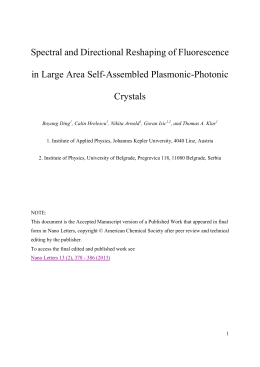
Provincia Autonoma di Trento
European Union – 7th research framework programme
Marie Curie Actions – COFUND
“TRENTINO”
- The research, training and mobility programme in Trentino PCOFUND-GA-2008-226070
FOTO
Ricercatore:
Soggetto
ospitante:
Bando:
Soggetto partner
(solo per outgoing):
e-mail:
Laura
Tosatto
ISTITUTO DI BIOFISICA DEL CONSIGLIO
NAZIONALE DELLE RICERCHE
Outgoing post-doc
2009
Department of Chemistry, Univesity of
Cambridge, UK
[email protected]
Ai sensi della Decreto Leg. n. 196 del 30/06/2003 – Codice in materia del trattamento dei dati
personali - autorizzo la pubblicazione sul Sito internet della PAT dell’abstract e delle immagini
(foto o filmati) relativi al mio progetto.
Area di ricerca:
Acronimo
Titolo
Data inizio
GENOMICS AND BIO-TECHNOLOGIES / GENOMICA E BIOTECNOLOGIE
SINGLESYN
SINGLE MOLECULE FLUORESCENCE APPROACH TO STUDY
PROTEIN OLIGOMER FORMATION
15 ottobre,
Durata 36
Finanziamento: € 180.000,00
2010
Abstract 1° anno di progetto
Single molecule techniques are useful methods in the study of rare species in a solution of analytes.
These methods can overcome limitations due to bulk techniques, in which the large majority of the
species hide signals of target molecules present in low amounts. Rare species in solution hence can
be investigated using methods that screen a sample at single species level and classify species in
agreement with their relative abundance. Single molecule fluorescence (SMF) is used for the study
of amyloid fibrils formation, which is linked to several neurodegenerative pathologies like
Alzheimer’s disease, Parkinson’s disease, Lateral Amyotrophic Sclerosis and Huntington’s disease.
All these ailments share a progressive loss of a selective population of neurons and the presence of
amyloid fibrils formed by different proteins. Amyloid fibrils are formed by proteins that lose their
native conformation to acquire one that is able to self-interact. This process seems to be proper of
every polypeptide chain upon specific non physiologic conditions, but it can occur in some cases in
specific parts of the human body leading to severe consequences. Protein aggregation is
thermodynamically unfavored until the formation of a critic number of oligomeric intermediates,
then it ends with the assembly of inert insoluble fibrils. Cell toxicity seems to be caused by these
rare oligomeric intermediates, which can bind to specific targets inducing damage even at low
concentration. The low amount of these species, their heterogeneity and their transient nature make
them a challenging object to study, nonetheless, unravelling the mechanism of fibril formation can
give the possibility to design specific therapeutic strategies.
The focus on my research deals with alpha-synuclein (syn) aggregation process. This protein is
linked to Parkinson’s disease by two lines of evidence: first, amyloid fibrils of the protein
accumulates in Lewy bodies, proteinaceous aggregates found in patients’ neurons; second, three
Pagina 1 di 2
Provincia Autonoma di Trento
European Union – 7th research framework programme
Marie Curie Actions – COFUND
“TRENTINO”
- The research, training and mobility programme in Trentino PCOFUND-GA-2008-226070
point mutation in the gene of syn and the gene triplication itself cause autosomic dominant early
onset forms of the disease. Several papers proposed syn oligomers to be culprit of cell death as
they can bind to membranes and induce destabilization. SMF comes to help characterizing these
oligomers. Syn is labelled with a single fluorophore in a specific position; the sample is prepared
as 50% labelled with Alexa Fluor 488 and 50% labelled with Alexa Fluor 594. The sample is put
under aggregation condition. At defined timepoint an aliquot is taken and analysed using single
molecule Foster Resonance Energy Transfer (FRET). The technique uses a confocal microscope
and it is based on the detection of coincident events passing through a tiny confocal volume (few
femtoliters). The sample is diluted and carried to the detection volume with a microfluidic device.
Sampling rate, sample dilution and flow rate are set to make only one molecule at time to pass
through the detection volume. In this way, whether a signal containing both dyes emission is
detected, it means that at least two molecules of syn are interacting. Moreover, as the dyes pair can
do FRET, some structural indications can be obtained from the detected species. Therefore, a set of
aliquots can be analysed and yield the kinetic of the formation of different soluble species in
solution. Rare oligomers species can be detected and classified in agreement with their apparent
mass and FRET efficiency. This technique then is very promising for what concern the
understanding of oligomer formation process. Two advantages can be earned: first, the comparison
of kinetics obtained in the presence of every compound able to modify/convert/accelerate/decrease
oligomers formation, in order to understand the detailed mechanism and design possible drugs;
second, once characterized, different oligomers ensembles can be incubate with cells and other
targets, to investigate the effects different oligomers can do; as an alternative, they can be analysed
to try to obtain further information. Finally, this method can be applied to several topics, including
fast protein folding or rare interaction events studies, to overcome limitations of bulk methods.
Principle of the TCCD method to detect oligomeric aggregates. (A) Detection of oligomer events
in a single molecule fluorescence detection for protein aggregation. The coincident fluorescent
bursts on both channels show the presence of oligomers (marked as asterisks). (B) Expansion of
fluorescence bursts in A. Comparison of the intensity of bursts from monomers and oligomers: The
monomer events are not coincident and are much less intense than those due to oligomers. (Figure
and caption adapted from Orte A., Birkett N. R., Clarke R. W., Devlin G. L., Dobson C. M. and
Klenerman D. (2008) PNAS 105, 14424–14429)
Pagina 2 di 2
Scaricare