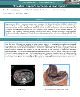
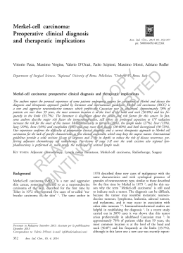
Synchronous Interdigitating Dendritic Cell Sarcoma and B-Cell Small Lymphocytic Lymphoma in a Lymph Node Antonio Cossu, MD; Angelo Deiana, MD; Amelia Lissia, MD; Maria Filomena Dedola, MD; Lucia Cocco, MD; Giuseppe Palmieri, MD; Francesco Tanda, MD ● A gradually enlarging axillary mass in a 79-year-old man was excised. The specimen was processed for light microscopy, immunohistochemical studies, and electron microscopy; gene rearrangement studies were also performed. A diagnosis of an interdigitating dendritic cell tumor of the lymph node and a B-cell small lymphocytic lymphoma occurring in the same anatomic location was made. We found that although rare cases of interdigitating dendritic cell tumor with an associated secondary malignancy have been described in the literature, to our knowledge, this is the first report of interdigitating dendritic cell tumor and synchronous neoplasm diagnosed at the same site. A possible relationship between the 2 disorders is also discussed. (Arch Pathol Lab Med. 2006;130:544–547) D endritic cells (DCs) constitute a complex system, made up of several distinct cell populations occupying discrete portions of lymphoid and nonlymphoid organs that are interconnected by defined pathways of movement. In each site, the DCs share morphologic, immunophenotypic, and functional characteristics, the most notable being the ability to capture antigens in an immunogenic form in situ and initiate B-cell– and T-cell– mediated immunity.1 The DC compartment comprises several subtypes: follicular DC of the germinal center of lymph node; Langerhans cell (LC) of the skin; interstitial DC, representing the counterpart of LC in parenchymal organs; indeterminate cell (veiled cell), derived from LC or interstitial DC and migrating into local lymphoid tissue after antigen capture; and interdigitating DC (IDC) of the T-zone of the lymph node. Follicular DCs are characterized by the presence of desmosomes and the expression of the following phenotype: CD21⫹, CD35⫹, CD1a⫺, and S100 protein positive or negative. Characteristically, LCs and interstitial DCs show intracytoplasmic Birbeck granules ultrastructurally and display the following immunohistochemical profile: Accepted for publication December 16, 2005. From the Institutes of Anatomic Pathology and Histopathology (Drs Cossu, Deiana, Lissia, Cocco, and Tanda), and Radiology, Radiotherapy section (Dr Dedola), University of Sassari, and C.N.R., Istituto di Chimica Biomolecolare (Dr Palmieri), Sassari, Italy. The authors have no relevant financial interest in the products or companies described in this article. Reprints: Antonio Cossu MD, Institute of Anatomic Pathology and Histopathology, via Matteotti 58, 07100 Sassari, Italy (e-mail: cossu@ uniss.it). 544 Arch Pathol Lab Med—Vol 130, April 2006 CD1a⫹, S100 protein positive, CD21⫺, CD35⫺, and CD86⫺. Indeterminate cells display the same immunophenotype as LCs, but do not have Birbeck granules.2 The IDCs are present within lymph nodes, tonsils, the spleen, and other lymphoid tissue. They resemble LCs morphologically, except for the absence of intracytoplasmic Birbeck granules. They are S100 protein positive, CD1a⫺, and reveal complex interdigitating cellular junctions ultrastructurally. The IDCs are characteristically located in T-cell domains, that is, the paracortex and interfollicular areas of lymph nodes and tonsils, and the periarteriolar lymphoid sheath of the spleen. They are typically surrounded by helper T cells.1 Tumors derived from DCs share morphologic, immunohistochemical, and microscopic features similar to the nonneoplastic counterparts; they are extremely rare, representing less than 1% of neoplasms present in the lymph nodes.3 Among tumors arising from the DCs system, to our knowledge, only a few cases (39 cases) of IDC sarcoma/ tumor (IDCT) have been reported in the literature; on analyzing their main clinical pathologic data, occurrence of a secondary solid or hematologic neoplasm was seen in 7 cases. We report an IDCT that was found in a lymph node biopsy that also showed a synchronous B-cell small lymphocytic lymphoma. To our knowledge, this report seems to be the first case of IDCT and B-cell small lymphocytic lymphoma simultaneously diagnosed, and our review of the literature seems to suggest that the association of IDCT and malignant secondary neoplasm, particularly B-cell small lymphoproliferative disorders, is more than coincidental. REPORT OF A CASE A 79-year-old man who had severe dilated cardiomyopathy was admitted into a local clinic with a history of inguinal hernia and painless, gradually enlarging axillary lymphadenopathy. On admission, he did not present systemic symptoms, and results from laboratory tests were within the normal limits. A complete blood count revealed a hemoglobin level of 10.2 g/dL, a platelet count of 280.2 ⫻ 103 cells/L, and a white blood cell count of 9800 cells/L. Peripheral blood smear was reported as normal. Physical examination and radiographic examination, including a total body computed tomography scan, showed no other lesions elsewhere. Manual reduction of the hernia was successfully performed. At the same time, the axillary lymph nodes were excised, and the specimen consisted of a solid, firm, whitish gray, capsulated mass Synchronous Lymph Node Sarcoma and Lymphoma—Cossu et al Figure 1. Microscopic features of interdigitating dendritic cell sarcoma/tumor (IDCT) of the lymph node: proliferation of atypical spindle cells with irregular nuclei and abundant cytoplasm. Numerous mitotic figures are also visible (hematoxylin-eosin, original magnification ⫻250). Figure 2. Immunohistochemistry for S100 protein shows immunoreactivity of the proliferating spindle cells in IDCT (original magnification ⫻250). Figure 3. Ultrastructure of IDCT. The spindle cells show a deeply indented irregular nucleus and scattered intracytoplasmic organelles. Birbeck granules and desmosomes are not appreciable (original magnification ⫻7000). Figure 4. B-cell small lymphocytic lymphoma shows a mixture of atypical lymphoid cells and larger nucleolated cells (hematoxylin-eosin, original magnification ⫻125). Figure 5. IDCT and B-cell small lymphocytic lymphoma. Positive immunoreactivity for CD20 in lymphoma cells (original magnification ⫻150). Figure 6. Polymerase chain reaction testing for immunoglobulin (Ig) H gene rearrangement showing clonal IgH rearrangement. Lanes: A, molecular weight marker; B, positive control from a patient with B cell lymphoma (frozen tissue); C, reactive lymph node, as a negative control; and D, the present case. Arch Pathol Lab Med—Vol 130, April 2006 Synchronous Lymph Node Sarcoma and Lymphoma—Cossu et al 545 Summary of Interdigitating Dendritic Cell Sarcoma/Tumors (IDCTs) Associated With Secondary Malignancy Source, y Age, y/Sex Hammar et al,5 1991 67/M Horschowski et al,4 1993 Nakamura et al,8 1994 8/M 70/M Site Associations Cervical lymph node Lymph nodes, colomesenteric masses Submandibular lymph node Vasef et al,6 1995 55/F 56/F Gaertner et al,7 2001 70/M Cervical lymph node Cervical and axillary lymph nodes Axillary lymph node 77/F Tonsil 79/M Axillary lymph node Present case (4.5 cm in diameter). The specimen was processed for light microscopy and electron microscopy. Microscopically, most of the lymph node was replaced by a spindle-cell proliferation intermingled with mature lymphocytes and few plasma cells. The neoplastic cells formed loosely compact bundles arranged in a storiform pattern, showed abundant eosinophilic cytoplasm, and had round-to-ovoid, vesicular nuclei, often with deeply cleaved, irregular nuclear membranes (Figure 1). Nucleoli were generally prominent. The spindle cells were large, exceedingly atypical, and frequently multinucleated. Mitotic activity, including atypical mitoses, was high (mean ⫽ 4–5 ⫻ 10 high-power fields), and focally heavy collagen deposition was observed, with some neoplastic cells encased within dense fibrous tissue. Immunohistochemistry performed on paraffin-embedded tissue showed the neoplastic cells to be immunoreactive for S100 protein (Figure 2), vimentin, HLA-DR, ␣1-antichymotrypsin, and weakly immunoreactive for actin and CD34; negative for HMB-45, cytokeratins (AE1/AE3), CD1a, CD45 (LCA), CD20 (L26), CD45RO (UCHL-1), CD21, CD35, CD68, CD30, epithelial membrane antigen, and desmin. Ultrastructurally (Figure 3), the tumor cells showed oval to irregularly shaped nuclei with deep indentations. The chromatin was finely granular and one or more large nucleoli were seen. The cytoplasm was abundant, with numerous mitochondria and scattered lysosomes. Neoplastic cells showed elongated interdigitating cytoplasmic processes and absence of desmosomes, melanosomes, or Birbeck granules. A portion of this lymph node and 2 adjacent lymph nodes of 2 ⫻ 1 cm each showed replacement of the architecture by a predominantly small-cell population intermingled with smaller numbers of larger nucleolated cells (Figure 4). The small cells seemed to have a striking subsinusal pattern of distribution, and had little cytoplasm and small, somewhat irregular nuclei. Scattered mitoses were observed. This proliferation was made up of lymphoid cells with the B phenotype (CD45⫹ and CD20⫹; Figure 5) and was associated with a minor component of small CD3⫹, CD45RO⫹ T cells. The vast majority of proliferating cells expressed CD5 and CD23; CD10 and nuclear cyclin D1 were negative. A meshwork of CD23⫹ follicular DCs was focally present among the atypical lymphoid cells. A clonal immunoglobulin (Ig) heavy chain gene rearrangement was detected by means of a polymerase chain reaction assay of deoxyribonucleic acid extracted and amplified from the formalin-fixed, paraffin-embedded lymphoid tissue (Figure 6). The final diagnosis was IDCT associated with B-cell small lymphocytic lymphoma. The patient refused postoperative hospitalization and further clinical investigations and treatment. Subsequently, his condition worsened because of progressive dilated cardiomyopathy, and 2 months after surgery he died from heart failure. Postmortem examination was not performed. 546 Arch Pathol Lab Med—Vol 130, April 2006 Interval B follicular center cell lymphoma T lymphoblastic lymphoma 13 y before IDCT Adenocarcinoma of the stomach and hepatocellular carcinoma of the liver Carcinoma of the breast B-cell small lymphocytic lymphoma Carcinoma of the colon 4 y after IDCT B-cell small lymphocytic lymphoma B-cell small lymphocytic lymphoma 5 mo before IDCT 13 y before IDCT 9 mo before IDCT 6 y before IDCT 1 y before IDCT Synchronous COMMENT This report, based on morphologic and immunophenotypic features, describes a rare case of IDCT associated with a synchronous B-cell small lymphocytic lymphoma. The IDCT is a neoplastic proliferation of spindle-to-oval– shaped cells with phenotypic features similar to those of IDCs found in normal tissues. The designation sarcoma/ tumor in the literature is used because of the variable cytologic grade and clinical behavior encountered in these lesions.3 This malignancy is very uncommon and only 39 cases have previously been documented in the literature, to our knowledge. IDCT usually occurs in adults (age range, 2– 86 years), with a slight male predominance (male-female ratio, 1.16). Most IDCTs are found in the lymph nodes. Rare cases of this neoplasm occur in extranodal lymphoid tissue, such as tonsil, nasopharynx, skin, intestine, testis, spleen, bladder, and salivary gland. Approximately half of the patients die of the disease within an average period of 6 to 7 months (range, 1 week–16 months). The response to therapy is also generally poor, although some patients experience either long disease-free survival or complete remission. One very interesting fact regarding this tumor, although not surprising, is the existence of a concomitant tumor in the clinical history of some patients. Such an association has been well documented for DC-related disorders, and, in our review, we found that a diagnosis of a malignant secondary neoplasm that may follow or precede IDCT was reported in 7 patients4–8 (Table). Four of the 7 cases described are malignant hematologic tumors, consistent with a non-Hodgkin lymphoma (NHL), either of a T-cell line (1 case)4 or a small B-cell type (3 cases).5–7 Although the number of these cases is low, the occurrence (17%) is greater than expected. Our case also represents an IDCT synchronous to B-cell small lymphocytic lymphoma in the same lymph node; to our knowledge, such an occurrence simultaneously diagnosed in the same biopsy has not previously been reported. Indeed, the high frequency of association with NHL (12% including the present case and the previous reports) suggests a clear relationship and raises interesting questions concerning the pathogenesis of a secondary malignancy (particularly, a small B-cell lymphoproliferative disorder) in IDCT. Synchronous Lymph Node Sarcoma and Lymphoma—Cossu et al It is generally accepted that escape from immune surveillance is a fundamental feature of tumors that contributes to their uncontrolled growth.9 Antigen-presenting cells are one of the most efficient vehicles for the delivery of tumor antigens; dysfunction of DCs, showing a morphologically and phenotypically immature population with a reduced response against neoplastic cells, has already been reported in different malignancies, including hepatocellular carcinoma10 and chronic lymphocytic leukemia.11 In IDCT, a specific defect of transformed neoplastic DCs in immune surveillance could also contribute to the onset or development of concurrent malignant tumors. Interestingly, IgD⫹ naı̈ve B cells in T-cell areas of the human mesenteric lymph nodes attach directly to IDCs by 1 or 2 cytoplasmic projections, and a small number of IDCs are present in B-cell–rich areas, such as the periphery of the mantle zones or primary follicles.12 The functional role of these IDCs in B-cell–rich areas remains unclear, but these data suggest that IDCs are more deeply involved in the B-cell–mediated immunologic responses than was previously thought, and that DCs may directly modulate differentiation and proliferation of small B-cell lymphocytes. Clearly, much remains to be investigated beyond these tentative studies to define the molecular mechanism involved in the interactions between DC and B cells and their possible role in human diseases involving B cells, such as lymphomas. Some authors have postulated that therapeutic agents in a patient with NHL play a role in the development of DC neoplasms,13 but this hypothesis has not been confirmed and, in our case, the patient had not previously been treated and IDCT was found with a B-cell small cell lymphoma. In conclusion, our review confirms that IDCT is both a well-defined and extremely rare entity; moreover, the more than coincidental association with other NHLs is of great interest, and our report of an IDCT occurring in the Arch Pathol Lab Med—Vol 130, April 2006 same lymph node simultaneously with a B-cell small lymphocytic lymphoma further supports such an association. However, additional studies should be performed to clarify the exact relationship between IDCT and small B-cell NHL and the possible pathogenetic factors. References 1. Knowles DM. Immunophenotypic markers useful in the diagnosis and classification of hematopoietic neoplasms. In: Knowles DM, ed. Neoplastic Hematopathology. Baltimore, Md: Williams & Wilkins; 2001:179–182. 2. Pileri SA, Grogan TM, Harris NL, et al. Tumours of histiocytes and accessory dendritic cells: an immunohistochemical approach to classification from the International Lymphoma Study Group based on 61 cases. Histopathology. 2002;41: 1–29. 3. Jaffe ES. Histiocytic and dendritic cell neoplasms: introduction. In: Jaffe ES, Harris NL, Stein H, Vardiman JW, eds. Pathology and Genetics of Tumours of Hematopoietic and Lymphoid Tissues. Lyon, France: IARC Press; 2001:275–277. World Health Organization Classification of Tumours; vol 3. 4. Horschowski N, Guitard AM, Arnoux I, et al. Interdigitating cell sarcoma: occurrence during incomplete remission of a lymphoblastic lymphoma. Pathol Biol (Paris). 1993;41:255–259. 5. Hammar SP, Rudolph RH, Bockus DE, Remington FL. Interdigitating reticulum cell sarcoma with unusual features. Ultrastruct Pathol. 1991;15:631–645. 6. Vasef MA, Zaatari GS, Chan WC, Sun NCJ, Weiss LM, Brynes RK. Dendritic cell tumors associated with low grade B-cell malignancies: report of three cases. Am J Clin Pathol. 1995;104:696–701. 7. Gaertner EM, Tsokos M, Derringer GA, Neuhauser TS, Arciero C, Andriko JAW. Interdigitating dendritic cell sarcoma: a report of four cases and review of the literature. Am J Clin Pathol. 2001;115:589–597. 8. Nakamura S, Koshikawa T, Kitoh K, et al. Interdigitating cell sarcoma: a morphologic and immunologic study of lymph node lesions in four cases. Pathol Int. 1994;44:374–386. 9. Ioannides C, Whiteside T. T cell recognition of human tumors: implications for molecular immunotherapy of cancer. Clin Immunol Immunopathol. 1993;66: 91–106. 10. Ninomiya T, Akbar SMF, Masumoto T, Horiike N, Onji M. Dendritic cells with immature phenotype and defective function in the peripheral blood from patients with hepatocellular carcinoma. J Hepatol. 1999;31:323–331. 11. Orsini E, Guarini A, Chiaretti S, Mauro FR, Foa R. The circulating dendritic cell compartment in patients with chronic lymphocytic leukemia is severely defective and unable to stimulate an effective T-cell response. Cancer Res. 2003; 63(15):4497–4506. 12. Takahashi K, Kenji A, Norihiro T, et al. Morphological interactions of interdigitating dendritic cells with B and T cells in human mesenteric lymph nodes. Am J Pathol. 2001;159:131–138. 13. de Camargo B, Alves AC, Gorender EF, Bianchi A. Association of malignancy and Langerhans’ cell histiocytosis: report of three cases. Med Pediatr Oncol. 1993;21:451–453. Synchronous Lymph Node Sarcoma and Lymphoma—Cossu et al 547
Scaricare