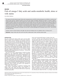
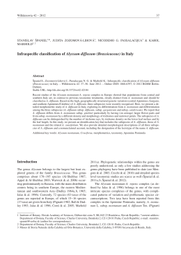
Int Microbiol (2002) 5: 3–9
DOI 10.1007/s10123-002-0051-6
R EV IE W A RT I C L E
Jesús L. Romalde
Photobacterium damselae subsp. piscicida : an integrated view
of a bacterial fish pathogen
Received: 10 September 2001 / Accepted: 10 October 2001 / Published online: 16 April 2002
Ó Springer-Verlag and SEM 2002
Abstract Pasteurellosis, or pseudotuberculosis, is a
bacterial septicaemia caused by the halophilic bacterium
Photobacterium damselae subsp. piscicida (formerly
Pasteurella piscicida). Although this disease was first
described in wild populations of white perch and striped
bass, currently the natural hosts of the pathogen are a
wide variety of marine fish. The disease has great
economic impact both in Japan, where it affects mainly
yellowtail cultures, and in the Mediterranean area, due
to the losses it causes in seabream and seabass farms.
This microorganism serves as a perfect model to study a
bacterial fish pathogen, either at an applied level, to
resolve or to mitigate the high economic losses of fish
farmers, or at a basic level, for a better understanding of
P. damselae subsp. piscicida biology. This article discusses the methods employed in our laboratory to study
the causative agent of pasteurellosis. It reviews important aspects, from the diverse procedures for the detection and isolation of the pathogen to the latest molecular
studies that have allowed its correct taxonomic allocation. Characterization of some virulence mechanisms
and the available methods to prevent the disease are also
presented.
Keywords Photobacterium damselae subsp. piscicida Æ
Taxonomy Æ Virulence Æ Detection Æ Vaccine Æ
Fish pathology
Lecture given in Alicante in the 18th SEM National Congress
(Alicante, September 16-20, 2001), on the occasion of receiving the
ninth BIennial Prize of the Spanish Society for Microbiology (see
Table 1)
J.L. Romalde
Departamento de Microbiologı́a y Parasitologı́a,
Facultad de Biologı́a,
Universidad de Santiago de Compostela,
15782 Santiago de Compostela, Spain
E-mail: [email protected]
Tel.: +34-981563100
Fax: +34-981596904
Introduction
Pasteurellosis is a bacterial septicaemia also referred to
as pseudotuberculosis because, in chronic cases, diseased
fish show, in several internal organs, whitish tubercles
consisting of bacterial accumulations. The halophilic
bacterium Photobacterium damselae subsp. piscicida
(formerly Pasteurella piscicida) is the aetiological agent
of pasteurellosis. Due to this change in the taxonomic
position of the causative agent, some authors have renamed this septicaemia as photobacteriosis. However, to
avoid possible confusion with other fish diseases caused
by other species of the genus Photobacterium, this review
will maintain the name pasteurellosis.
The disease was first described in wild populations of
white perch (Morone americanus) and striped bass
(Morone saxatilis) in 1963, when a massive epizootic
occurred in Chesapeake Bay (USA) [52]. Sniezsko and
coworkers placed the microorganism isolated during this
first epizootic within the genus Pasteurella on the basis
of its morphological and biochemical properties. Later,
Jansen and Surgalla [11] examined the microorganism
and found sufficient distinctions in physiological and
serological characteristics from other species in the genus to propose the new species name Pasteurella piscicida. A few years later, pasteurellosis became a problem
in yellowtail (Seriola quinqueradiata) and ayu (Plecoglosus altivelis) cultures in Japan [13, 16, 17,34]. Soon
thereafter, the disease spread to other fish species in
Japan, and several epizootics were described in red
seabream (Acanthopagrus schlegeli), red grouper (Epinephelus akaara), oval file fish (Navodan modestus), and
hybrid striped bass [M. saxatilis (M. chrysops)] [see
review 28].
In Europe, isolation of the bacterium from rudd
(Scardinius erythrophthalmus), chub (Coregonus zenithicus), Atlantic salmon (Salmo salar), brown trout (Salmo
trutta), or sheat fish (Silurus glanis) was reported in
different countries such as England, Norway and Hungary, but the infectious agent was later confirmed as
4
Aeromonas salmonicida isolates. No cases of pasteurellosis were noted on the continent until 1990; Toranzo
et al. [55] described the first outbreak of the disease in
Spain, which affected juvenile gilthead seabream (Sparus
aurata) cultures in the northwestern area. Almost
simultaneously, outbreaks of pasteurellosis also occurred in southwestern Spain, France, Italy, Israel,
Greece and Portugal, mainly in populations of seabream
and seabass [see review 28]. Since then, pasteurellosis has
been a major limiting factor in the culture of these fish
species in the Mediterranean region.
Today, the natural hosts of the pathogen are a wide
variety of marine fish. Pasteurellosis has had a great
economic impact in Japan, where it affects mainly yellowtail cultures, and in the Mediterranean area, because
of the losses it causes in seabream and seabass farms.
For these reasons, many aspects of the disease and its
causative agent P. damselae subsp. piscicida have been
extensively studied, resulting in a quite complete picture
of fish pasteurellosis. This article reviews the current
state of knowledge of P. damselae subsp. piscicida, focusing on important aspects such as the phenotypic and
genetic characterization of the bacterium, its main virulence mechanisms, and effective strategies to prevent
the disease.
Isolation and phenotypic characterization of isolates
P. damselae subsp. piscicida can be isolated from internal organs of diseased fish after 2–4 days incubation at
22 °C by using media such as trypticase soy agar (TSA)
and blood agar, both supplemented with 1–2% NaCl, or
marine agar 2216E. The presumptive diagnosis is based
on the isolation of a gram-negative, non-motile bipolar
rod that is oxidase- and catalase-positive, fermentative
without gas production, sensitive to the vibriostatic
agent O/129, and has strict salt requirements. However,
diagnosis can be hampered by the slow growth of this
bacterium in laboratory media, which is easily obscured
by other fast-growing bacteria. This fact greatly encourages biochemical, serological and genetic characterization studies in order to develop an accurate and
specific diagnostic procedure.
Based on such exhaustive characterization, a complete
description of the pathogen was obtained [9,28].
P. damselae subsp. piscicida is a pleomorphic bacterium
changing from coccoidal to long rods depending on the
culture conditions. The etiological agent of fish pasteurellosis shows positive responses for arginine dihydrolase,
lipase and phospholipase activities, and is negative for
indole, nitrate reduction, urease, gelatinase, amylase and
hydrogen sulfide production [28]. Regarding the use of
carbohydrates, this bacterium produces acid only from
glucose, mannose, galactose and fructose. Variable results
were reported for methyl-red, Voges-Proskauer, sucrose
and maltose reactions [41]; as suggested by Koike et al.
[15], the employment of different basal media could account for this variability. Hemolytic activities against
trout and sheep erythrocytes were not detected.
P. damselae subsp. piscicida can grow at temperatures
from 15 to 32.5 °C, although its optimum growth temperature is 22.5–30 °C. It is able to produce capsular
material [1] composed of 99.6% carbohydrate and 0.4%
protein. These overall phenotypic results show that
P. damselae subsp. piscicida constitutes an homogeneous
group that differs from the other subspecies – P. damselae
subsp. damselae (formerly Vibrio damsela) – in important
biochemical and physiological traits, such as motility, gas
production from glucose, nitrate reduction, urease, lipase,
amylase, and hemolysin production [5,24]. The homogeneity of P. damselae subsp. piscicida facilitates use of the
miniaturized system API-20E for its identification. In
agreement with Kent [12], although the pathogen is not
included in the API-20E code index, the system is valuable
for a rapid presumptive identification of the bacterium,
because all strains have a similar pattern (2005004), with
neither false-positive nor false-negative reactions being
detected [47].
From a serological standpoint, P. damselae subsp.
piscicida is also a highly homogeneous group [24,46],
which makes it impossible to establish serotypes. Its
serological homogeneity was supported by lipopolysaccharide (LPS) profiles. In fact, all strains analyzed,
regardless of their origin or source of isolation, had the
same electrophoretic pattern: O side-chains consisting of
high-molecular-mass bands in a ladder-like pattern [24].
In addition, similar profiles of total and outer membrane
proteins (OMPs) were also observed for all isolates. The
LPS and OMP profiles of all the P. damselae subsp.
piscicida strains proved to be immunologically related in
the respective Western blot assays [24,28]. This antigenic
uniformity encouraged the development of serological
techniques for the detection and identification of
P. damselae subsp. piscicida isolates. Slide agglutination
and latex agglutination tests, direct immunofluorescent
assays, and enzyme-linked immunosorbent assay
(ELISA) procedures are among the methods that have
been employed and even commercialized for this purpose [14, 41, 43,45].
Photobacterium damselae subsp. piscicida showed a
different plasmid content depending on the strain analyzed. Thus, whereas European and USA strains share
plasmids of 20 and 7 MDa, the Japanese isolates have a
common 37-MDa plasmid. In addition, a high-molecular-mass plasmid band of approximately 50 MDa has
been detected in the majority of European isolates
studied [24]. Restriction endonuclease analysis demonstrated that the plasmid bands shared by most European
strains are genetically homogeneous.
Genetic studies and the design of molecular
detection methods
Despite the fact that pasteurellosis has long been recognized as a
disease of fish, the taxonomic position of its etiological agent,
Photobacterium damselae subsp. piscicida, was a matter of contro-
5
versy that has only recently been clarified. On the basis of its
physiological characteristics, such as Gram staining, oxidase activity, absence of motility, and the typical bipolar rod shape, the
bacterium was first assigned to the genus Pasteurella [52]. Shortly
after, Janssen and Surgalla [11] proposed the name Pasteurella
piscicida to designate this pathogen, although some traits did not
match with the description of the genus (i.e. its inability to reduce
nitrate reduction and its sensitivity to low pH and temperatures).
P. piscicida was at that time the only halophilic member of the
genus. In the early 1970s, some Japanese authors suggested different genera, such as Corynebacterium [13] and Arthrobacter [48],
as better taxonomic positions for this fish pathogen on the basis of
diverse characteristics including carbohydrate metabolism, pleomorphism, and sensitivity to penicillin. Immunological techniques
showed that P. piscicida had no common antigens either with
Corynebacterium or with Arthrobacter [21]. However, a positive
relationship was observed with Pasteurella plecoglosicida isolated
from ayu, which supported the first assignation of the bacterium to
the genus Pasteurella.
It has been reported that P. piscicida shares major phenotypic
characteristics with other fish pathogens including Vibrio anguillarum, Aeromonas salmonicida, and the clinically important
pasteurellae, Pasteurella multocida and Pasteurella haemolytica
[24]. A study of the fatty acid methyl ester (FAME) profiles by gas
chromatography [44] revealed significant homogeneity in the major
fatty acids among the different P. piscicida strains. In addition,
comparison of these results with those obtained from representative
strains of V. anguillarum, A. salmonicida and Pasteurella species of
clinical origin suggested that the agent of fish pasteurellosis should
be closer to Pasteurella than to Vibrio or Aeromonas.
On the other hand, De Ley et al. [2], employing DNA-rRNA
hybridization techniques, suggested that P. piscicida should be a
member of the Vibrionaceae strongly related to V. parahaemolyticus. Unfortunately, this study included only a presumptive isolate
of P. piscicida, which, according to the culture conditions specified
by the authors, could be the result of cross-contamination. Later,
Nicolas et al. [36], on the basis of rRNA studies, reallocated
P. piscicida as a new subspecies of Vibrio damsela. Due to this long
uncertainty and controversy, the name Pasteurella piscicida was
never included in the Approved List of Bacterial Names [50]. In
1994, Ruimy et al. [8] carried out an exhaustive study of the family
Vibrionaceae and related organisms by means of 16S rRNA sequencing and DNA-DNA hybridization. One year later [9], they
proposed the reassignment of P. piscicida as a new subspecies of the
recently created Photobacterium damsela comb. nov. (formerly
Vibrio damsela)[51], since the two organisms differ only in one
nucleotide in the 16S rRNA sequence and showed a DNA-DNA
hybridization of approximately 80%. These results were later
confirmed by other authors [39], who also demonstrated the high
similarity in rRNA sequence of other members of the genus Photobacterium (98.1% for P. histaminum) with both subspecies of
P. damsela. Therefore, the new, and finally approved, name for the
causal agent of fish pseudotuberculosis was established as Photobacterium damsela subsp. piscicida. Truper and De’Clari [56] corrected the specific epithet ‘‘damsela’’ (substantive) to ‘‘damselae’’
(genitive) giving the current name of the bacterium, Photobacterium
damselae subsp. piscicida.
Once the taxonomic position of the bacterium responsible of
fish pasteurellosis had been clarified, and new molecular procedures for genetic analysis became available, studies to obtain
better knowledge of the pathogen’s genome were carried out.
Ribotyping and random amplification of polymorphic DNA
(RAPD) showed the existence of two clonal lineages within
P. damselae subsp. piscicida (Fig. 1), one including the European
strains and the other the Japanese isolates [31, 33,53]. These
results constituted the first evidence of genetic heterogeneity
within this fish pathogen.
In addition, the number of sequenced genes from P. damselae
subsp. piscicida has increased in recent years, not only to further
characterization of the bacterium, but also to find a sequence on
which the design of a subspecies-specific detection method could be
based. The high degree of similarity in the 16S rRNA sequence of
Fig. 1. RAPD fingerprints (A), achieved after DNA amplification
using primer 4 (Amersham Pharmacia), and ribotype patterns (B),
obtained employing the endonuclease PvuII, for the P. damselae
subsp. piscicida isolates. Lanes: a–g European isolates, h–j Japanese
isolates, M molecular ladder (50–2,000 bp; Bio-Rad). The molecular sizes (in kb) are indicated on the right
the two subspecies of P. damselae has allowed only the development of a PCR-based detection method at a species level [39]. We
have compared the sequences of the two other ribosomal genes, 5S
and 23S, in both subspecies [38] and found similarities also higher
than 99%. The intergenic spacer regions ITS-1 (located between the
16S and 23S genes) and ITS-2 (between the 23S and 5S genes),
which are believed to evolve between lineages at a higher ratio than
rRNA genes, showed again similarity percentages of 98–99.5%.
Note that these genomic regions have a peculiar mosaic-like
structure, and there is a possibility that there is some inter-subspecies heterogeneity in terms of differential reorganizations of the
sequence pieces [38].
The gyrB gene, coding for the B subunit of the DNA gyrase, as
well as four regulatory genes, toxR, rpoN, luxS, and a sigma-54dependent activator, were also analyzed as alternative molecular
chronometers. In all cases, the results supported those deduced
from the ribosomic operon study, i.e. high similarities between both
subspecies [38].
Furthermore, we investigated at a DNA level one of the main
phenotypic differences between P. damselae subsp. piscicida and
subsp. damselae [40], namely, urease activity. Whereas subsp.
damselae harbors the ureC gene, included in the ure operon, subsp.
piscicida lacks this gene, which is in accordance with its ureasenegative phenotype. Although these results raise new questions on
the speciation processes and the dramatic phenotypic divergence of
two subspecies so closely related at the ribosomal operon level, they
were decisive for the design of a subspecies-specific detection
method. This method, based on multiplex-PCR for 16S rRNA and
ureC genes [40], is currently the only PCR assay that can successfully discriminate between subspecies of P. damselae (Fig. 2), and is
therefore a serious candidate to become a useful tool for diagnosis
of pasteurellosis in the field. In fact, the assay is being used in our
laboratory for routine detection of P. damselae subsp. piscicida
(unpublished results).
6
Fig. 2. Agarose electrophoresis of the multiplex-PCR products
obtained for the different Photobacterium strains studied. Lanes:
M, Molecular mass markers (100-bp molecular ladder; Bio-Rad);
a–d, P. damselae subsp. damselae strains; e, P. histaminum isolate;
f–p P. damselae subsp. piscicida strains. Numbers on the right
indicate the size of the amplification products, corresponding to the
448- and 267-bp internal fragments of the ureC and 16S rRNA
genes, respectively
Virulence mechanisms
Photobacterium damselae subsp. piscicida is a highly
pathogenic bacterium that does not seem to have host
specificity. Therefore, pasteurellosis can be a risk even
for marine fish species in which the disease has not been
yet described. Some authors have pointed out differences in susceptibility of some fish species (i.e. gilthead
seabream and seabass) to pasteurellosis on the basis of
fish age [37]. Thus, whereas small seabream (less than
5 g) are susceptible to the disease, fish above 50 g
become resistant. This is due to the functionality of
macrophages and neutrophils in larger seabream, which
can efficiently phagocytize and kill the bacteria [37,49].
In any case, the fact that this bacterium is highly
pathogenic indicates that it must have strong virulence
mechanisms.
Adherence and invasive capacities are essential in the
first stages of infection. Although P. damselae subsp.
piscicida has been described as adhering weakly to different cell lines, it showed significant binding to fish intestines, with values ranging from 104–105 bacteria per
gram of tissue [29]. In addition, despite being only
moderately invasive, the pathogen has been demonstrated to survive for a few days within host cells and
even to spread to adjacent cells [29].
Extracellular products (ECPs) secreted by a variety of
bacterial fish pathogens are important virulence factors,
since they can contribute to the development of the
disease in terms of bacterial nutrition or aggresins, enabling the bacteria to counteract the host’s defence
mechanisms [54]. The ECPs of P. damselae subsp.
piscicida strains were shown to be lethal for different fish
species and for mice, and had phospholipase, cytotoxic
and hemolytic activities as major characteristics [25].
Histological studies have implicated these activities, in
particular the phospholipases, in the pathogenesis of
pasteurellosis [37].
Today it seems clear that the main virulence factors
of P. damselae subsp. piscicida consist of polysaccharide
capsular material (Fig. 3) [1, 23, 30, 42] and a high-affinity siderophore-mediated iron-sequestering system.
Fig. 3A, B. Transmission electron micrographs of thin sections of
cells of P. damselae subsp. piscicida. A Avirulent strain EPOY8803-II; B virulent strain DI
Implication of the capsular material in virulence was
clearly demonstrated in experimental fish infections using virulent constitutively capsulated and avirulent noncapsulated forms of P. damselae subsp. piscicida. The
induction of capsular expression in the non-capsulated
forms increased their resistance to the bactericidal action
of the fish serum and resulted in a 50% lower lethal dose
(LD50), around 2–3 log units [30].
The ability to acquire iron is essential for the growth
of pathogenic bacteria within the host; therefore, it is
also essential to cause infections. The existence in
P. damselae subsp. piscicida of a chromosomally located
iron uptake system of has been described. It consists of a
siderophore, chemically and biologically related to
multicidin produced by Pasteurella multocida, and, at
least three iron-regulated high-mass OMPs [26]. In addition, it has also been reported that P. damselae subsp.
piscicida can utilize hemin directly as the sole iron source
by means of constitutive and additional inducible surface protein receptors, and that intraperitoneal injection
of hemin before experimental infection increased the
lethality of this pathogen [26].
Further studies [3,4] demonstrated the relationship
between both virulence mechanisms. First, it was evidenced that capsular polysaccharides play a minor role
in the binding of hemin and, second, that the expression
of capsular material is dependent on iron availability
and growth phase. Regarding this second point, cells
grown under iron-limited conditions always had a significantly lower amount of capsular material than ironsupplemented cells [3]. This finding can be explained by
the need of the bacterium to express its siderophore and/
or iron receptors during its time within the host circulatory system. Once the microorganism reaches the
different tissues, the amount of capsular material
probably increases in response to host cellular defence
mechanisms (i.e. phagocytosis by macrophages). In
addition, the role of iron in the expression of enzymatic
activities has been reported, and some proteolytic
enzymes, such as gelatinase and caseinase, are only
synthesized when strains are cultured under ironrestricted conditions [26].
It is obvious that the pathogenesis of P. damselae
subsp. piscicida is a complex, multifactorial process not
7
yet fully understood, although the considerable work
carried out in the last few years has contributed to
clarifying it to a great extent.
OMPs [26] as protective antigens. Both formulations
have shown promising results in preliminary studies.
Conclusions
Vaccination: an effective measure of prevention
Chemotherapy was effective for the treatment of fish
pasteurellosis until the late 1980s. Since then, plasmidor chromosome-coded resistance to ampicillin, tetracycline, and other drugs has been observed [25,36];
therefore, new chemotherapeutants, such as florfenicol
and phosphomycin, have been employed to control
pasteurellosis outbreaks. We have used proteomic
analysis to study the mechanisms of resistance. Comparison of resistant and susceptible isolates by two-dimensional polyacrylamide gels and analysis of the
protein sequences have yielded promising results which
will aid in understanding the biology of resistance acquisition. Moreover, it has been described that
P. damselae subsp. piscicida has a period of intracellular parasitism within macrophages during infection
[20], which can explain the ineffectiveness of chemotherapy in the treatment of some outbreaks of the
disease. Therefore, immunoprophylaxis has become the
best way to prevent pasteurellosis.
Throughout the last 20 years, there have been
a variety of studies analyzing the effectiveness of
immunization in preventing pasteurellosis [42]. Most
vaccines tested consisted of heat- or formalin-killed
cells [6, 10, 18,19]. Although some protection was
achieved with these preparations, the results were not
reproducible [20]. Better results were obtained using
formulations based on LPS and ribosomal fractions of
the bacteria [7,22]. However, these formulations presented not only problems of reproducibility, but also
difficulties of production on a large scale. Passive
immunization has also been evaluated [6], but, due to
the short period of protection achieved, it was effective
only if the vaccine was administered up to 24 h prior
to challenge.
The best protection against pasteurellosis was obtained with an ECP-enriched bacterin developed in our
laboratory, achieving relative percent survival (RPS)
values higher than 75% when vaccinating fish between
0.5 and 2 g [27]. Further studies demonstrated that
vaccination at the larval stage (50-day old fish) is also
effective, with RPS values of 84–90% [31]. Therefore,
the vaccination program recommended consists of a
first immunization when larvae are around 50 mg and
a booster immunization when fish reach 2 g body
weight. This vaccine is currently commercially available and has been successfully employed in several
European countries including Spain, Portugal and
Greece.
Future trends in vaccine formulations against
P. damselae subsp. piscicida are the use of live attenuated
bacteria [19], although their utilization in the field is
currently not allowed, and the use of iron-regulated
Great effort has been invested in the last few years in the
study of Photobacterium damselae subsp. piscicida, a
bacterium that can serve as a model for the study of any
bacterial fish pathogen. Although some aspects of this
complex disease and its etiological agent are not yet fully
understood, the global extent of this disease and the
enormous economic losses it causes will constitute an
important impetus to further our knowledge of the
pathobiological characteristics of this microorganism.
Acknowledgments The author is indebted to the Spanish Society
for Microbiology (SEM) for presenting him with the ninth Biennal
Prize and the invitation to deliver the Closing Lecture at the 18th
SEM National Congress in Alicante (Table 1). Thanks are due to
Dr. Alicia E. Toranzo for critical reading of the manuscript.
References
1. Bonet R, Magariños B, Romalde JL, Simón-Pujol MD,
Toranzo AE, Congregado F (1994) Capsular polysaccharide
expressed by Pasteurella piscicida grown in vitro. FEMS Microbiol Lett 124:285–290
2. De Ley J, Mannheim W, Mutters R, Piechulla K, Tytgat R,
Segers, P, Bisgaar M, Frederiksen W, Hinz KH, Vanhoucke M
(1990) Inter- and intrafamilial similarities of rRNA cistrons of
the Pasteurellaceae. Int J Syst Bacteriol 40:126–137
3. Do Vale A, Ellis AE, Silva MT (2001) Electron microscopic
evidence that expression of capsular polysaccharide by Photobacterium damselae subsp. piscicida is dependent on iron
availability and growth phase. Dis Aquat Org 44:237–240
4. Do Vale A, Magariños B, Romalde JL, Lemos ML, Ellis AE,
Toranzo AE (2002) Binding of haemin by the fish pathoghen
Photobacterium damselae subsp. piscicida. Dis Aquat Org
48:109-115
5. Fouz B, Larsen JL, Nielsen B, Barja JL, Toranzo AE (1992)
Characterization of Vibrio damsela strains isolated from
turbot Scophthalmus maximus in Spain. Dis Aquat Org
12:155–166
6. Fukuda Y, Kusuda R (1981) Efficacy of vaccination for
pseudotuberculosis in cultured yellowtail by various routes of
administration. Bull Japan Soc Sci Fish 47:147–150
7. Fukuda Y, Kusuda R (1985) Vaccination of yellowtail against
pseudotuberculosis. Fish Pathol 20:421–425
8. Gauthier G, Lafay B, Ruimy R, Breittmayer V, Nicolas JL,
Gauthier M, Christen R (1994) Phylogenetic analysis and assessment of the genera Vibrio, Photobacterium, Aeromonas, and
Plesiomonas deduced from small-subunit rRNA sequences. Int
J Syst Bacteriol 44:416–426
9. Gauthier G, Lafay B, Ruimy R, Breittmayer V, Nicolas JL,
Gauthier M, Christen R (1995) Small-subunit rRNA sequences
and whole DNA relatedness concur for the reassignment of
Pasteurella piscicida (Sniezsko et al.) Janssen and Surgalla to
the genus Photobacterium as Photobacterium damsela subsp.
piscicida comb. nov. Int J Syst Bacteriol 45:139–144
10. Hamaguchi M, Kusuda R (1989) Field testing of Pasteurella
piscicida formalin killed bacteria against pseudotuberculosis in
cultured yellowtail, Seriola quinqueradiata. Bull Mar Sci Fish,
Kochi Univ. 11:11–16
11. Janssen WA, Surgalla MJ (1968) Morphology, physiology, and
serology of Pasteurella species pathogenic for white perch.
J Bacteriol 96:1606–1610
8
12. Kent ML (1982) Characteristics and identification of Pasteurella
and Vibrio pathogenic to fishes using API-20E (Analytab
Products) multitube test trips. Can J Fish Aquat Sci 39:1725–
1729
13. Kimura T, Kitao T (1971) On the causative agent of tuberculoidosis of yellowtail. Fish Pathol 6:8–14
14. Kitao T, Kimura M (1974) Rapid diagnosis of pseudotuberculosis in yellowtail by means of the fluorescent antibody
technique. Bull Jpn Soc Sci Fish 40:889–893
15. Koike Y, Kuwahara A, Fujiwara, H (1975) Characterization of
Pasteurella piscicida isolated from white perch and cultivated
yellowtail. Jpn J Microbiol 19:241–247
16. Kubota, S, Kimura M, Egusa S (1970) Studies of a bacterial
tuberculoidosis of the yellowtail. I. Symptomatology and histopathology. Fish Pathol 4:11–18
17. Kusuda R, Miura W (1972) Characteristics of a Pasteurella sp.
pathogenic for pond cultured ayu. Fish Pathol 7:51–57
18. Kusuda R, Hamaguchi M (1987) A comparative study on
efficacy of immersion and a combination of immersion and oral
vaccination methods against pseudotuberculosis in yellowtail.
Nippon Suisan Gakkaishi 53:1005–1008
19. Kusuda R, Hamaguchi M (1988) The efficacy of attenuated live
bacterin of Pasteurella piscicida against pseudotuberculosis in
yellowtail. Bull Eur Ass Fish Pathol 8:51–53
20. Kusuda R, Salati F (1993) Major bacterial diseases affecting
mariculture in Japan. Ann Rev Fish Dis 3:69–85
21. Kusuda R, Kawai K, Matsui M (1978) Etiological studies on
bacterial pseudotuberculosis in cultured yellowtail with
Pasteurella piscicida as the causative agent. II. On the serological properties. Fish Pathol 13:79–83
22. Kusuda R, Ninomiya M, Hamaguchi M, Muraoka A (1988)
The efficacy of ribosomal vaccine prepared from Pasteurella
piscicida against pseudotuberculosis in cultured yellowtail. Fish
Pathol 23:191–196
23. Magariños B (1995) Pasteurella piscicida: aspectos taxonómicos, serológicos y de patogenicidad. Ph.D. Thesis. University of
Santiago de Compostela, Spain
24. Magariños B, Romalde JL, Bandı́n I, Fouz B, Toranzo AE
(1992a) Phenotypic, antigenic, and molecular characterization
of Pasteurella piscicida strains isolated from fish. Appl Environ
Microbiol 58:3316–3322
25. Magariños B, Santos Y, Romalde JL, Rivas C, Barja JL, Toranzo AE (1992b) Pathogenic activities of live cells and extracellular products of the fish pathogen Pasteurella piscicida.
J Gen Microbiol 138:2491–2498
26. Magariños B, Romalde JL, Lemos, ML, Barja JL, Toranzo AE
(1994) Iron uptake by Pasteurella piscicida and its role
in pathogenicity for fish. Appl Environ Microbiol 60:2990–
2998
27. Magariños B, Noya M, Romalde JL, Pérez G, Toranzo AE
(1994) Influence of fish size and vaccine formulation on the
protection of gilthead seabream against Pasteurella piscicida.
Bull Eur Ass Fish Pathol 14:120–122
28. Magariños B, Toranzo AE, Romalde JL (1996) Phenotypic and
pathobiological characteristics of Pasteurella piscicida. Annu
Rev Fish Dis 6:41–64
29. Magariños B, Romalde JL, Noya M, Barja, JL, Toranzo, AE
(1996) Adherence and invasive capacities of the fish pathogen
Pasteurella piscicida. FEMS Microbiol Lett 138:29–34
30. Magariños B, Bonet R, Romalde JL, Martı́nez MJ, Congregado F, Toranzo AE (1996) Influence of the capsular layer on
the virulence of Pasteurella piscicida for fish. Microb Pathogen
21:289–297
31. Magariños B, Osorio CR, Toranzo AE, Romalde JL (1997)
Applicability of ribotyping for intraspecific classification and
epidemiological studies of Photobacterium damsela subsp. piscicida. System Appl Microbiol 20:634–639
32. Magariños B, Romalde JL, Barja JL Núñez S, Toranzo AE
(1999) Protection of gilthead seabream against pasteurellosis at
the larval stages. Bull Eur Assoc Fish Pathol 19:159–161
33. Magariños B, Toranzo AE, Barja JL, Romalde JL (2000) Existence of two geographically-linked clonal lineages in the
34.
35.
36.
37.
38.
39.
40.
41.
42.
43.
44.
45.
46.
47.
48.
49.
50.
51.
52.
53.
bacterial fish pathogen Photobacterium damselae subsp. piscicida evidenced by random amplified polymorphic DNA analysis. Epidemiol Infect 125:213–219
Muroga K, Sugiyama, T, Uek, N (1977) Pasteurellosis in cultured black seabream, Mylio macrocephalus. J Fac Fish Anim
Hiroshima Univ 16:17–21
Nakano S, Aoki T, Kitao T (1989) In vitro antimicrobial activity of pyridonecarboxilyc acid against fish pathogens.
J Aquat An Health 1:43–50
Nicolas JL, Gauthier G, Orsini L, Corre S (1994) Use of 16S
rRNA sequences for taxonomy and detection of pathogenic
bacteria. 6th International Colloquium on Pathology in Marine
Aquaculture. Montpellier, France, p. 54
Noya M, Magariños B, Toranzo AE, Lamas J (1995) Sequential pathology of experimental pasteurellosis in gilthead
seabream, Sparus aurata. A light-and electron microscopic
study. Dis Aquat Org 21:177–186
Osorio CR (1999) Caracterización genética de Photobacterium
damselae: estudio del operón ribosómico, microevolución y
diagnóstico molecular. Ph.D. Thesis. University of Santiago de
Compostela, Spain
Osorio CR, Collins MD, Toranzo AE, Barja JL, Romalde JL
(1999) 16S rRNA gene sequence analysis of Photobacterium
damselae and nested PCR method for rapid detection of the
causative agent of fish pasteurellosis. Appl Environ Microbiol
65:2942–2946
Osorio CR, Toranzo AE, Romalde JL, Barja JL (2000) Multiplex PCR assay for ureC and 16S rRNA genes clearly discriminates between both subspecies of Photobacterium
damselae. Dis Aquat Org 40:177–183
Robohm RA (1983) Pasteurella piscicida. In: Anderson DP,
Dorsonand M, Dubourget P (eds) Antigens of fish pathogens.
Collection Foundation Marcel Merieux, Lyon, France, pp 161–175
Romalde JL, Magariños B (1997) Immunization with bacterial
antigens: pasteurellosis. In: Gudding R, Lillehaug A, Midtlyng
PJ, Brown F (eds) Fish vaccinology. Karger, Basel, pp 167–177
Romalde JL, Magariños B, Fouz B, Bandı́n I, Núnez S, Toranzo, AE (1995) Evaluation of BIONOR mono-kits for rapid
detection of bacterial fish pathogens. Dis Aquat Org 21:25–34
Romalde JL, Magariños B, Turnbull, KD, Baya AM, Barja
JM, Toranzo AE (1995) Fatty acid profiles of Pasteurella piscicida. Comparison with other gram-negative fish pathogens.
Arch Microbiol 163:211–216
Romalde JL, Magariños B, Lores F, Toranzo AE (1999) Assessment of a magnetic bead-EIA based kit for rapid diagnosis
of fish pasteurellosis. J Microbiol Methods 38:147–154
Salati F, Giorgetti G, Kusuda R (1994) Comparison among
strains of Pasteurella piscicida from Japan, Italy and USA.
Rivista Italiana Acquacoltura 29:133–139
Santos Y, Romalde JL, Bandı́n I, Magariños B, Núnez S, Barja
JL, Toranzo AE (1993) Usefulness of the API-20E system for
the identification of bacterial fish pathogens. Aquaculture
116:111–120
Simidu U, Egusa, S (1972) A re-examination of the fishpathogenic bacterium that had been reported as a Pasteurella
species. Bull Jpn Soc Sci Fish 38:803–812
Skarmeta, AM, Bandı́n I, Santos Y, Toranzo AE (1995) In
vitro killing of Pasteurella piscicida by fish macrophages. Dis
Aquat Org 23:51–57
Skerman VBD, McGowan V, Sneath PHA (1989) Approved
list of bacterial names, amended edition. American Society for
Microbiology, Washington DC, p. 72
Smith SK, Sutton DC, Fuerst JA, Reichelt JL (1991) Evaluation of the genus Listonella and reassignment of Listonella
damsela (Love et al.) MacDonell and Colwell to the genus
Photobacterium as Photobacterium damsela comb. nov. with an
emended description. Int J Syst Bacteriol 41:529–534
Snieszko SF, Bullock GL, Hollis E, Boone JG (1964) Pasteurella sp. from an epizootic of white perch (Roccus americanus)
in Chesapeake Bay tidewater areas. J Bacteriol 88:1814–1815
Thyssen A, Goris J, Pedersen K, Swings J, Larsen JL, Ollevier
F (1999) Phenotypic and genotypic characterization of Photo-
9
bacterium damselae subsp. piscicida. Proceedings of the 9th
International Conference on Diseases of Fish and Shellfish.
European Association of Fish Pathologists. Rhodes, O-153
54. Toranzo AE, Barja JL (1993) Virulence factors of bacteria
pathogenic for coldwater fish. Ann Rev Fish Dis 3:5–36
55. Toranzo AE, Barreiro S, Casal JF, Figueras A, Magariños B,
Barja JL (1991) Pasteurellosis in cultured gilthead seabream
(Sparus aurata): first report in Spain. Aquaculture 99:1–15
56. Trüper HG, De’Clari L (1997) Taxonomic note: necessary
correction of epithets formed as substantives (nouns) ‘‘in
apposition’’. Int J Syst Bacteriol 47:908–909
Table 1. SEM Biennial Prize. The Spanish Society for Microbiology (SEM) Biennial Prize dates back to 1983, when the SEM decided that a lecture should be given by a young researcher at each
SEM National Congress. The nominees are selected from among
the SEM membership; they must be under 40 years of age, and
carrying out research of excellence in a field of microbiology
The following researchers have been awarded the SEM Biennial
Prize (the centers indicated are those where the scientists worked
when they received the prize):
First: Juan Ortı́n Montón, Center for Molecular Biology (CBM),
Autonomous University of Madrid (10th SEM National Congress, Valencia, 1985)
Second: Enrique Herrero Perpiñán, Department of Microbiology,
University of Valencia (11th SEM National Congress, Gijón,
1987)
Third: Ernesto Garcı́a López, Center for Biological Research
(CIB), CSIC, Madrid (12th SEM National Congress, Pamplona,
1989)
Fourth: Antonio Ventosa Ucero, Department of Microbiology,
University of Sevilla (13th SEM National Congress, Salamanca,
1991)
Fifth: Alicia Estévez Toranzo, Department of Microbiology,
University of Santiago de Compostela (14th SEM National
Congress, Zaragoza, 1993)
Sixth: Sergio Moreno Pérez, Department of Microbiology, University of Salamanca (15th SEM National Congress, Madrid,
1995)
Seventh: Daniel Ramón Vidal, Department of Biotechnology,
Institute for Agrochemistry and Food Technology (IATA),
CSIC, Valencia (16th SEM National Congress, Barcelona, 1997)
Eighth: José Antonio Vázquez Boland, Department of Animal
Pathology, Complutense University of Madrid (17th SEM National Congress, Granada, 1999)
Ninth: Jesús L. Romalde, Departament of Microbiology and
Parasitology. University of Santiago de Compostela (18th SEM
National Congress, Alicante, 2001)
Scaricare