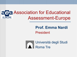
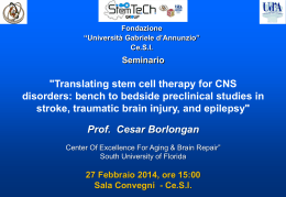
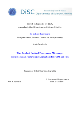
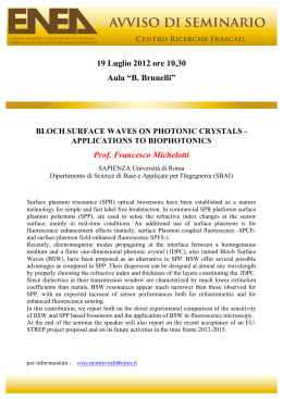
VALUTAZIONE DI UN NUOVO AUSILIO OTTICO PER L’IDENTIFICAZIONE DI LESIONI DELLE MUCOSE ORALI
EVALUATION OF A NEW VISUAL AID FOR THE IDENTIFICATION
OF ORAL MUCOSAL DISEASES AND CONDITIONS
Silvio ABATI
1 DISS
1,
Paolo CASTELLARIN
1,
Chiara MORESCHI
e UO Diagnosi Orale, Università degli Studi e AO San Paolo, Milano
Fig. 1 – oral mucosal autofluorescence detection with Goccles®
Fig. 2 – the use of Goccles® in clinical setting
Fig. 3 – devices for examination and photographic recording
2,
2 Ateneo
Giorgio GASTALDI
2
Vita Salute San Raffaele, Milano
INTRODUCTION
Recent systematic reviews, expert opinions and best current practices confirm that the clinical examination
with accurate visual and tactile inspection of oral tissues could improve the detection and early diagnosis of
oral and oropharyngeal cancers and therefore the overall survival rate of affected patients. In the last fifty
years various methods have been proposed to enhance the ability of clinicians in detecting benign and
malignant oral mucosal conditions with visual oral examination.
It was observed that the autofluorescence of tissues or tissue fluorescence could potentially be used for
cancer screening and detection for a number of anatomic sites including the oral cavity. The fluorescence
imaging of oral mucosa involves the exposure of tissues to a rather specific wavelength of light in the blueviolet range, which results in the excitation of autofluorescence of cellular and extracellular fluorophores with
the emission of visible light in the green range. The presence of cellular alterations will change the
concentrations of fluorophores, affecting the scattering and absorption of light in the tissue, thus resulting in
changes in color that can be observed visually, mainly represented by the loss of autofluorescence greenish
color with the darkening of the diseased areas and enhancement of their visual detection (fig.1). In recent
years, various appropriate instruments have been developed and tested to improve the visual recognition of
oral mucosal diseases with different and inconsistent results.
SCOPE
The aim of this preliminary study was to evaluate a new filtered eyeglasses device to reveal the loss of tissue
autofluorescence, in a case series of patients with mucosal diseases and conditions.
PATIENTS and METHODS
In 15 consecutive patients treated in the Oral Diagnosis Unit the Goccles® eyeglasses system has been tested
and evaluated as a method to improve the detection of oral mucosal inflammation, precancer and cancer
during the standard oral visual examination performed by four untrained dentists and dental hygienists
assisted by two trained experts in oral medicine and pathology (Goccles® patent licensed to Univet srl, Italy)
(fig.2). The autofluorescence has been disclosed lighting up the oral mucosa with a standard dental curing
light and the loss of fluorescence classified in relation to the darkness of the involved areas; the relevant fields
were recorded with a Panasonic Leica LX-100 digital camera set for oral macro pictures with and without the
custom photographic filter supplied by the manufacturer; the manufacturer supplied an additional phographic
filter to be used with cellular phones cameras, and we employed it for comparison; figure 3 shows the
eyeglasses and other devices employed in the study. The visual and photographic findings were immediately
related and compared.
RESULTS
15 patients were examined: 3 affected by oral cancer, 5 with potentially malignant conditions (PVL, FL, OLP),
5 with inflammatory benign conditions (mucosal trauma, MG, candidiasis), 2 with BRONJ. In all patients the
diagnosis have been clinically and or histologically confirmed.
Fig. 4 – clinical case # 1
Fig. 5 – clinical case # 2
Fig. 6 – clinical case # 4
Fig. 7 – clinical case # 5
Fig. 8 – clinical case # 9
pt #
age
sex
clin. Dx
histo. Dx
loss of FL
score
picture
1
75
F
PVL
severe dysplasia
Y
3
4
2
62
M
PVL + EP
mild dysplasia
Y
3-4
5
3
55
F
FL
benign keratosis
N
-
-
4
29
F
previous SCC
mild dysplasia
Y
1-2
6
5
48
F
previous SCC
mild dysplasia
Y
3
7
6
65
F
acute candidiasis
-
N
-
-
7
24
M
mucosal trauma
-
Y/N
1
-
8
34
F
OLP
OLP
Y/N
1
-
9
41
M
OLP – previous in situ SCC
OLP
Y/N
1
8
10
79
M
oral metastasis
mesothelioma
Y
3
-
11
43
F
frictional ulcer
-
Y
2
-
12
64
M
chronic candidiasis
-
Y
1
-
13
39
M
migratory glossitis
-
Y
2
-
14
73
F
BRONJ
ON
bright fluorescence
-
-
15
65
M
BRONJ
ON
bright fluorescence
-
9
DISCUSSION and CONCLUSIONS
The Goccles® device has been able to detect areas of mucosal alteration due to inflammation or malignancy
with loss of tissue autofluorescence and different levels of dark appearance; a relevant finding in the study
was the self-evidence of mucosal areas with bone exposure due to BRONJ that always showed a vivid and
bright appearance. The photographic system and filter are essential to record and archive the relevant clinical
pictures of patients and could be improved to simplify the photographic taking procedure and ensure a better
reproducibility of results. Another critical issue to be improved is the standardization of the distance and
position of the curing light to obtain a correct lighting of the different intraoral areas.
With the innovative Goccles® system it is possibile to enhance the results of a simple, non-invasive and
painless clinical oral examination improving the abilities of the clinicians to detect mucosal alterations and
diseases of the oral cavity, and perform a screening aimed to the early recognition of oral cancer and
premalignant conditions. The autofluorescence examination of oral tissues, in addition to the naked eye
examination, contributes to the detection of the actual margins of precancerous and cancerous lesions even in
the initial stage. The system works with common curing lights available in every dental setting, it is simply,
accessible and easy to use by general dental practitioners, dental hygienists, surgeons, ENT and other
specialists in medical fields.
REFERENCES
(i) Ingrams DR, Dhingra JK, Roy K, Perrault Jr DF, Bottrill ID, Kabani S, et al. Autofluorescence characteristics of oral
mucosa. Head Neck 1997;19(1):27–32; (ii) Lingen MW1, Kalmar JR, Karrison T, Speight PM. Critical evaluation of
diagnostic aids for the detection of oral cancer. Oral Oncol. 2008 Jan;44(1):10-22; (iii) Moyer VA. Screening for oral
cancer: U.S. Preventive Services Task Force recommendation statement. Ann Intern Med 2014;160(1):55-60.
Fig. 9 – clinical case # 15
Acknowledgements The authors wish to thank Univet srl (Rezzato, BS, Italy - [email protected]) for the supplied
equipments – Goccles® patent and trademark belong to Pierrel Pharma srl, exclusively licensed to Univet srl
GOCCLES
(Glasses for Oral Cancer – Curing Light Exposed – Screening)
A new medical device for the prevention of oral cancer
1
Nardo
2
Moro
1
Waure
3
Spadari
4
Mignogna
Authors: Francesco Di
Alessandro
Chiara de
Francesco
Michele D
Michele
6
7
2
4
3
8
Luigi Califano Aldo B Giannì Lorenzo Cardarelli Antonio Celentano Gianpaolo Bombeccari Sandro Pelo
5
Giuliani
Affiliations: 1:Istituto di Sanità Pubblica, Università Cattolica del Sacro Cuore (Roma); 2:UO Chirurgia Maxillo-facciale, Nuovo Ospedale San Giovanni Battista (Foligno);3:Dipartimento di scienze
biomediche, chirurgiche ed odontoiatriche, Università di Milano; 4:UOC Medicina Orale, AOU Federico II, Università di Napoli Federico II; 5:UOC Odontoiatria Operativa e Pedodonzia, Policlinico
Gemelli, Università Cattolica del Sacro Cuore (Roma); 6:UOC Chirurgia Maxillo Facciale, AOU Federico II, Università di Napoli Federico II; 7:UOC Chirurgia Maxillo Facciale e Odontostomatologia,
Fondazione IRCCS Ca' Granda Ospedale Maggiore Policlinico, Università di Milano; 8:UO Chirurgia Maxillo-facciale, C.I. Columbus, Università Cattolica del Sacro Cuore (Roma)
Background: oral cancer is responsible for 128,000 deaths per year, and nearly
all of them are preventable. There are currently no recommended screening
tests, but studies on low-cost examinations are ongoing. These include the
examination of oral cavity autofluorescence (loss of fluorescence may be
related to dysplasia or cancer). The GOCCLES device (fig. 1), patented at the
Catholic University of the Sacred Heart of Rome, consists of filters that show
oral mucosal autofluorescence abnormalities when used together with any
dental curing light. The aim of the study is to demonstrate that GOCCLES
allows an effective examination of autofluorescence in the dental practice.
Methods: data from two trials (including a multicenter clinical trial, not yet
published) on consecutive patients at risk for oral cancer (heavy
smokers/heavy alcohol drinkers/history of oral cancer/presenting with
potentially malignant lesions of the oral cavity). All patients underwent naked
eye inspection followed by autofluorescence examination with GOCCLES and
four curing lights with different technical characteristics. All fluorescence loss
areas and all potentially malignant lesions persisting for three weeks
underwent excisional biopsy, regardless of being visible to the naked eye.
AF-
AF+
NE-
NE+
-
1
(2.3%)
4
(9.1%)
39
(88.6%)
AF margins
Not visible
AF margins
not
to AF
infiltrated
infiltrated
Not visible to the NE
NE margins not inf.
NE margins infiltrated
-
0 (0%)
1 (3.1%)
1 (3.1%)
3 (9.4%)
6 (18.8%)
0 (0%)
4 (12.5%) 17 (53.1%)
Results: overall, 78 lesions were sampled. The device allowed the visualization
of fluorescence loss in all moderate to severe dysplasias and cancers and in
93,3% of mild dysplasias (fig. 2). In 4 cases the device allowed to identify
otherwise invisible lesions (including one oral cancer, tab 1). In 10 cases the
device allowed complete resections of lesions with infiltrated naked-eyevisible margins (tab. 2). False positives at naked eye examination: 37.2%. False
positives with GOCCLES: 39,7%. The device worked properly with all tested
curing lights. Of nineteen patients at risk of oral cancer excluded from the
study because showed no lesions at both autofluorescence and naked eye
examination, no one developed oral cancer during the follow up (at one year).
Figure 1 (left). The GOCCLES medical device. Figure 2 (right). Oral cancer in an edentulous
patient in follow-up after surgical resection of a malignant lesion. Autofluorescence
examination (left) vs. conventional visual examination (right). The lesion is barely visible if
the oral examination is performed with superficiality. Loss of fluorescence increased
contrast making it easier to see the tumor. Also visible in this figure is a clear difference in
the extension of the margins of the lesion: fluorescence loss (margins highlighted) extended
beyond the margins which were visible to the naked eye. Arrow points to the main lesion.
Conclusions: GOCCLES has high sensitivity and allows an easy and low-cost
oral cancer screening in the dental practice. It is recommended as a
complementary inspection following the naked eye examination on patients at
risk and in follow-up for oral cancer. The device may also be used in the
surgical setting (excision of dysplasias/tumors of the mouth).
Table 1 (left). True positive lesions identified by autofluorescence examination with
GOCCLES (AF) and naked eye inspection (NE). Table 2 (right). Margins correctly identified by
AF and NE in true positive lesions (margins were studied on 32 true positive patients).
References: Moro A, Di Nardo F, Boniello R, Marianetti TM, Cervelli D, Gasparini G, Pelo S. Autofluorescence and early detection of mucosal lesions in patients at risk for oral cancer. J Craniofac
Surg. 2010;21(6):1899-903. Monici M. Cell and tissue autofluorescence research and diagnostic applications. Biotechnol Annu Rev. 2005;11:227-56.
Corresponding Author: Francesco Di Nardo: [email protected]
Istituto di Sanità Pubblica, Sezione di Igiene
Università Cattolica del Sacro Cuore, Largo F. Vito 1, 00168 Rome, Italy
This study was funded by Pierrel Pharma srl
Patent and trademark belong to Pierrel Pharma srl
Exclusively licensed to Univet Srl
ACTA otorhinolaryngologica italica 2015;35:449-454; doi: 10.14639/0392-100X-922
Clinical techniques and technology
The GOCCLES® medical device is effective
in detecting oral cancer and dysplasia in dental
clinical setting. Results from a multicentre clinical trial
Il dispositivo medico GOCCLES® è in grado di individuare displasie e cancro orale
se impiegato nel setting odontoiatrico. Risultati da uno studio multicentrico
A. Moro1,2, C. de Waure3, F. Di Nardo3, F. Spadari4, M.D. Mignogna5, M. Giuliani6, L. Califano7,
A.B. Giannì8, L. Cardarelli1, A. Celentano5, G. Bombeccari4, S. Pelo2
1
Unit of Maxillofacial Surgery, Nuovo Ospedale San Giovanni Battista. Foligno, Italy; 2 Unit of Maxillofacial Surgery,
Policlinico Gemelli, C.I. Columbus, Università Cattolica del Sacro Cuore, Rome, Italy; 3 Institute of Public Health,
Università Cattolica del Sacro Cuore, Rome, Italy; 4 Department of Reconstructive and Diagnostic Surgical
Sciences, Unit of Oral Pathology and Medicine, Fondazione IRCCS Ca’ Granda Ospedale Maggiore Policlinico,
University of Milan, Italy; 5 Department of Integrated Activities Head-Neck, Unit of Oral Medicine, Università di Napoli
Federico II. Naples, Italy; 6 Department of Dentistry, Università Cattolica del Sacro Cuore, Rome, Italy; 7 Department
of Integrated Activities Head-Neck, Unit of Maxillofacial Surgery, Università di Napoli Federico II. Naples, Italy;
8 Department of Reconstructive and Diagnostic Surgical Sciences, Complex Unit of Maxillofacial Surgery,
Fondazione IRCCS Ca’ Granda Ospedale Maggiore Policlinico, University of Milan, Italy
SUMMARY
The purpose of this study is to demonstrate that the GOCCLES® medical device allows proper autofluorescence examination of the oral
mucosa in a dental care setting. This is a non-randomised multicentre clinical trial on consecutive patients at risk for oral cancer. Patients
underwent a classical naked eye inspection of the oral cavity followed by autofluorescence examination wearing the GOCCLES® spectacles
while the light from a dental curing light irradiated the oral mucosa. Lesions were defined as visible potentially malignant lesions and/or
fluorescence loss areas. All persisting lesions underwent excisional or incisional biopsy. Sixty-one patients were enrolled. Data from 64
biopsies were analysed. Of the 62 lesions identified by the device, 31 were true positives. The device identified 31 of 32 true positive lesions. One lesion (an invasive carcinoma) was not visible to the naked eye. The device identified all lesions classified as moderate dysplasia
to invasive cancer. In 56.7% of cases, true positive lesions showed greater extension when observed through the device. The GOCCLES®
medical device allowed the direct visualisation of fluorescence loss in patients suffering from mild to severe dysplasia and in situ to invasive
oral cancer. It allowed autofluorescence examination with each source of light used during the study. These results suggest that the role of
the autofluorescence visualisation is that of a complementary inspection following naked eye examination when dealing with patients at
risk for oral cancer. The device allows detection of otherwise invisible lesions and otherwise impossible complete resections.
KEY WORDS: Oral cancer • Early Detection of Cancer • Dentistry • Fluorescence • Curing Lights • Dental
RIASSUNTO
Scopo di questo studio è dimostrare che il dispositivo medico GOCCLES® permette di condurre l’esame dell’autofluorescenza del cavo
orale nel setting odontoiatrico. Si tratta di uno studio multicentrico non randomizzato su pazienti consecutivi a rischio di cancro orale. I
pazienti sono stati sottoposti ad ispezione del cavo orale ad occhio nudo seguita dall’esame dell’autofluorescenza condotto indossando gli
occhiali GOCCLES® mentre una lampada fotopolimerizzante illuminava la mucosa orale. Le lesioni sono state definite come qualunque
lesione precancerosa del cavo orale visibile ad occhio nudo o area di perdita di fluorescenza visibile con GOCCLES®. Tutte le lesioni
persistenti sono state sottoposte a biopsia escissionale o incisionale. Sono stati reclutati 61 pazienti e analizzati i dati da 64 lesioni. Delle
62 lesioni identificate dal dispositivo, 31 erano veramente positive. Il dispositivo ha identificato 31 delle 32 lesioni veramente positive.
Una lesione (un carcinoma invasivo) non era visibile ad occhio nudo. Tutte le lesioni classificate come displasia tra moderata e severa e
ogni carcinoma sono stati correttamente identificati dal dispositivo. Nel Il 56,7% delle lesioni identificate dal dispositivo mostrava margini
più ampi rispetto a quelli visibili ad occhio nudo. Il dispositivo medico GOCCLES® permette di osservare il fenomeno della perdita di
fluorescenza in pazienti affetti da displasia o cancro del cavo orale. Ha permesso di effettuare l’esame dell’autofluorescenza con ciascuna
lampada fotopolimerizzante testata. I risultati suggeriscono di impiegare GOCCLES® come esame complementare rispetto all’ispezione
ad occhio nudo del cavo orale su pazienti a rischio per cancro orale. Il dispositivo permette di identificare lesioni altrimenti visibili o i cui
margini sono sottostimati dall’ispezione ad occhio nudo.
PAROLE CHIAVE: Cancro orale • Diagnosi precoce del cancro • Odontoiatria • Fluorescenza • Lampade fotopolimerizzanti
Acta Otorhinolaryngol Ital 2015;35:449-454
449
A. Moro et al.
Introduction
No oral cancer screening test on large populations is currently recommended for oral cancer. However, studies on
low-cost oral cancer diagnostic techniques are currently
ongoing 1. The autofluorescence examination is among
these techniques.
Autofluorescence of the oral mucosa consists of a dim
light coming from oxidised flavin adenine dinucleotide
(FAD) and other fluorophores when excited by blue-violet and ultra-violet (UV) light 2. Healthy tissues produce a 515 nm (green) light. Conversely, tissues with
disorders of cell metabolism (such as dysplastic mucosa) appear as dark spots on a green background 2. The
disruption of the extracellular matrix, hyperemia and
neo-angiogenesis also contribute to reduce fluorescence
emission 2.
Persistent areas of decreased fluorescence can therefore
be a sign of dysplasia or cancer and should be treated accordingly. Today, it is commonly accepted that potentially
malignant lesions of the oral cavity must be treated if they
persist for more than 2 weeks, and many researchers are
trying to find a role for autofluorescence not only in the
early detection, but also in the complete surgical resection
of cancerous or pre-cancerous lesions 3.
The GOCCLES® (Glasses for Oral Cancer – Curing Light
Exposed – Screening) medical device was created in order to provide comfortable, easy and low cost direct visualisation of abnormalities of the oral cavity tissue fluorescence.
This device has already been tested in a clinical trial, with
promising results 4. However, that study suffered from
several limitations, and was a pilot study on just 32 patients. Two-thirds of enrolled patients were in follow-up
after the surgical resection of an oral cancer, and therefore
at high risk of showing dysplastic tissues at histological
examination regardless of the results of autofluorescence
examination because of disease relapse or incomplete resection. Moreover, during this trial only a prototype of the
device was studied, and it was tested with only one dental
curing light (an Elipar 2500 3M ESPE). Furthermore, the
study was conducted only by the research team responsible for the creation of the device, with obvious conflicts
of interest.
In order to demonstrate the capability of the GOCCLES®
medical device to allow proper autofluorescence examination of the oral mucosa, which is the aim of this multicentre trial, it is therefore necessary to involve more patients, multiple research groups and curing lights, and to
test the final model of the device.
Materials and methods
The GOCCLES® device (Pierrel S.p.A, Italy) consists of a
pair of glasses equipped with filters (Fig. 1) that highlight
450
Fig. 1. The GOCCLES (Glasses for Oral Cancer – Curing Light
Exposed – Screening) medical device.
autofluorescence when the oral mucosa is illuminated by
the light emitted by any dental curing light.
The device was studied in a non-randomised multicentre
clinical trial in which eligible consecutive patients underwent oral examination with GOCCLES®.
Subjects above the age of 17 years were eligible for the
study if showing potentially malignant lesions of the oral
mucosa or if they were in follow-up after surgical resection for oral cancer. Patients who underwent radiotherapy
for oral or head and neck cancer in the previous three
months were excluded from the study.
All eligible subjects were informed in detail on the study
protocol and participants joined the study voluntarily
after signing an informed consent form. All eligible
subjects showing lesions at naked eye oral inspection or
on autofluorescence analysis were asked to participate
in the study. For ethical reasons, subjects with totally
negative naked eye and autofluorescence examinations
did not undergo biopsy of the oral mucosa and were
excluded.
Enrollment took place at the following six Centers: Unit
of Maxillofacial Surgery, C.I. Columbus, Università Cattolica del Sacro Cuore (Rome); Department of Reconstructive and Diagnostic Surgical Sciences, Unit of Oral
Pathology and Medicine and Complex Unit of Maxillofacial Surgery, Fondazione IRCCS Ca’ Granda Ospedale Maggiore Policlinico, University of Milan (Milan);
Department of Integrated Activities Head-Neck, Unit of
Maxillofacial Surgery and Unit of Oral Medicine, Università di Napoli Federico II (Naples); Unit of Maxillofacial Surgery, Nuovo Ospedale San Giovanni Battista
(Foligno). Enrollment lasted one year and started in September 2013.
Patients underwent a naked eye classical inspection of
the oral cavity followed by the autofluorescence examination wearing the GOCCLES® medical device while
the light from a dental curing light irradiated the oral
mucosa. All examinations were performed by skilled
physicians and surgeons with patients lying in a dental
chair in a setting similar to that of the dental practice.
All operators were asked to hold the light at 20-40 cm
GOCCLES® is effective in detecting oral cancer in a dental clinical setting
distance from the oral mucosa and to direct the light
perpendicularly to the inspected area. The naked eye inspection and the autofluorescence examination had to be
performed by the same operator. All fluorescence loss
areas were regarded as potentially malignant lesions of
the oral mucosa. The following dental curing lights were
used during the study: Elipar S10 3M ESPE (used by the
Units of Milan); Led.B Carlo de Giorgi (Units of Rome
and Foligno); Optilux 501 Kerr Corporation (Units of
Naples). After examinations, all lesions persisting for at
least two weeks (detected by at least one between the
naked eye inspection and the autofluorescence examination) underwent excisional biopsy. If the excisional
biopsy was not feasible, patients underwent incisional
biopsy. All biopsies were properly oriented and showed
the margins detected by both examinations. If an incisional biopsy including both identified margins was not
feasible, two incisional biopsies of the same lesion on
different margins were allowed. Different incisional biopsies of the same lesion were considered as a single biopsy. All biopsies underwent histological examination.
No blinding on pathologist assessment or data analysts
was planned.
For each lesion the following data were recorded: if visible to the naked eye and/or on autofluorescence analysis;
which examination showed the greater extension if results
were non-overlapping; which margins identified by the
two techniques were infiltrated; the histological report.
True positive lesions were defined as any dysplasia or
cancer of the oral cavity. False positive lesions included
negative histological findings and any other disorder of
the oral mucosa not related to cancer.
Primary outcomes of the study were: proportion of visible lesions; proportion of infiltrated margins; proportion of true positive, false positive and false negative
lesions. Given the study design, it was not possible to
assess true negative lesions (no proper follow-up was
planned).
The study protocol was approved by the respective ethics
committees of the Institutions involved in the study.
Statistical analysis
Differences in terms of diagnostic performance between
the autofluorescence analysis and naked eye inspection
of the oral mucosa were assessed using the two-tailed
McNemar test. Data analysis was performed with the
IBM SPSS 22 Statistics Software for Windows. Statistical
significance was set at p = 0.05.
Sample size calculation was based on the results of the
previous study 4. Assuming that the examination of autofluorescence showed larger margins compared to the naked eye in 25% of the lesions and that the opposite occurs
in about 5% of cases, a sample of 100 patients was set
in order to observe a significant difference between the
two examinations with a probability of 95% (accepting a
probability of type I error of 5%).
An interim analysis was planned at one year. After analysis of the first results the study was discontinued given the
low probability of achieving more conclusive results with
the preset sample of 100 patients.
Results
Sixty-one patients were enrolled and all underwent both
naked eye inspection and autofluorescence examination
of oral cavity mucosa. Data from all 61 patients entered
the analysis. Main characteristics of the patients are summarised in Table I.
Autofluorescence of the oral mucosa was analysed using
a Led.B curing light on 29 patients (47.6% of the sample),
an Elipar S10 on 21 patients (34.4%) and an Optilux 501
on 11 patients (18.0%).
Naked eye inspection of the oral cavity detected 60 suspect lesions, while autofluorescence examination with the
GOCCLES® device detected 62 suspect lesions. A total of
65 lesions were detected: in 59 cases (90.8% of observed
suspected lesions) they were visible to both naked eye and
autofluorescence, while 2 suspected lesions (3.1%) were
only visible during the naked eye inspection and 4 (6.2%)
were only visible on the autofluorescence examination.
No significant differences in terms of detected suspect
lesions were observed between the naked eye inspection
Table I. Main demographic and clinical characteristics of the patients enrolled in the GOCCLES study, by center
Rome
Milan
Naples
Patients [N]
Age [mean, (SD)]
Gender [N, (%)]
Group [N, (%)]
Biopsies/patient
[N, (%)]
Female
A*
B†
1
2
3
14
67 (14)
8 (57.1)
11 (78.6)
3 (21.4)
14 (100)
0 (0)
0 (0)
21
66 (16)
15 (71.4)
16 (76.2)
5 (23.8)
21 (100)
0 (0)
0 (0)
11
66 (13)
8 (72.7)
11 (100)
0 (0)
10 (90.9)
1 (9.1)
0 (0)
Foligno
TOTAL
15
63 (11)
7 (46.7)
14 (93.3)
1 (6.7)
13 (86.6)
1 (6.7)
1 (6.7)
61
66 (14)
39 (63.9)
52 (85.2)
9 (14.8)
58 (95.1)
2 (3.3)
1 (1.6)
* Patients suffering from suspected dysplastic lesions of the oral mucosa.
†
Patients in follow-up after surgical resection of oral cancer.
451
A. Moro et al.
Table II. Autofluorescence examination.
Lesion description
False positive† [N (%)]
True positive [N (%)]
Of which:
Mild dysplasia [N (%)]
Moderate dysplasia [N (%)]
Severe dysplasia [N (%)]
Carcinoma in situ [N (%)]
Invasive cancer [N (%)]
TOTAL [N (%)]
Detected by naked
eye inspection only
Detected by both
techniques
Detected by
AF examination only
TOTAL*
1 (3.1)
1 (3.1)
28 (87.5)
30 (93.8)
3 (9.4)
1 (3.1)
32 (50.0)
32 (50.0)
1 (9.1)
0 (0)
0 (0)
0 (0)
0 (0)
2 (3.1)
10 (90.9)
4 (100)
3 (100)
2 (100)
11 (91.7)
58 (90.6)
0 (0)
0 (0)
0 (0)
0 (0)
1 (8.3)
4 (6.3)
11 (17.2)
4 (6.3)
3 (4.7)
2 (3.1)
12 (18.8)
64 (100)
* The percentages under the “total” column relate to the whole sample. † Includes other non-precancerous and non-cancerous disorders of the oral mucosa.
Definitive diagnoses are based on histological findings. No patients were negative for both naked eye and autofluorescence analysis because no subject with totally negative
physical examination underwent biopsy of the oral mucosa.
and autofluorescence examination of oral mucosa (McNemar test p = 0.687).
Sixty-five biopsies of the oral mucosa were thus taken. Fifty-eight patients underwent one oral mucosa biopsy, while two underwent two biopsies and another
patient underwent three biopsies because of multiple
lesions. An invalid sample was excluded from statistical analyses and data from 64 of 65 biopsies (98.5%
of all biopsies) were analysed. Thirty-two of 64 samples (50.0% of the valid samples) were classified as
false positives. Of the 32 truly positive samples, 11
(17.2% of the valid samples) were classified as mild
dysplasia, 4 (6.3%) as moderate dysplasia, 3 (4.7%)
as severe dysplasia, 2 (3.1%) as carcinoma in situ and
12 (18.8%) as invasive cancer.
Of the 60 suspected lesions detected by naked eye inspection of the oral cavity, 31 were true positive lesions
(51.6%), while of the 62 lesions identified on autofluorescence examination, 31 (50.0%) were true positives.
In particular, autofluorescence examination identified all
the lesions classified as moderate to severe dysplasia and
all lesions classified as cancer. Main results of the study
are summarised in Table II. Both techniques identified 31
of 32 (96.9%) actual lesions. One lesion (a carcinoma)
was not visible to the naked eye, while one lesion (a mild
dysplasia) was not visible on the autofluorescence examination. No significant differences in terms of diagnostic
performance were observed between the two techniques
(McNemar test p = 1.000).
Thirty true positive lesions were visible by both classical inspection and autofluorescence analysis. In 17 cases
(56.7% of the 30 true positive lesions), extension of the
lesion detected on the autofluorescence examination was
greater than that observed at naked eye inspection, while
in two cases (6.7%) the margins overlapped and in nine
cases (30.0%) autofluorescence examination showed a
smaller lesion. In two cases (6.7%), the operator was unsure whether the margins were overlapping or not.
The resection margins identified during the naked eye
inspection were infiltrated in 21 cases (67.7% of the 31
lesions identified). Of the 31 true positive lesions identified by autofluorescence examination, 24 (77.4%) showed
infiltrated margins. No significant differences were observed between the two techniques in terms of free resection margins (McNemar test p = 0.754, see Tab. III).
No statistically significant differences were observed in
the performance of the autofluorescence test using different curing lights (Tab. IV).
Discussion
The GOCCLES® medical device allowed visualisation of
fluorescence loss in patients suffering from mild to severe
dysplasia and oral cancer (Fig. 2). The device worked
Table III. Proportions of free and infiltrated margins of the lesions identified during naked eye inspection of the oral cavity and on autofluorescence examination.
Not visible
AF margins
AF margins
TOTAL
to AF
not infiltrated
infiltrated
Not visible to the NE
NE margins not infiltrated
NE margins infiltrated
TOTAL
1 (3.1%)
0 (0%)
1 (3.1%)
0 (0%)
3 (9.4%)
4 (12.5%)
7 (21.9)
1 (3.1%)
6 (18.8%)
17 (53.1%)
24 (75.0%)
1 (3.1%)
10 (31.3%)
21 (65.6%)
32 (100%)
NE: naked eye inspection; AF: auto fluorescence examination.
No patients showed lesions invisible to both techniques because no subject with totally negative physical examination underwent the biopsy of the oral mucosa.
452
GOCCLES® is effective in detecting oral cancer in a dental clinical setting
Table IV. Comparison of the performance of the GOCCLES device with different curing lights.
Auto fluorescence examination results
Led.B [N (%)]
Elipar S10 [N (%)]
Optilux 501 [N (%)]
Lesion margin detection
TP lesion
FP lesion
Not detected
p value*
Free margins
Infiltrated
margins
p value*
15 (46.9)
8 (38.1)
8 (72.7)
16 (50.0)
13 (61.9)
3 (27.3)
1 (3.1)
0 (0)
0 (0)
0.488
4 (26.7)
0 (0)
3 (37.5)
11 (73.3)
8 (100)
5 (62.5)
0.174
TP: true positive; FP: false positive. * Chi square test.
properly with each source of light used in this study,
which have different technical characteristics reproducing
the variety of available dental curing lights. The Led.B
is a LED lamp with a wavelength of 440-490 nm and an
intensity of 1,000-1,200 mW/cm2; the Elipar S10 LED
lamp has a 430-480 nm wavelength and an intensity of
1,000 mW/cm2; the Optilux 501 is a halogen lamp with a
wavelength of 400-505 nm and an intensity of 850-1,000
mW/cm2. The GOCCLES® device was also previously
successfully tested with another halogen lamp with different characteristics (400-500 nm wavelength and 600
mW/cm2 intensity).
The study was performed with patients lying in dental
chairs in order to reproduce the settings of common dental practice. The intended purpose of the device was to
allow any dentist equipped with a curing light to perform
low-cost autofluorescence examination. According to the
scientific literature, autofluorescence examination shows
otherwise invisible characteristics of the oral mucosa that
are associated with oral cancer 2 4. However, the current
evidence suggests that the role of the autofluorescence
examination is that of a complementary inspection following the naked eye examination, which should not be
replaced by any screening test. Moreover, given the high
risk of false positive findings (50% in this study), it is recommended that every dentist equipped with the device
is properly trained before using it and that only patients
at risk for oral cancer or showing potentially malignant
lesions undergo examination with GOCCLES®. Remarkably, however, in one case GOCCLES® allowed the detection of an otherwise invisible lesion, and in four cases it
allowed otherwise impossible complete resections of lesions with infiltrated margins.
We hope that the availability of additional low cost
screening techniques encourages more careful and more
frequent inspections of the oral cavity among dentists,
and further promotes a much needed culture of oral cancer prevention: naked eye inspection of the oral cavity
alone could, in fact, save about 37,000 lives worldwide
each year 5.
Furthermore, data on its diagnostic performance are
lacking because of the small sample size and study de-
Fig. 2. Oral cancer in an edentulous patient in follow-up after surgical resection of a malignant lesion. Autofluorescence
examination (on the left) vs. conventional visual examination (on the right). The lesion is barely visible if the oral examination
is performed with superficiality. Loss of fluorescence increased contrast making it easier to see the tumour. Also visible in
this figure is a clear difference in the extension of the margins of the lesion: fluorescence loss extended beyond the margins,
which were visible to the naked eye. The arrow points to the main lesion. The margins of the lesion (as visible on the autofluorescence examination) are also highlighted.
453
A. Moro et al.
sign (it was impossible to assess the proportion of false
negatives in this study as no follow-up of negative patients was planned). Further studies on much larger
samples (possibly randomised clinical trials comparing
the device with other techniques) are needed to define
its diagnostic performance. The previous pilot study
(in which patients underwent a follow-up of one year)
showed 100% sensitivity, 95% specificity, 93% positive
predictive value and 100% negative predictive value.
However, the device was studied on patients at very high
risk, and most had a history of oral cancer. It is likely
that the diagnostic performance is heavily affected by
the population tested, being poorer in the general population and improving greatly when dealing (as appropriate) with subjects at risk.
Conflict of interests
This study was funded by Pierrel S.p.A., owner of the
rights on the GOCCLES medical device. Three research-
ers involved in this study (SP, AM, FDN) have royalty
percentages on the sales of the device.
References
1
Petersen PE. Oral cancer prevention and control--the approach of the World Health Organization. Oral Oncol
2009;45:454-60.
2
Monici M. Cell and tissue autofluorescence research
and diagnostic applications. Biotechnol Annu Rev
2005;11:227-56.
3
Succo G, Garofalo P, Fantini M, et al. Direct autofluorescence during CO2 laser surgery of the larynx: can it really
help the surgeon? Acta Otorhinolaryngol Ital 2014;34:17483
4
Moro A, Di Nardo F, Boniello R et al. Autofluorescence and
early detection of mucosal lesions in patients at risk for oral
cancer. J Craniofac Surg 2010;21:1899-903.
5
Sankaranarayanan R, Ramadas K, Thomas G, et al. Effect of
screening on oral cancer mortality in Kerala, India: a cluster-randomised controlled trial. Lancet 2005;365:1927-33.
Received: September 1, 2015 - Accepted: October 1, 2015
Address for correspondence: Francesco Di Nardo, Istituto di Sanità
Pubblica, Sezione di Igiene. Università Cattolica del Sacro Cuore,
largo Francesco Vito 1, 00168 Rome, Italy. E-mail: [email protected]; [email protected]
454
Scaricare