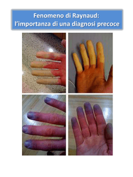
Il fenomeno di Raynaud e la diagnosi precoce delle malattie del connettivo Marco Matucci Cerinic Raynaud Fenomenus, Quo Vadis… ? Table II: Raynaud’s phenomenon classification: Primary Raynaud’s phenomenon (fig 1): young women below 30 years, attacks are symmetric and limited in time, no tissue lesions (digital pitting scars, teleangectasia) and no complications (digital ulcers, necrosis/gangrene) on the extremities, capillaroscopy normal, antinuclear antibodies negative, history and clinical examination negative, erythrocyte sedimentation rate normal Secondary Raynaud’s phenomenon: age over 30 years, attacks are asymmetric, extremely painful and prolonged in time, associated with digital lesions and some time with complications, auto-antibodies positive, capillaroscopic changes, clinical features characteristic of a connective tissue disease or another disease (fig …). The following are the possible causes of secondary RP: Autoimmune/Connective tissue diseases: Systemic sclerosis , Systemic lupus erythematosus, Dermatomyositis, polymyositis, Siogren’s syndrome, Primary biliary cirrhosis Joint diseases: Rheumatoid arthritis Arterial diseases: Thromboangiitis obliterans (Buerger’s disease), Takayasu aortitis, Giant cell arteritis Brachiocephalic atherosclerosis, Migraine or vascular headaches, Prinzmetal angina Mechanical : Vibration (Hand arm vibration syndrome), Crutch pressure, Thoracic outlet syndrome, scalenus anticus syndrome, cervical rib, carpal tunnel syndrome. Temperature: Frost bite Endocrine disorders: Carcinoid syndrome, Pheochromocytoma, Hypothyroidism Neoplasias: ovarian carcinoma, angiocentric lymphoma Rheological and coagulation disorders: cryoglobulins, cryofibrinogenaemia, cold agglutinins, paraproteinaemia, plasmacytoma, polycythaemia, micro thromboembolism, Protein C, Protein S & Antithrombin III deficiency, Factor V Leiden Infections: Parvovirus B19 , Helicobacter pylori , Hepatitis C and B, mycoplasma (agglutinins) Chemicals : Polyvinyl chloride, toluene, xylene, acetone Drugs: Bleomycin, Vinblastine, b blockers, Ergots, Methysergide, Interferon a & b, Tegafur, amphetamines, cyclosporine A, cocaine. Matucci Cerinic M in press 2013 Raynaud’s ph Classification Phase/colour simmetricity pain limb Primary/secondary Bi-triphasic/ white,blue,red Primary-bilateral & symmetrical Secondary-mono/bilateral asymmetric ++ Hands, feet, nose, ears, tongue T° extremities Cold Precipitated by Cold Erythromelalgia Livedo Reticularis Primary/secondary familial, (autosomal dominant )/sporadic juvenile/adult Primary/secondary Mono/ dark blue Mono/erythematous macular, violaceous, netlike rings Bilateral Bilateral ++++ ----- Acral Cyanosis Mono/bilateral uncommon Hands, feet Cold Cold & lowering of the limb Feet, (may extend to hand, ears and nose) Upper and lower limbs Hot Cold Hot Cold Phase/duration Paroxistic/lasts from minutes to hours hyperhydrosis Modification of the position of the limb Complications + No change Ulcers, gangrene,amputation Chronic/ seasonal (amelioration in summer) +++ Normalizes when limb is elevated None Intermittent/ may last from minutes to days Significant reduction No change None Primary:chronic Secondaryacute/intermittent/chronic ---No change None in primary in secondary necrosis and gangrene Ascertain that what the patient is complaining for is Raynaud’s phenomenon with adequate questions Obtain a detailed patient’s history and drugs (chemotherapy et al) and working exposures (chemical, vibration) as well as quality of attacks of RP (symmetricity, frequency) Perform a basic clinical assessment in search for any sign or symptom that may indicate an underlying disease Heart (arrhytmias, fibrillation,murmurs) & lung (shortness of breath, crackles and velcro) ,; vascular ( ); Muscles (weakness, atrophy) & joints (synovitis, deformities); Liver, thryroid & lymphonodal palpation After minimal lab work up ( esr, crp,ANA, platelets, clotting) move to more lab investigations. if signs and symptoms indicate any specific abnorrmality Inspections of the extremities: TSH, tpoAtb,tgAtb HCV +, Crioglobuline + Thyroiditis Hypothiroidism Crioglobulinemia Hepatologist Sclerodactily, proximal scleroderma, puffy fingers, teleangectasias, vasculitis Endocrinologist ANA + Topo I/ ACA + Hyperviscosity s. Topo I/ ACA - Hematologist smoke Echodoppler IgG k/l, hiperg Bence Jones protiduria Myeloma Plasmacytoma digital ulcers, DPS, necrosis Digital paresthesias NC + VEDOSS Angiography Angio MRI EMG Neck xray Wrist US Carpal tunnel s. Burger s. Follow up Skin fibrosis Organ involv. SSc Atherosclerosis Outlet syndrome Figure 3 Raynaud’s phenomenon in SSc • • • • 12% of patients with RP develop SSc1 Commonly the first clinical sign of SSc2 Occurs in ~96% of SSc patients3 Precedes the first non-RP clinical feature of SSc by several years3 – 4.8 years in lcSSc – 1.9 years in dcSSc 1. Koenig M, et al. Arthritis Rheum 2008; 58:3902-12. 2. Korn JH. Cleve Clin J Med 2003; 70:954-68. 3. Walker UA, et al. Ann Rheum Dis 2007; 66:754-63. . Raynaud’ ph e “puffy fingers” The disease evolution lung, heart, GI, kidney intermediate skin thickness late early DIFFUSE SSc pulmonary hypert intermediate late 5 10 disease duration (years) 20 early 2 Medsger T & Steen V, Systemic Sclerosis, 1995, p 51,Williams & Wilkins ., malabsorption LIMITED SSc Changes in causes of SSc-related deaths over time 50 Scleroderma renal crisis PAH Interstitial lung disease Frequency (%) 40 30 20 10 0 1972-1976 1977-1981 1982-1986 1987-1991 1992-1996 1997-2001 Time period Slide courtesy of Virginia Steen. Window of Opportunity Very Early SSc Puffy fingers ANA NVC ACA/ATA Raynaud ph Early SSc Established SSc fibrotic & atrophic Skin Fibrosis & Atrophy Esophageal/Anal Heart, Lung, Kidney Digital Ulcers Matucci Cerinic et al ARD 2012 Vasospasm vs Obstructive Vascculopathy 13 Lidia T. January 2005 The disease evolution lung, heart, GI, kidney intermediate skin thickness late early DIFFUSE SSc pulmonary hypert intermediate late 5 10 disease duration (years) 20 early 2 Medsger T & Steen V, Systemic Sclerosis, 1995, p 51,Williams & Wilkins ., malabsorption LIMITED SSc Mean time to diagnosis (years) Diagnosis of SSc is typically delayed 5 4 3 2 1 0 1970-79 1980-89 1990-99 > 2000 Decade No significant change in time to diagnosis of SSc in past 3 decades Improvements are needed Johnson SR, et al. J Rheumatol 2006; 33:1123-7. It is not easy to recognise an early SSc patient because… ANA RP PUFFY FINGERS SLE, Sjogren’s Sindrome, MCTD, UCTD, SSc, Antiphospholipid syndrome and others autoimmune diseases Very Early Systemic Sclerosis Raynaud’s phenomenon Pre-SSc Puffy Fingers UCTD MCTD Anti-nuclear antibodies Matucci Cerinic et al , Ann Rheum Dis 2009 Capillaroscopy Autoantibodies and microvascular damage are independent predictive factors for the progression of Raynaud's phenomenon to systemic sclerosis: a twenty-year prospective study of 586 patients, with validation of proposed criteria for early systemic sclerosis. Of the 586 patients who were followed up for 3,197 person-years, 74 (12.6%) developed definite SSc. In RP evolving to definite SSc, microvascular damage is dynamic and sequential, while SSc specific autoantibodies are associated with the course and type of capillary abnormalities. Abnormal findings on NCM at baseline together with an SSc-specific autoantibody indicate a very high probability of developing definite SSc, whereas their absence rules out this outcome. At followup, 79.5% of patients with 1 of these autoantibodies and abnormal findings on NCM at baseline had developed definite SSc. Koenig M, et al Arthritis Rheum. 2008 ;58:3902-12 Sclerosi Sistemica Il quadro clinico molto precoce e precoce VEDOSS: Criteria to trigger early referral VEDOSS red flags Raynaud’s phenomenon Puffy fingers Positive antinuclear antibodies Avouac J, et al. Ann Rheum Dis 2010 normal active early late From Raynaud‘s to SSc: Early damage Loss of red blood cells from the damaged wall Enlarged and damaged vessel wall Slide courtesy of Maurizio Cutolo. Serological subsets in SSc Diffuse Limited 50-60% have 3 common Ab Anti-centromere, anti-SCL70, Anti-RNA pol III Anti- Scl-70 = pulmonary fibrosis Anti-Centromere = PAH Anti-RNA pol III = renal crisis Anti-PM-Scl = SSc-myositis overlap Anti-U3RNP and Anti-Th= nucleolar pattern on ANA Anti-U3RNP Anti-U1RNP Overlap Clements PJ and Furst DE. Systemic Sclerosis. Lippincott, Williams & Wilkins, Baltimore, 2004. VEDOSS- Very Early Diagnosis Of SSc Presence of red flags raises suspicion of very early SSc 1. Capillaroscopy 2. Serology (TOPO-I, ACA) If either one is positive, diagnosis of very early SSc & further investigations ? 1st level: Suspicion 2nd level: Diagnosis of very early SSc 3nd level: Diagnosis of early SSc Avouac J, et al. Ann Rheum Dis 2010; Signs of pre-clinical organ involvement in early SSc Patients with early SSc* demonstrated signs of preclinical SSc-related internal organ involvement: Pre-clinical internal organ involvement Definition Occurrence (n/N) Early SSc cardiac involvement Inverted mitral E : A ratio 1/19 Early lung involvement Diffusing lung capacity for CO < 80% of predictive value 7/19 Early oesophageal involvement Basal low oesophageal sphincter pressure < 15 mmHg 4/18 *As defined by Koenig M, et al. Arthritis Rheum 2008; 58:3902-12. n = no. of patients with organ involvement; N = no. of patients investigated. Valentini G, et al. Rheumatology (Oxford) 2010 VEDOSS- Very Early Diagnosis Of SSc Presence of red flags raises suspicion of very early SSc 1. Capillaroscopy 2. Serology (TOPO-I, ACA) If either one is positive, diagnosis of very early SSc & further investigations 1. Oesophageal manometry 2. Chest HRCT & PFTs 3. echocardiography If either one is positive, diagnosis of early SSc & further investigations 1st level: Suspicion 2nd level: Diagnosis of very early SSc 3nd level: Diagnosis of early SSc Avouac J, et al. Ann Rheum Dis 2010; VEDOSS patients 25/110 pz reported history and/or showed the presence of DU 9/25 DU previous 2/25 DU active + previous 14/25 DU active previo us previo us+actDU ive active Bruni et al (submitted) Data from a VEDOSS Clinic Digital Ulcers a «sentinel sign» of organ involvement in vedoss patients…!!! Digital Ulcers DU 110 VEDOSS patients • 4 pts with DPS (3.6%) • 16 pts active DU (14.5%) of whom 14 also had a previous history of DU • 9 pts a history of DU (8.2%) 2 groups: • With lung/GI involv.- 25 pts with DU or history DU 105 pts(95.4%)!!! ( 22.7 %) • Without organ involv.- No DU or history DU ( 0%) Organ Involvement Bruni et al 2013 ARD in press Reversible and irreversible patterns Normal Myocardial fibrosis Myocardial oedema irreversible reversible MRI provides information on heart and lung disease activity in the early phase of SSc. Pingitore et al Matucci Cerinic Rheumatology 2013 myocardial oedema, 2 pts abnormal Myocardial oedema normal After 30 days of steroid therapy normal After 2 months FIG. 1 T2-weighted fast spin-echo images with fat suppression of the basal, middle and distal myocardial segments before (a) and after (b) corticosteroid therapy. Pingitore and Matucci Cerinic Rheumatology 2013 DE 15.4.04 RP ANA/ACA pos NVC active Elvira D. 15.4.04 1. 2. 3. 4. Finger edema & Raynaud NVC- Active pattern Anticentromere pos LES Dysfunction Limited cutaneous SSc 2009 RP ANA/Topo I pos NVC active RP ANA/ACA pos NVC early Simona C. December 2004 Claudia P. 2005 RP ANA/Topo I pos NVC active RP ANA/ACA pos NVC early Diffuse SSc- Six months Simona C. December 2004 Limited SSc- five years Claudia P. 2005 Matucci Cerinic M. Kahaleh B, Wigley F: «Scleroderma is a vascular disease», A&R 2013 Asymptomatic SSc Very Early Early Established Advanced Silent phase years to decades ??? Lung GG reticulation Skin Puffy fingers Sclerodactily (fibrosis) Vascular PAH Raynaud Autoantibodies Ulcers ANS Esophageal/anal Intestine Honey combing Retraction (Atrophy) Fatti… 1. 2. 3. 4. 5. 6. 7. Diagnosi è difficile e speso ritardata F Raynaud’s ph è il primo segno della fase precoce della malattia “red flags” devono sempre indurre sospetto di una malattia molto precoce ! Di fronte ad un F Raynaud deve essere condotta una attenta DD per differenziare una forma primaria da una secondaria L’indagine sulla condizione degli organi interni è fondamentale per comprendere se la malattia è evoluta verso una forma precoce Il follow up dei pazienti è necessario per comprendere l’evoluzione della malattia The window of opportunity…!!! Paul Klee La finestra Very early versus early disease: the evolving definition of the many faces of Systemic Sclerosis M Matucci Cerinic , S Bellando Randone, G Lepri, C Bruni, S Guiducci ARD 2013 Paul Klee «The window» Very Early SSc Puffy fingers ANA NVC ACA/ATA Raynaud ph Early SSc Established SSc fibrotic & atrophic Skin Fibrosis & Atrophy Esophageal/Anal Heart, Lung, Kidney Digital Ulcers Early Phase Oedema Strike the iron when it is hot !! Fibrosis Many Faces of SSc !!!! Advanced phase Atrophy VEDOSS Very Early Diagnosis Of SSc • EUSTAR initiative to identify criteria for VEDOSS • Experts in field of SSc from 171 EUSTAR centres asked to participate in a Delphi exercise • Experts proposed and rated preliminary criteria for VEDOSS based on clinical relevance and their importance in leading to an early referral • Validation of criteria ongoing in prospective EUSTAR observational cohort www.eustar.org Dept Rheumatology AVC Dept Biomedicine & Div Rheumatology AOUC Dept Medicine & DENOthe Centre University of Florence Dr F Galluccio Dr. ML Conforti Dr S Cappelli Dr A Righi Dr V Denaro Dr G Baccano Dr T Barskova Dr. S Maddali Bongi Dr R De Luca Clinical Trial Unit Dr L Giovannini Dr. A Del Rosso Dr. F Nacci Dr A Calabrò Dr. D Melchiorre Dr. F Bartoli Dr E Bellucci Dr. M Maresca Digital Ulcers Unit Dr M Orlandi Dr F Peruzzi Dr. F Bandinelli Dr. G Fiori Dr C Bruni Dr. S Bellando Randone Signora F Braschi Dr. S Guiducci Regional Reference Centre for Dr. G Salvadorini Systemic Sclerosis Dr. F Porta Dr. J Blagojevic Dottssa P Cerboni Dr. G Carnesecchi Laboratory Unit Young Adults Clinic Dr M Manetti Prof F Falcini Dr C Ceccarelli Dr E Romano
Scaricare