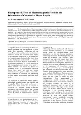
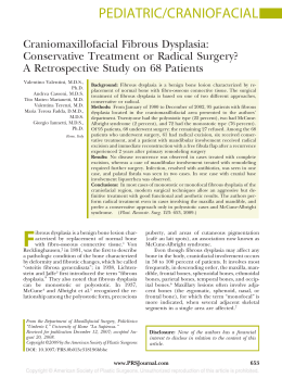
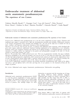
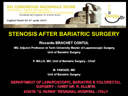
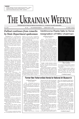
ACTA OTORHINOLARYNGOL ITAL 24, 188-192, 2004
Personal experience in the repair of microtic ear
Esperienza personale nella ricostruzione auricolare totale
D. DI MASCIO, F. CASTAGNETTI
Department of Surgery, Plastic Surgery and Burn Unit, Hospital Maggiore of Parma, Parma, Italy
Key words
External ear malformations • Microtia • Surgical treatment •
Rib Cartilage Graft
Summary
Parole chiave
Malformazioni dell’orecchio esterno • Microtia • Trattamento chirurgico • Lembo di cartilagine costale
Riassunto
Aim of this report is to share with colleagues, the senior
author’s experience in total auricular reconstruction with
autogenous rib cartilage, based on Burt Brent’s technique.
The method has been personalized in the sequence of the
surgical procedures. There are three stages: first, the cartilage carved framework graft is performed; secondly, the
retro-auricular sulcus is reconstructed and thirdly, the
earlobe is transposed onto a three-dimensional frame.
Lo scopo di questo articolo è condividere l’esperienza acquisita dal primo autore nella ricostruzione auricolare totale,
con cartilagine costale autologa, basata sui principi di tecnica chirurgica descritti da Brent. Tali tecniche hanno subito alcune personalizzazioni. Gli interventi sono tre e vengono effettuati nella seguente successione cronologica: 1° tempo –
innesto di cartilagine costale autologa scolpita; 2° tempo –
elevazione del neopadiglione e ricostruzione del solco auricolo-cefalico; 3° tempo – trasposizione del lobo.
Introduction
posterior two thirds. Consequently, in the first stage
of surgery, the rib cartilage is harvested from the contra-lateral chest side, carved into the pattern of the
unaffected ear, and finally grafted inside a cutaneous
pocket 4, (Case 2, Figs. 6-8). In the second procedure,
the reconstructed auricle, still bidimensional, is separated from the mastoid area. The raw retro-auricular
sulcus is repaired with a full-thickness skin graft, taken from homolateral groin area, plus scalp advancement. The auricle under reconstruction is now a
three-dimensional structure, free in the posterior two
thirds.
The lobe joins the cauda helicis in the free part of the
pinna. Up to now, all methods have described lobule
transposition when the auricle is still bidimensional
and attached to the head. The first author considers it
is much more difficult to judge the physiological position of the earlobe and define the position of the
precise meeting point between the lobule and the helix when the auricle is still bidimensional and
attached to the head. For this reason, since 1998, the
author has carried out transposition of the earlobe on
a three-dimensional auricle as the third and last stage
of reconstruction (Case 3, Figs. 9, 10; Case 4, Figs.
11, 12).
This surgical sequence presents two advantages: the
first is anatomical, since only on a three-dimensional
frame is it possible to understand where exactly the
helix terminates and where the earlobe starts, and
Total auricular reconstruction, on account of the
complex morphology and small size of the auricle,
remains, still to-day, a serious problem, even for the
most able reconstructive surgeon.
In the last 45 years, an incredible evolution has been
witnessed, with improvement leading to standardization of the various surgical approaches 1-5.
In the past, numerous materials, such as homogenous
and heterogeneous cartilage, bone, and a variety of
implants were used. Today, the autogenous cartilage
is widely accepted as the most successful long-term
material and is much less susceptible to trauma 3 6.
Materials and methods
The first Author’s experience is based on observations, since 1993, of 90 patients with microtia, as described by Tanzer 7. At present, 35 patients have completed reconstruction with autogenous rib cartilage.
Five patients had previously undergone functional
surgery.
Since 1998, keeping in mind Brent’s technique 3 8
(Case 1, Figs. 1, 2), the first Author 9 10, has personalized the sequence into 3 stages (Figs. 3-5). He argues
that the auricle is a laminar three-dimensional frame,
joined to the head in its anterior third, and free in the
188
REPAIR OF MICROTIC EAR
Fig. 1. A 20-year-old female with unilateral microtia: preoperative appearance.
Fig. 3. Design after first reconstructive stage: cartilage framework has
been grafted in cutaneous pocket.
Fig. 2. Post-operative appearance, according to Brent’s
technique.
Fig. 4. Design of second surgical
stage: reconstruction of retroauricular sulcus.
189
Fig. 5. Design of new pinna after
earlobe transposition.
D. DI MASCIO, F. CASTAGNETTI
Fig. 7. Carved framework.
Fig. 6. Adult male with unilateral microtia: pre-operative
lateral view.
how deep the concha really is. The second advantage
is surgical, since when earlobe transposition is the
last procedure, it is possible to have three anatomical
subunits, Concha, Lobule and Tragus “surgically
open”. This makes it easy to improve the conchal
depth and the Tragus, or to reconstruct the Tragus
with other techniques 3 11.
Results
A total of 35 patients (22 male, 13 female, aged range
8-40 years) with microtia, were treated. Unilateral
microtia was present in 31 patients and bilateral in 4.
In 25 patients, the right side was affected, and in 6 the
left.
In this group, the first 15 patients were treated according to Brent’s classic technique. The other 20
have been treated with the author’s sequence, since
1998. Complete ear reconstruction takes one year. A
period of 6 to 8 months will elapse between the first
and second procedure and a mean of 3 months before
Fig. 8. Appearance 3 years post-operatively: satisfactory
result has been achieved.
190
REPAIR OF MICROTIC EAR
Fig. 9. Adult female with right microtia: pre-operative lateral view.
Fig. 10. Result 2 years post-operatively.
Fig. 11. An 11-year-old female with left microtia.
Fig. 12. Result 2 years after end of reconstruction.
191
D. DI MASCIO, F. CASTAGNETTI
earlobe transposition. Complications, in the present
series, were rare, but present: one infection not responding to antibiotic treatment and one cartilage
graft loss; 2 drained haematomas; one framework exposure repaired with partial earlobe transposition;
one hypertrophic scar treated with 2 triamcinolone
infiltrations, and, finally, two stitch exposures,
promptly removed.
Discussion
Total auricle reconstruction is considered crucial for
the psychological development of patients, thus,
clearly, a poor result can be devastating.
Over these years, the first Author has developed a
personal sequence to reduce the time interval between the 3 stages by joining the stages together, attempting to be very conservative, from the anatomical point of view, and to avoid, if possible, the use of
fascial flaps or implants. For the last 5 years, the first
surgical procedure has always been a fine sculpted
rib cartilage graft, the second, the construction of the
retro-auricular sulcus with a full-thickness skin graft,
and the last, lobule transposition. At this point, two
considerations are necessary. First of all, in our opinion, the current tendency for retro-auricular sulcus
reconstruction is too aggressive and too expensive in
elevating the construct by placing a further piece of
cartilage beneath the framework in a fascial pocket,
closing the defect with skin graft. This approach
gives a much more rigid ear and also destroys the fascial layer that could be useful at some time in the future 12 13. The second consideration is the advantage
gained from earlobe transposition, as the last operation, on a three-dimensional auricle. It becomes
much easier to join the cauda helicis exactly with the
lobule. Moreover, having 3 opened subunits makes it
possible to plan the surgery of adjustment earlier. In
conclusion, thanks to Tanzer1, Brent 14, Nagata4 and
Firmin 5 we have gained so much knowledge that,
nowadays, it is possible to achieve very good results
while keeping the technique as simple as possible.
genital ear defects: Report of 110 cases with Brent’s technique. Plast Reconstr Surg 1999;104:1951-62.
References
1
2
3
4
5
6
7
8
Tanzer RC. Total reconstruction of the external ear. Plast
Reconstr Surg 1959;23:1-15.
Rueckert F, Brown FE, Tanzer RC. Overview of experience
of Tanzer group with microtia. Clin Plast Surg 1990;17:22340.
Brent B. The correction of microtia with autogenous cartilage graft: 1 the classic deformity. Plast Reconstr Surg
1980;66:1-12.
Nagata S. A new method of total reconstruction of the auricle for microtia. Plast Reconstr Surg 1993;92:187-202.
Firmin F. Microtie reconstruction par la technique de Brent.
Ann Chir Plast Esthet 1992;37:119-31.
Brent B. Ear reconstruction with an expansible framework
of autogenous rib cartilage. Plast Reconstr Surg
1974;53:619-28.
Bocchiotti G, et al. La correzione chirurgica delle malformazioni del padiglione auricolare. Turin: Edizioni Minerva
Medica; 1992. p. 28.
Osorno G. Autogenous rib cartilage reconstruction of con-
9
Di Mascio D, Costa P, Trapassi S, Papadia F. Ricostruzione
auricolare con cartilagine costale autologa. Proceedings of
the 48th Congresso Nazionale SICPRE, Gubbio, Italy. 2530 September 1999. p. 664-6.
10
Di Mascio D, Papadia F. “Timing” personale nella ricostruzione auricolare totale. 49° Congresso Nazionale
SICPRE, Torino 28.10-1.11, 2000 (Abstract Book). p. 62-3.
11
Kirkham HJD. The use of preserved cartilage in ear reconstruction. Ann Surg 1940;111:896-901.
12
Brent B, Upton J, Acland RD, Shaw WW, Finseth FJ,
Rogers C, et al. Experience with the temporoparietal fascial
free flap. Plast Reconstr Surg 1985;76:177-88.
13
Heitmann C, Felmerer G, Ingianni G. Donor area morbidity after flaps: clinical applications of the superficial temporal fascial flap. Eur J Plast Surg 1999;22:353-6.
14
Brent B. Technical advances in ear reconstruction with autogenous rib cartilage grafts: personal experience with
1200 cases. Plast Reconstr Surg 1999;104:319-34.
n Received: April 15, 2003
Accepted: May 14, 2004
n Address for correspondence: Dr. D. Di Mascio, Assistant
Professor in Plastic Surgery, Via Garibaldi 46/ter, 43030
Basilicanova, Parma, Italy. Fax +39 0521 683039. E-mail:
[email protected]
192
Scaricare