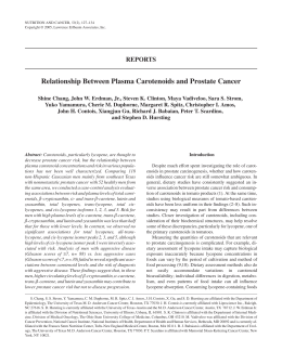
Priority Report
Combined Inhibitory Effects of Curcumin and Phenethyl
Isothiocyanate on the Growth of Human PC-3 Prostate
Xenografts in Immunodeficient Mice
1,2
1,2
1,2
1,2
1,2
Tin Oo Khor, Young-Sam Keum, Wen Lin, Jung-Hwan Kim, Rong Hu, Guoxiang Shen,
1,2
1,2
1,3
1,3
Changjiang Xu, Avanthika Gopalakrishnan, Bandaru Reddy, Xi Zheng,
1,3
1,2
Allan H. Conney, and Ah-Ng Tony Kong
1,2
1
Center for Cancer Prevention Research, 2Department of Pharmaceutics, and 3Susan Lehman Cullman Laboratory of Cancer Research,
Department of Chemical Biology, Ernest Mario School of Pharmacy, Rutgers, The State University of New Jersey, Piscataway, New Jersey
not decreased in past decades, because prostate cancer cells are
barely responsive to the cytotoxic effects of high concentrations
of chemotherapeutic agents or radiotherapy. In addition, although
patients with metastatic prostate cancer can benefit from androgen
ablation, most of them will die of prostate cancer progression to an
androgen-refractory state. Therefore, an effective prevention strategy against prostate cancer is needed to spare the burden of the
patients (1). Hence, much attention has been paid to the discovery
of chemopreventive substances from various edible and medicinal
plants, and a large number of naturally occurring chemopreventive
compounds have been identified (2). Actually, a majority of patients
with prostate cancer are now combining the conventional therapies
with these compounds as alternative, adjuvant, and/or complementary medication. Numerous epidemiologic studies have shown that
consumption of cruciferous vegetables is protective against various
types of human cancers (3). Cruciferous vegetables are a group
of vegetables named by the cross-shaped flowers, which include
broccoli, Brussel sprouts, watercress, cabbage, kale, cauliflower,
kohlrabi, and turnip. Compared with other families of vegetables,
they contain a significant amount of isothiocyanates, and strong
anticarcinogenic activities of cruciferous vegetables have thus been
attributed to the high abundance of isothiocyanates (4). Phenethyl
isothiocyanate (PEITC), a naturally occurring isothiocyanate
particularly abundant in watercress, has received much attention
as a possible chemopreventive agent. The mechanisms by which
PEITC protects against cancer have been shown to involve the
deletion of preneoplastic cells through induction of cell cycle arrest
and apoptosis (5, 6) and inhibition of carcinogen activation via
modulation of cytochrome P450-dependent monooxygenases
and enhancement of the antioxidant response element–dependent
carcinogen detoxification enzymes (7). In contrast to the high
incidence of prostate cancer in the United States, the incidence of
this disease is very low in India, which has been attributed to the
dietary consumption of large amounts of plant-based foods rich in
phytochemicals (8). The powdered root of turmeric (Curcuma longa
L., Zingeberaceae) has been widely used as a spice and a source of
yellow coloring agent (9). Curcumin (diferuloylmethane), a major
constituent of turmeric, has been shown to be highly effective
in affording protection against cancer in experimental animals
induced by a variety of chemical carcinogens (10–12). Treatment
of curcumin caused apoptosis and cell cycle arrest but inhibited
cell growth, activation of signal transduction, and transforming
activities in both androgen-dependent and androgen-independent
prostate cancer cells in culture (13). Curcumin also exerted strong
antioxidant and anti-inflammatory activities by suppressing
both constitutive and inducible nuclear factor-nB (NF-nB) and activator protein-1 activation (14). Moreover, combined treatment of
Abstract
Earlier studies using prostate cancer cells in culture showed
that phenethyl isothiocyanate (PEITC) and curcumin have
significant chemopreventive and possibly chemotherapeutic
effects. However, their in vivo effects are still lacking. Hence,
this study was undertaken to determine the possible in vivo
efficacy of prostate cancer-prevention as well as cancertherapeutic treatment by PEITC and curcumin alone or in
combination. We evaluated the effects on tumor growth
in vivo, using NCr immunodeficient (nu/nu) mice bearing
s.c. xenografts of PC-3 human prostate cancer cells. Molecular
biomarkers representing proliferation and apoptosis were
determined. Continued i.p. injection of curcumin or PEITC
(6 and 5 Mmol; thrice a week for 28 days), beginning a day
before tumor implantation significantly retarded the growth
of PC-3 xenografts. Combination of i.p. administration of
PEITC (2.5 Mmol) and curcumin (3 Mmol) showed stronger
growth-inhibitory effects. Next, we evaluated the cancertherapeutic potential of curcumin and PEITC in mice with
well-established tumors, and the results showed that PEITC
or curcumin alone had little effect, whereas combination of
curcumin and PEITC significantly reduced the growth of
PC-3 xenografts. Immunohistochemistry staining and Western
blot analysis revealed that the inhibition of Akt and nuclear
factor-KB signaling pathways could contribute to the inhibition of cell proliferation and induction of apoptosis. Taken
together, our results show that PEITC and curcumin alone or
in combination possess significant cancer-preventive activities in the PC-3 prostate tumor xenografts. Furthermore,
we found that combination of PEITC and curcumin could
be effective in the cancer-therapeutic treatment of prostate
cancers. (Cancer Res 2006; 66(2): 613-21)
Introduction
Prostate cancer is one of the most prevalent types of malignancy
in the United States. Prostate carcinogenesis has been viewed as a
multistage and complex process consisting of initiation, promotion,
and progression. Despite tremendous efforts and resources devoted
to treatment, the incidence and mortality of prostate cancer have
Note: T.O. Khor and Y-S. Keum contributed equally to this work.
Requests for reprints: Ah-Ng Tony Kong, Department of Pharmaceutics, Rutgers,
The State University of New Jersey, 160 Freilinghuysen Road, Piscataway, NJ 088548020. Phone: 732-445-3831; Fax: 732-445-3134; E-mail: [email protected].
I2006 American Association for Cancer Research.
doi:10.1158/0008-5472.CAN-05-2708
www.aacrjournals.org
613
Cancer Res 2006; 66: (2). January 15, 2006
Cancer Research
Figure 1. Effects of i.p. injections of curcumin or PEITC alone or in combination on the growth of PC-3 prostate tumors in immunodeficient mice (experiment 1).
Male NCr mice received injections with vehicle (100 AL), curcumin (6 Amol; 100 AL vehicle), PEITC (5 Amol; 100 AL vehicle), or curcumin (3 Amol) + PEITC (2.5 Amol)
in 100 AL vehicle. A, growth curve of PC-3 tumors in each group. B, tumor volume for each group after 28 days of treatment. C, body weight changes of mice during the
28 days of study. Statistical analysis of the percentage of initial body weight changes showed no significant differences between the treatment and control groups.
D, relationship between tumor volume and tumor weight. Points and columns , mean; bars, SE. *, P < 0.05, statistically significant. **, P < 0.01, statistically significant.
10% fetal bovine serum that was supplemented with penicillin (100 units/mL)
and streptomycin (100 Ag/mL). Cells were cultured at 37jC in a humidified
atmosphere of 5% CO2 and passaged twice a week.
Growth of PC-3 tumors in immunodeficient mice. Six-week-old
male NCr immunodeficient mice were obtained from Taconic Farms, Inc.
(Germantown, NY). The animals were housed in sterile filter-capped
microisolator cages and provided with sterilized AIN76A diet (Research
Diet, New Brunswick, NJ) and water. Prostate cancer PC-3 cells (2 106/
0.2 mL/mouse) were suspended in 50% Matrigel in RPMI 1640 and injected
s.c. into the right flank of the mice. Curcumin and PEITC were dissolved in
vehicle containing polyethylene glycol, benzyl alcohol, ethanol, and water
(40:0.1:10:49.1). For experiment 1, i.p. injections of the compounds were
given a day before the injection of tumor cells and continued for another 28
days. For experiment 2, i.p. injections of the compounds were given thrice
a week for 28 days starting after the injected tumor cells grew to a size of
6.5 to 10 mm (f3 weeks). Tumor size and body weight were measured
before i.p. injections of curcumin, PEITC, and the combination at various
time intervals throughout the study. At the end of study, mice were
sacrificed at 24 to 48 hours after the last administration of compounds. The
tumors were excised and weighed. Part of the tumor was snap-frozen for
protein assay, whereas the remaining portion was placed in phosphatebuffered formalin and then ethanol before preparing paraffin sections for
immunohistochemistry. All animal experiments were carried out under an
Institutional Animal Care and Use Committee–approved protocol.
curcumin sensitized and thus enhanced the cytotoxicity of prostate
cancer cells by various chemotherapeutic agents (15). Despite plenty
of convincing data from cell culture–based experiments, little is
known about the roles of PEITC and curcumin in vivo, especially on
prostate cancer. In line with this notion, the present study was
undertaken to evaluate the potential efficacy of PEITC and curcumin
and their combination as prostate cancer–preventive and possibly
prostate cancer–therapeutic agents, using NCr immunodeficient
mice (nu/nu) bearing s.c. xenografts of PC-3 androgen-independent
human prostate cancers.
Materials and Methods
Cell culture and reagents. PC-3 cells were purchased from the American
Type Culture collection (Manassas, VA). Curcumin and PEITC were obtained
from Sigma (St. Louis, MO). Matrigel was obtained from BD Biosciences
(Bedford, MA). RPMI 1640 tissue culture medium, penicillin-streptomycin,
and fetal bovine serum were from Life Technologies (Grand Island, NY).
Rabbit polyclonal antibodies against cleaved poly(ADP-ribose) polymerase
(PARP), cleaved caspase-3, phosphorylated Akt (p-Akt; Ser473), p-BAD
(Ser136), p-GSK3ha, p-IKKha, and p-InBa were purchased from Cell
Signaling (Beverly, MA). Anti-actin was a product of Santa Cruz Biotechnology (Santa Cruz, CA). PC-3 cells were maintained in RPMI 1640 containing
Cancer Res 2006; 66: (2). January 15, 2006
614
www.aacrjournals.org
Combined PEITC and Curcumin and Prostate Cancer
4jC. After three 5-minute washes with PBST, the membrane was then
incubated with horseradish peroxidase–conjugated secondary antibody in
5% nonfat dry milk/PBST for 1 hour at room temperature and then washed
with PBST thrice. The transferred proteins were visualized with the Super
Signal chemiluminescent substrate (Pierce). The intensity of visualized
bands was measured with Quantity One software (ver 4.4.0, Bio-Rad,
Richmond, CA).
Statistical analysis. Tumor volumes, body weights, and percentage of
apoptotic and proliferative cells were represented as mean F SE. Relative
fold activation/suppression was represented as mean F SD. Student’s t test
was used for the statistical analysis. Ps < 0.05 were considered significant.
Apoptosis and proliferation. The apoptotic cells were detected using
an ApopTag In Situ Apoptosis Detection kit (Chemicon, Temecula, CA). The
assay was done according to the manufacturer’s manual. After deparaffinization, the tissue sections were incubated in proteinase K for 15 minutes
at room temperature. The sections were then incubated with terminal
deoxynucleotidyl transferase enzyme at 37jC for 1 hour, washed in
three changes of PBS, and incubated with anti-digoxigenin conjugate in a
humidified chamber at room temperature for 30 minutes. The color was
developed by incubating the sections with peroxidase substrate and then
counterstained with hematoxylin for 30 seconds. For detection of
proliferative cells, proliferating cell nuclear antigen (PCNA) antibody (1:50;
DAKO, Carpinteria, CA) was used. The assay was done following the
manufacturer’s protocols. The scoring of apoptotic and proliferative cells
was done at 400. A positive control slide of rat mammary glands provided
by the manufacturer was used as positive control for the in situ apoptosis
detection assay. For the PCNA staining, mouse intestinal crypt cells were
used as a positive control.
Western blotting. Tumors dissected from mice of each treatment group
were weighed, pooled, and treated with radioimmunoprecipitation buffer
[50 mmol/L NaCl, 0.5% Triton X-100, 50 mmol/L Tris-HCl (pH 7.4),
25 mmol/L NaF, 20 mmol/L EGTA, 1 mmol/L DTT, 1 mmol/L Na3VO4,
protease inhibitor cocktail tablet (Roche, Mannheim, Germany)] at a
concentration of 10 Ag/mL for 40 minutes on ice followed by centrifugation
at 14,800 g for 15 minutes. The protein concentrations of the supernatant
were measured by using the bicinchoninic acid solution (Pierce, Rockford,
IL). Protein (20 Ag) was loaded onto NuPAGE 4% to 12% electrophoresis gel
(Invitrogen, Carlsbad, CA) and, after electrophoresis, transferred onto
polyvinylidene difluoride membrane. The membrane was blocked with 5%
nonfat dry milk in 0.1% Tween 20 in PBS (PBST) for 1 hour and incubated
with primary antibody in 5% bovine serum albumin/0.1% PBST overnight at
Results
Effects of i.p. Injections of Curcumin or PEITC Alone
or in Combination on the Growth of PC-3 Prostate
Tumors in Immunodeficient Mice
Experiment 1. Male NCr mice received injections with vehicle
(100 AL), curcumin (6 Amol; 100 AL vehicle), PEITC (5 Amol; 100
AL vehicle), or curcumin (3 Amol) + PEITC (2.5 Amol) in 100 AL
vehicle thrice a week for 28 days starting a day before the injection
of PC-3 prostate cancer cells. Tumor growth was measured once
a week, and tumor volume (V ) was calculated as V = (L W 2) 0.52, where L is the length and W is the width of a xenograft
(Fig. 1A). The tumor volumes were significantly different between
the treatment groups and the control (P = 0.02 for test between
PEITC and control; P = 0.03 for test between curcumin and
control and P < 0.001 for PEITC + curcumin versus control group).
Figure 2. Effects of i.p. injections of curcumin or PEITC alone or in combination on the growth of PC-3 prostate tumors in immunodeficient mice (experiment 2).
Male NCr mice received injection with vehicle (100 AL), curcumin (6 Amol; 100 AL vehicle), PEITC (5 Amol; 100 AL vehicle), or curcumin (3 Amol) + PEITC (2.5 Amol)
in 100 AL vehicle. A, growth curve of PC-3 tumors in each group. B, tumor volume for each group after 28 days of treatment. C, body weight changes of mice
during the 28 days of study. D, relationship between tumor volume and tumor weight. Points and columns, mean; bars, SE. *, P < 0.05, statistically significant.
www.aacrjournals.org
615
Cancer Res 2006; 66: (2). January 15, 2006
Cancer Research
The mean F SE for tumor volumes was 990 F 78 mm3 for the
control group, 624 F 49, 711 F 61, and 234 F 49 mm3 for PEITC,
curcumin, and PEITC + curcumin group, respectively (Fig. 1B).
The effect of various treatments on body weight is shown in
Fig. 1C. Statistical analysis of the percentage of initial body weight
changes showed no significant differences between the treatment
and control group. A good relationship between tumor volumes
(as calculated using measurement from live animals) and tumor
weights obtained in individual mice at the time of sacrifice was
observed (r = 0.877; Fig. 1D).
Experiment 2. Male NCr mice received injections with vehicle
(100 AL), curcumin (6 Amol; 100 AL vehicle), PEITC (5 Amol; 100 AL
vehicle), or curcumin (3 Amol) + PEITC (2.5 Amol) in 100 AL vehicle
thrice a week for 28 days starting after the xenografts were well
established (6.5-10 mm). Tumor growth was measured twice a
week, and the tumor volume was calculated as V = (L W 2) 0.52, where L is the length and W is the width with shorter
diameter of a xenograft (Fig. 2A). The tumor volumes were only
significantly different between the PEITC + curcumin and the
control group (P = 0.026). The mean F SE for tumor volumes was
568 F 50 mm3 for the control group, 469 F 43, 775 F 220, and
271 F 60 mm3 for PEITC, curcumin, and PEITC in combination
with curcumin group, respectively (Fig. 2B).
The effect of various treatments on body weight is shown in
Fig. 2C. Statistical analysis of the percentage of initial body weight
changes showed statistically significant differences between the
treatment and control group (P < 0.05 for control versus curcumin,
PEITC alone, and combination). An excellent relationship between
tumor volumes (as calculated using the measurement from live
animals) and tumor weight in individual mice obtained at the time
of sacrifice was observed (r = 0.900; Fig. 2D).
Effect of i.p. Injections of Curcumin or PEITC Alone
or in Combination on Apoptosis and Proliferation in
PC-3 Prostate Tumors in Immunodeficient Mice
Experiment 1. The apoptotic and proliferative cells in tumors
were evaluated as described in Material and Methods. The
percentage of apoptotic cells in tumors of control mice was
significantly lower compared with tumors from the treatment
groups (P = 0.029, 0.048, and 0.005 for PEITC versus control,
curcumin versus control, and PEITC + curcumin versus control,
respectively). The mean F SE for percentage of apoptotic cells in
control tumors was 1.5 F 0.2%, whereas 3.1 F 0.3%, 3.0 F 0.4%,
and 5.3 F 0.6% of apoptotic cells were detected in tumors from
the PEITC, curcumin, and PEITC + curcumin groups, respectively
(Fig. 3A). In contrast, the percentage of proliferative cells was
Figure 3. Effect of i.p. injections of curcumin
or PEITC alone or in combination on apoptosis
and proliferation in PC-3 prostate tumors in
immunodeficient mice. The presence of apoptotic
(A and C ) and proliferative cells (B and D ) in
tumors from the different groups was evaluated
using an ApopTag In Situ Apoptosis Detection kit
and PCNA labeling, respectively. A and B,
experiment 1; C and D, experiment 2. Columns,
mean; bars, SE. *, P < 0.05, statistically
significant. **, P < 0.01, statistically significant.
Cancer Res 2006; 66: (2). January 15, 2006
616
www.aacrjournals.org
Combined PEITC and Curcumin and Prostate Cancer
for PEITC versus control, curcumin versus control, and PEITC +
curcumin versus control, respectively). The mean F SE for
percentage of proliferative cells in tumors from the control, PEITC,
curcumin, and PEITC + curcumin group was 79.7 F 4.4%, 76.8 F
4.2%, 74.9 F 2.8%, and 46 F 4.3%, respectively (Fig. 3D).
found to be significantly higher in tumors from the control group
compared with the treatment groups (P = 0.001, 0.002, and <0.001
for PEITC versus control, curcumin versus control, and PEITC +
curcumin versus control, respectively). The mean F SE for the
percentage of proliferative cells in tumors from the control,
PEITC, curcumin, and the PEITC + curcumin group was 86 F
0.8%, 66.1 F 3.9%, 64.8 F 4.4%, and 44.6 F 4.1%, respectively
(Fig. 3B).
Experiment 2. The percentage of apoptotic cells in tumors of
control mice was significantly lower compared with tumors from
the PEITC + curcumin–treated mice but not in the PEITC- nor
curcumin-treated mice (P = 0.5, 0.11, and <0.001 for PEITC versus
control, curcumin versus control, and PEITC + curcumin versus
control, respectively). The mean F SE for the percentage of
apoptotic cells in control tumors was 1.1 F 0.2%, whereas 1.4 F
0.2%, 1.8 F 0.3%, and 4.9 F 0.7% of apoptotic cells were detected in
tumors from PEITC, curcumin, and PEITC + curcumin groups,
respectively (Fig. 3C). The percentage of proliferative cells was
found to be significantly higher in tumors from the controls
compared with the PEITC + curcumin–treated mice, but neither
the PEITC- nor curcumin-treated mice were found to be
statistically different from the controls (P = 0.7, 0.5, and <0.001
Effect of i.p. Injections of Curcumin or PEITC Alone or
in Combination on the Expression Levels of Cleaved
Caspase-3, Cleaved PARP, p-Akt (Ser473), p-GSK3BA,
p-BAD (Ser136), p-IKBA, and p-IKKBA in PC-3
Prostate Tumors in Immunodeficient Mice
To gain insights into the mechanisms for increased apoptosis in
tumors of treated mice, the tumors were analyzed for the
expression of cleaved caspase-3 and PARP that is known to be
activated during programmed cell death. Biomarkers from the
prosurvival/proliferation Akt and NF-nB signal transduction pathways were also examined.
Experiment 1. Western blots of control and treated mice are
shown in Fig. 4A. Densitometric scanning of the immunoreactive
bands followed by normalization with actin for differences in
protein loading indicated that a 7.3-, 9.4-, and 13.8-fold induction of
cleaved caspase-3 was observed in tumors of PEITC-, curcumin-,
Figure 3 Continued .
www.aacrjournals.org
617
Cancer Res 2006; 66: (2). January 15, 2006
Cancer Research
mice and P = 0.01 for the combination versus control group). In
addition, suppression of p-IKKha and p-InBa was also observed in
the treatment groups compared with the control counterparts.
Experiment 2. Western blots of cleaved caspase-3 and PARP
for control and treated mice are shown in Fig. 4C. A 1.2-, 2.2-,
and 9.3-fold induction of cleaved caspase-3 was observed in
tumors of PEITC-, curcumin-, and PEITC + curcumin–treated
mice, respectively, compared with the controls (P = 0.08, 0.07, and
0.01, respectively; Fig. 4D). In addition, there was a 1.7-, 2.2-, and
5.8-fold induction of PARP detected in the tumor of PEITC-,
curcumin-, and PEITC + curcumin–treated mice, respectively,
compared with the controls (Fig. 4D). A marked reduction in the
expression of p-Akt, p-GSK3ha, p-BAD, p-IKKha, and p-InBa
was only observed in the combination group versus the controls
and PEITC + curcumin–treated mice, respectively, compared with
the controls (P = 0.024, <0.01, and 0.047, respectively). In addition,
there was a 2.7-, 4-, and 8.1-fold induction of PARP detected in the
tumors of PEITC-, curcumin-, and PEITC + curcumin–treated mice,
respectively, compared with the controls (P = 0.118, 0.032, and 0.05,
respectively; Fig. 4A). In addition, we also examined the expression
level of p-Akt, p-GSK3ha, p-BAD, p-IKKha, and p-InBa, which
are known to be important regulator of the Akt and NF-nB signal
transduction pathway. The result shows that expression of p-Akt
and its downstream targets p-GSK3ha as well as p-BAD was
suppressed in the treatment group (Fig. 4B), except p-BAD for
PEITC. Statistical analysis reveals that the expression of p-GSK3ha
was significantly suppressed in all the treatment groups compared
with the control group (P = 0.03 for PEITC- and curcumin-treated
Figure 4. Effect of i.p. injections of
curcumin or PEITC alone or in combination
on expression level of cleaved caspase-3,
cleaved PARP, p-Akt (Ser473), p-GSK3ha,
p-BAD (Ser136), p-IKKha, and p-InBa in PC-3
prostate tumors in immunodeficient mice. A
and B, experiment 1; C and D, experiment 2.
Columns, mean from two separate
experiments; bars, SD. *, P < 0.05,
statistically significant. **, P < 0.01,
statistically significant.
Cancer Res 2006; 66: (2). January 15, 2006
618
www.aacrjournals.org
Combined PEITC and Curcumin and Prostate Cancer
that daily administration of diets supplemented with PEITC-NAC
(8 Amol/g), a major metabolite of PEITC, inhibited the growth of
xenografted tumors of PC-3 prostate cancer cells (16). Xiao et al.
have also reported that oral administration of 9 and 12 Amol of
PEITC inhibited the growth of established TRAMP-C1 xenografts in
nude mice (6). Moreover, administration of other naturally
occurring isothiocyanates, such as sulforaphane and AITC, have
been shown to exhibit significant inhibitory effects on the growth
of PC-3 prostate tumor xenografts (5, 17). Together, these results
indicate that the growth inhibitory effect of PEITC on PC-3 tumor
xenografts may not be a unique property but rather a general event
(P = 0.16, 0.03, 0.03, 0.06, and 0.11, respectively; Fig. 4D). Statistical
significant suppression of p-BAD was also observed in the
curcumin treated group (P = 0.02).
Discussion
Our current study presents evidence that continued i.p. injection
of naturally occurring PEITC, starting 1 day ahead of implantation
of PC-3 prostate xenografts in nude mice, significantly retarded the
growth of tumors compared with that of mice treated with vehicle
(Fig. 1A-D). In agreement with our results, Chiao et al. have shown
Figure 4 Continued .
www.aacrjournals.org
619
Cancer Res 2006; 66: (2). January 15, 2006
Cancer Research
Although single administration of PEITC and curcumin failed to
affect the growth of PC-3 prostate tumor xenografts, combined i.p.
injections of PEITC and curcumin strongly inhibited the growth of
well-established PC-3 tumor xenografts. This finding is quite
intriguing, and it is tempting to speculate that a combination
of PEITC and curcumin might have efficiently influenced the cell
death–related signaling pathways, ultimately leading to the
suppression of well-established PC-3 tumor xenografts in vivo,
which might not possibly be achieved by a single administration of
PEITC or curcumin alone. In addition, it should be further noted
that the combined administration of nontoxic doses of PEITC and
curcumin inhibited the growth of this PC-3 tumor xenograft in
mice. This finding is significant, because it could provide the basis
for the development of combination dose regimens of PEITC and
curcumin in preclinical models against prostate carcinogenesis.
Continued i.p. injection of curcumin, PEITC (6 and 5 Amol), and
the combination (3 and 2.5 Amol of curcumin and PEITC,
respectively) thrice a week for 28 days in the present study caused
some loss in body weight (Fig. 1C and Fig. 2C). However, gross
autopsy of these animals failed to reveal any abnormalities.
Therefore, it is very unlikely that the weight loss observed in the
present study is due to the nonspecific toxicity.
Recent studies have shown that PEITC and curcumin are strong
inducers of apoptosis in PC-3 cells in vitro. Xiao et al. have shown
that PEITC-induced apoptosis involved extracellular signalregulated kinase (ERK) activation and the cleavage of procaspase-3,
procaspase-8, and procaspase-9 in PC-3 cells (6, 24), whereas
apoptotic induction of PC-3 cells by curcumin involved p21WAF1/CIP1
and CCAAT/enhancer binding protein h and inhibition of cell
survival signal protein kinase B/Akt (13, 15). From earlier studies,
it is still unclear whether apoptosis of prostate cancer cells occurs
in vivo in response to treatment with these compounds and/or
whether the mode of actions occurred in vitro during apoptosis is
identical to those in vivo. In the present study, we have shown that
suppression of proliferation and induction of apoptosis are indeed
occurred in vivo, which could be responsible for the inhibitory
effects on tumor growth by PEITC and curcumin. In particular,
Western blot analysis showed that apoptosis induction of PC-3
tumor xenografts involved caspase-3 activation and PARP cleavage.
This fact suggests that apoptotic pathways involving caspase-3 and
PARP may likely be the major molecular target(s) in apoptotic
responses of PC-3 prostate xenografts to PEITC and curcumin
treatment. To dissect the possible mechanisms in which the
apoptotic and/or antiproliferation signaling were triggered, we also
examined the expression levels of the Akt/NF-nB signal transduction pathway–related biomarkers. Our result suggests that suppression of the p-Akt, p-GSK3ha, p-BAD, p-IKKha, and p-InBa was
closely correlated with the reduction of PC-3 tumor xenografts.
Because the inhibition of NF-nB and Akt are both associated with
suppression of angiogenesis, invasion, and metastasis in prostate
cancer cells (25, 26), it can be surmised that suppression of the
activity of these proteins might have also contributed to a delay of
the growth of PC-3 xenografts. A puzzling aspect of our study is,
however, that some observations previously made in vitro did not
occur in our current in vivo study. For example, although the
activation of mitogen-activated protein kinase was responsible for
the induction of apoptosis in PC-3 cells in vitro (24, 27), changes in
the phosphorylation level of ERK and c-Jun NH2-terminal kinase
were not observed in pooled extracts of PC-3 xenografts by PEITC
or curcumin (data not shown). In addition, although PEITC- or
curcumin-mediated inhibition of epidermal growth factor receptor
exerted by the isothiocyanate class of compounds. The inhibitory
effect of curcumin on carcinogenesis was first shown in a 12-Otetradecanoylphorbol-13-acetate (TPA)–, BP-, and 7,12-dimethylbenz(a)anthracene–induced mouse skin tumor model (10, 11).
Since then, inhibitory effects of dietary curcumin on colon,
duodenal, stomach, esophageal, and oral carcinogenesis were also
reported (12). However, the effects of curcumin on prostate cancer
cells in vivo are still lacking. In the present study, we have shown
that continued i.p. injection of curcumin before implantation of
PC-3 prostate cancer cells significantly retarded the growth of
these cells on a xenograft, implying that curcumin possesses inhibitory effects on the growth of prostate cancer in vivo. Although
the opinion about in vivo chemopreventive effects by curcumin
is generally held, there exist some reports that contradict this
view. For example, Imaida et al. have shown that curcumin failed
to prevent the formation of ventral prostate carcinogenesis in
rats, induced by 3,2V-dimethyl-4-aminobiphenol and 2-amino1-methylimidazo[4,5-b]pyridine (18). Somasundaram et al. also
have shown that administration of dietary curcumin did not
augment cyclophosphamide-mediated growth inhibition of BT-474
breast cancer xenografts but rather contributed to an increase
tumor size (19). Together, these facts suggest that the growthinhibitory effect of curcumin might vary, depending on the animal
tumor models, in vivo oral absorption, and/or target organs and
that chemopreventive efficacy of curcumin in vivo thus needs to
be further evaluated.
It is reasonable to expect that appropriate combinations of
chemopreventive agents might provide greater efficacy than the
administration of individual agents. For example, a combination of
piroxicam, a nonsteroidal anti-inflammatory agent, and difluoromethylornithine, a synthetic ornithine decarboxylase inhibitor,
produced synergistic inhibitory effects on colon cancer development in male F344 rats (20). Likewise, a combination of clinically
achievable concentrations of TPA and all-trans-retinoic acid
showed synergistic inhibitory effects on the growth of wellestablished LNCaP cells in athymic nude mice (21). Narayanan
et al. have also reported that a combination of docosahexaenoic
acid and 1,4-phenylene bis(methylene) selenocyanate suppressed
colon cancer cell growth and induced apoptosis more effectively
than high doses of individual agents (22). Consistent with these
observations, we also found that a combination of PEITC and
curcumin at lower doses inhibited the growth of PC-3 prostate
tumor xenografts more effectively than administration of PEITC or
curcumin alone at a higher dose.
As seen in Fig. 2A-D, i.p. injection of PEITC and curcumin
alone failed to inhibit the growth of fully established PC-3 tumor
xenografts in nude mice. This fact suggests that the effect of
PEITC or curcumin may be effective in the early stages of carcinogenesis (preventive effect) but may not inhibit tumor growth
at later stages, when tumors are fully established to certain sizes.
However, contrary to our results, Dorai et al. have observed
that administration of diet containing 2% curcumin right after
the implantation of LNCaP prostate cancer cells significantly
inhibited the growth of LNCaP tumor xenografts in nude mice
(23). The reason for this discrepancy is unclear at present. One
possibility is that because LNCaP cells are androgen-responsive
cell lines, reduced androgen level in mice due to aging might
contribute to the growth inhibition of implanted LNCaP tumor
xenografts, or it could be due to differences in the size of the
tumors and/or curcumin concentration levels achieved in the
tumors.
Cancer Res 2006; 66: (2). January 15, 2006
620
www.aacrjournals.org
Combined PEITC and Curcumin and Prostate Cancer
Acknowledgments
(EGFR) phosphorylation was clearly observed in cultured PC-3 cells
in our preliminary study, no change in EGFR phosphorylation was
observed in vivo (data not shown). Hence, it seems that further
in vivo and in vitro correlation studies are needed to better
understand the pharmacologic efficacy of PEITC and curcumin.
References
1. Denis L, Morton MS, Griffiths K. Diet and its
preventive role in prostatic disease. Eur Urol 1999;35:
377–87.
2. Surh YJ. Cancer chemoprevention with dietary phytochemicals. Nat Rev Cancer 2003;3:768–80.
3. Conaway CC, Yang YM, Chung FL. Isothiocyanates as
cancer chemopreventive agents: their biological activities and metabolism in rodents and humans. Curr Drug
Metab 2002;3:233–55.
4. Zhang Y. Cancer-preventive isothiocyanates: measurement of human exposure and mechanism of action.
Mutat Res 2004;555:173–90.
5. Singh AV, Xiao D, Lew KL, Dhir R, Singh SV.
Sulforaphane induces caspase-mediated apoptosis in
cultured PC-3 human prostate cancer cells and retards
growth of PC-3 xenografts in vivo . Carcinogenesis 2004;
25:83–90.
6. Xiao D, Zeng Y, Choi S, Lew KL, Nelson JB, Singh SV.
Caspase-dependent apoptosis induction by phenethyl
isothiocyanate, a cruciferous vegetable-derived cancer
chemopreventive agent, is mediated by Bak and Bax.
Clin Cancer Res 2005;11:2670–9.
7. Rushmore TH, Kong AN. Pharmacogenomics, regulation and signaling pathways of phase I and II drug
metabolizing enzymes. Curr Drug Metab 2002;3:481–90.
8. Sinha R, Anderson DE, McDonald SS, Greenwald P.
Cancer risk and diet in India. J Postgrad Med 2003;49:
222–8.
9. Sharma OP. Antioxidant activity of curcumin and
related compounds. Biochem Pharmacol 1976;25:1811–2.
10. Huang MT, Smart RC, Wong CQ, Conney AH.
Inhibitory effect of curcumin, chlorogenic acid, caffeic
acid, and ferulic acid on tumor promotion in mouse
skin by 12-O-tetradecanoylphorbol-13-acetate. Cancer
Res 1988;48:5941–6.
www.aacrjournals.org
Received 8/1/2005; revised 10/10/2005; accepted 10/25/2005.
The costs of publication of this article were defrayed in part by the payment of page
charges. This article must therefore be hereby marked advertisement in accordance
with 18 U.S.C. Section 1734 solely to indicate this fact.
11. Huang MT, Wang ZY, Georgiadis CA, Laskin JD,
Conney AH. Inhibitory effects of curcumin on tumor
initiation by benzo[a]pyrene and 7,12-dimethylbenz
[a]anthracene. Carcinogenesis 1992;13:2183–6.
12. Huang MT, Lou YR, Ma W, Newmark HL, Reuhl KR,
Conney AH. Inhibitory effects of dietary curcumin on
forestomach, duodenal, and colon carcinogenesis in
mice. Cancer Res 1994;54:5841–7.
13. Chaudhary LR, Hruska KA. Inhibition of cell survival
signal protein kinase B/Akt by curcumin in human
prostate cancer cells. J Cell Biochem 2003;89:1–5.
14. Han SS, Keum YS, Seo HJ, Surh YJ. Curcumin
suppresses activation of NF-nB and AP-1 induced by
phorbol ester in cultured human promyelocytic leukemia cells. J Biochem Mol Biol 2002;35:337–42.
15. Hour TC, Chen J, Huang CY, Guan JY, Lu SH, Pu YS.
Curcumin enhances cytotoxicity of chemotherapeutic
agents in prostate cancer cells by inducing p21(WAF1/
CIP1) and C/EBPh expressions and suppressing NF-nB
activation. Prostate 2002;51:211–8.
16. Chiao JW, Wu H, Ramaswamy G, et al. Ingestion of an
isothiocyanate metabolite from cruciferous vegetables
inhibits growth of human prostate cancer cell xenografts by apoptosis and cell cycle arrest. Carcinogenesis
2004;25:1403–8.
17. Srivastava SK, Xiao D, Lew KL, et al. Allyl isothiocyanate, a constituent of cruciferous vegetables, inhibits
growth of PC-3 human prostate cancer xenografts
in vivo . Carcinogenesis 2003;24:1665–70.
18. Imaida K, Tamano S, Kato K, et al. Lack of chemopreventive effects of lycopene and curcumin on experimental rat prostate carcinogenesis. Carcinogenesis
2001;22:467–72.
19. Somasundaram S, Edmund NA, Moore DT, Small GW,
Shi YY, Orlowski RZ. Dietary curcumin inhibits chemotherapy-induced apoptosis in models of human breast
cancer. Cancer Res 2002;62:3868–75.
20. Reddy BS, Nayini J, Tokumo K, Rigotty J, Zang E,
Kelloff G. Chemoprevention of colon carcinogenesis by
concurrent administration of piroxicam, a nonsteroidal
antiinflammatory drug with D L-a-difluoromethylornithine, an ornithine decarboxylase inhibitor, in diet.
Cancer Res 1990;50:2562–8.
21. Zheng X, Chang RL, Cui XX, et al. Inhibitory effect of
12-O -tetradecanoylphorbol-13-acetate alone or in combination with all-trans-retinoic acid on the growth of
LNCaP prostate tumors in immunodeficient mice.
Cancer Res 2004;64:1811–20.
22. Narayanan BA, Narayanan NK, Desai D, Pittman B,
Reddy BS. Effects of a combination of docosahexaenoic
acid and 1,4-phenylene bis(methylene) selenocyanate on
cyclooxygenase 2, inducible nitric oxide synthase and hcatenin pathways in colon cancer cells. Carcinogenesis
2004;25:2443–9.
23. Dorai T, Cao YC, Dorai B, Buttyan R, Katz AE.
Therapeutic potential of curcumin in human prostate cancer. III. Curcumin inhibits proliferation,
induces apoptosis, and inhibits angiogenesis of
LNCaP prostate cancer cells in vivo . Prostate 2001;
47:293–303.
24. Xiao D, Singh SV. Phenethyl isothiocyanate-induced
apoptosis in p53-deficient PC-3 human prostate cancer
cell line is mediated by extracellular signal-regulated
kinases. Cancer Res 2002;62:3615–9.
25. Suh J, Payvandi F, Edelstein LC, et al. Mechanisms of
constitutive NF-nB activation in human prostate cancer
cells. Prostate 2002;52:183–200.
26. Bonaccorsi L, Marchiani S, Muratori M, Carloni V,
Forti G, Baldi E. Signaling mechanisms that mediate
invasion in prostate cancer cells. Ann N Y Acad Sci
2004;1028:283–8.
27. Shenouda NS, Zhou C, Browning JD, et al. Phytoestrogens in common herbs regulate prostate cancer cell
growth in vitro . Nutr Cancer 2004;49:200–8.
621
Cancer Res 2006; 66: (2). January 15, 2006
Scaricare