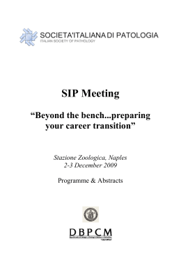
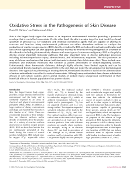
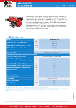
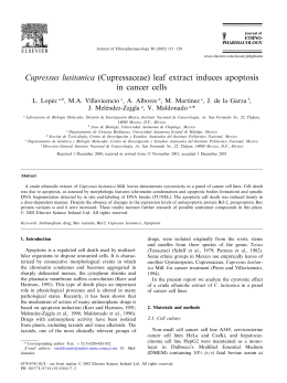
Molecular Cell 21, 509–519, February 17, 2006 ª2006 Elsevier Inc.
DOI 10.1016/j.molcel.2006.01.009
c-Myc Phosphorylation Is Required
for Cellular Response to Oxidative Stress
Barbara Benassi,1 Maurizio Fanciulli,2
Francesco Fiorentino,3 Alessandro Porrello,4,5
Giovanna Chiorino,6 Massimo Loda,7 Gabriella Zupi,1
and Annamaria Biroccio1,*
1
Experimental Chemotherapy Laboratory
2
‘‘B’’ Laboratory
Experimental Research Center
Regina Elena Cancer Institute
3
‘‘Genoma’’ Molecular Genetics Laboratory
Rome
Italy
4
Institute for Genome Sciences and Policy
Duke University
Durham, North Carolina 27708
5
Molecular Oncogenesis Laboratory
Experimental Research Center
Regina Elena Cancer Institute
Rome
Italy
6
Cancer Genomics Laboratory
Fondo Edo Tempia
Biella
Italy
7
Department of Medical Oncology
Dana Farber Cancer Institute
Harvard Medical School
Boston, Massachusetts 02115
Summary
Aside from the well-established roles of c-Myc in the
regulation of cell cycle, differentiation, and apoptosis,
a recent picture is beginning to emerge linking c-Myc
to the regulation of metabolic pathways. Here, we define a further function for c-Myc in determining cellular
redox balance, identifying glutathione (GSH) as the
leading molecule mediating this process. The link between c-Myc and GSH is g-glutamyl-cysteine synthetase (g-GCS), the rate-limiting enzyme catalyzing
GSH biosynthesis. Indeed, c-Myc transcriptionally
regulates g-GCS by binding and activating the promoters of both g-GCS heavy and light subunits. Exposure to H2O2 enhances c-Myc recruitment to g-GCS
regulatory regions through ERK-dependent phosphorylation. Phosphorylation at Ser-62 is required
for c-Myc recruitment to g-GCS promoters and determines the cellular response to oxidative stress induced by different stimuli. Thus, the c-Myc phosphorylation-dependent activation of the GSH-directed
survival pathway can contribute to oxidative stress resistance in tumor cells, which generally exhibit deregulated c-Myc expression.
Introduction
c-myc is the most investigated among the nonredundant myc gene family members and encodes a transcrip*Correspondence: [email protected]
tional factor implicated in many cellular processes such
as proliferation, differentiation, transformation, and apoptosis (Nilsson and Cleveland, 2003). The c-Myc protein
belongs to the basic helix-loop-helix leucine-zipper protein family (b/HLH/Z) and, by dimerizing with the ubiquitously expressed protein Max, binds to the specific
CAC(G/A)TG motif (E box) and regulates transcription
both positively and negatively. c-Myc can also bind to
DNA sites that differ from the palindromic hesanucleotide canonical sequence (Blackwell et al., 1993). A number of cellular target genes have been identified that are
directly or indirectly regulated by c-Myc, and this list
is rapidly increasing (Zeller et al., 2003; Adhikary and
Eilers, 2005). A c-Myc Target Gene Database assembling the c-Myc-responsive genes has been launched
(http://www.myccancergene.org/site/mycTargetDB.asp).
Thus far, most of the literature has focused on the relationship between modulation of c-Myc protein expression levels and induction or repression of target gene
transcription. Recently, a growing number of groups have
started to explore c-Myc posttranslational modifications,
which affect c-Myc transcription factor capabilities by
activating and stabilizing the protein (Sears et al., 2000;
Kamemura et al., 2002). In this context, many serine and
threonine sites have been identified along the c-Myc protein as phosphorylation target residues, although the
functional consequences of these modifications have
not been fully elucidated. The known effects include
changes in the stability of the protein and its ability to
elicit either transformation or apoptosis (Pulverer et al.,
1994; Noguchi et al., 1999; Gregory and Hann, 2000;
Sears et al., 2000; Chang et al., 2000; Yada et al., 2004).
Aside from the well-established involvement of c-Myc
in the regulation of cell cycle, differentiation, and apoptosis, a recent picture is beginning to emerge that identifies further functions of c-Myc in metabolic pathways
such as amino acid and nucleotide synthesis, regulation
of lipid metabolism, glycolysis, and mitochondrial homeostasis (Dang, 1999). Whether c-Myc regulates cellular homeostasis in response to oxidative stress remains
to be determined. Glutathione (GSH) is the most important low molecular thiol involved in cellular detoxification, redox balance, and stress response (Deneke and
Fanburg, 1989). g-glutamyl-cysteine synthetase catalyzes the first rate-limiting step in GSH biosynthesis.
g-GCS is a heterodimer composed of a heavy catalytic
(g-GCSH) and a light regulatory (g-GCSL) subunit, which
are encoded by two different genes in both rat and
human (Gipp et al., 1995). The g-GCS enzyme exerts a
key role in the maintenance of intracellular redox balance and in determining cellular response to several different stimuli, including oxidative stress, xenobiotic and
drug exposure, hormones, and growth factors (Wild and
Mulcahy, 2000). The 50 flanking regions of both g-GCS
subunits have been cloned and sequenced, identifying
putative NF-kB, Sp-1, AP-1, and AP-2 binding sites together with metal response (MRE), antioxidant response
(ARE), and electrophile-responsive (EpRE) elements
(Moinova and Mulcahy, 1998; Mulcahy et al., 1997;
Wild et al., 1998; Moinova and Mulcahy, 1999).
Molecular Cell
510
Previous work from our group has implicated c-Myc in
modulating the expression of g-GCS genes and, consequently, the intracellular GSH content (Biroccio et al.,
2002), but the mechanism(s) involved in this facet have
not been uncovered. Here, we demonstrate that c-Myc
oncoprotein transcriptionally regulates g-GCS genes
by binding and activating the promoters of both
g-GCS heavy and light subunits. Exposure to H2O2 enhances c-Myc recruitment to g-GCS regulatory regions
through ERK-dependent phosphorylation, thus activating a c-Myc-mediated response to oxidative stress.
Results
c-Myc Transcriptionally Regulates g-GCS
Expression and Determines Cellular Response
to Oxidative Stress
To investigate a possible involvement of c-Myc in the
regulation of redox balance and response to oxidative
stress, c-Myc levels were modulated in two different human melanoma lines (M14 and SbCl), which displayed
different endogenous c-Myc expression, and in immortalized human fetal fibroblasts (HFF), which showed almost no detectable c-Myc protein levels. Specific inhibition of c-Myc expression in the M14 line by RNA
interference strategy (siMyc) produced a decrease of intracellular GSH content, whereas an increase of GSH
was observed in SbCl and HFF cells when transfected
with the pcDNA-c-myc expression vector (Figure 1A).
Moreover, when cells were exposed to increasing doses
of H2O2, inhibition of c-Myc significantly enhanced H2O2induced reactive oxygen species (ROS) production and
cell death, whereas its overexpression protected cells
from H2O2-induced damage, indicating that c-Myc is
determinant in cellular response to oxidative stress (Figure 1B). The c-Myc-dependent stress response was
reverted by preincubating siMyc M14 cells with GSH
ethyl-ester (GSHest), an exogenous source of GSH,
and c-Myc overexpressing HFF/SbCl cells with L-buthionine-sulfoximine (BSO), a specific inhibitor of GSH synthesis (Figure 1B). To investigate the mechanism(s) underlying c-Myc-dependent regulation of cellular redox
balance, we analyzed whether c-Myc controlled the expression of g-GCS. mRNA expression and promoter activity of both heavy and light subunit genes were evaluated in melanoma and HFF cells after treatment with
H2O2. Inhibition of c-Myc in M14 cells decreased the
endogenous mRNA expression of both g-GCSH and
g-GCSL and abrogated the effect on g-GCS gene expression induced by H2O2 treatment (Figure 1C). Conversely,
overexpression of c-Myc in SbCl and HFF cells increased
transcript levels of both subunits and potentiated the
H2O2-induced g-GCS mRNA expression (Figure 1C).
Consistent with these results, changes in c-Myc protein
levels, by either knockdown or overexpression, led to
a modulation of both g-GCSH and g-GCSL promoter activity (Figure 1D). More interestingly, after oxidative
stress, g-GCS transcriptional activity was markedly
impaired in siMyc M14 cells and significantly enhanced
in c-Myc overexpressing SbCl and HFF lines (Figure 1D).
Oxidative Stress Recruits c-Myc to g-GCS Promoters
Upon analysis of the nucleotide sequence of both
g-GCS promoters, we identified five CACATG nonca-
nonical c-Myc binding sites. Two E boxes, mapping at
2559/2554 (E box 2) and 2500/2495 (E box 1), are
located in the region proximal to the transcription starting point of the g-GCSH promoter (Figure 2A), and three
E boxes, mapping at 22340/22335 (E box 3), 22039/
22034 (E box 2), and 21609/21604 (E box 1), are placed
on the g-GCSL gene promoter (Figure 2B). When either
the single or both E boxes on the g-GCSH promoter
were mutated (mut M1, mut M2, and mut M1-2), the
basal luciferase activity was significantly impaired (Figure 2A). More interestingly, the response to oxidative
stress was strongly affected and even more compromised upon double mutation of both E boxes (Figure
2A). Consistent with previously reported data, deletion
of the 50 distal portion of the g-GCSH promoter (D597
mutant), including several transcription factor binding
sites and four AREs, led to a dramatically decreased
basal and stress-induced transcription activity and, together with the double E box mutation (D597-mut M),
completely abolished the ability to respond to oxidative
damage (Figure 2A). Because the distal ARE4 site is
reported to be crucial in regulating both basal and
stress-induced promoter activity (Mulcahy et al., 1997),
we evaluated its contribution in relation to the c-Myc
binding sites. Upon single ARE4 mutation (mut A4), the
luciferase activity driven by the g-GCSH promoter significantly decreased after oxidative damage when compared to wild-type (wt), with the degree of reduction
being comparable to that observed in the double E
box mutant. Simultaneous disruption of both ARE4
and c-Myc binding sites (mut M-A) led to a more severe
impairment of the luciferase activity when compared to
each single mutant (mut A4 and mut M1-2), even if the
promoter still retained the ability to elicit a response to
stress. In the g-GCSL promoter, the deletion of the region containing both distal E boxes (D1633 mutant) did
not affect g-GCSL promoter activity (Figure 2B). When
mutating the c-Myc E box 1 (D1633 mut M), the luciferase activity decreased and the relevance of the putative
c-Myc binding site was more evident after oxidative
stress (Figure 2B). A similar effect was observed by
introducing a single base mutation in the ARE element
located in the proximal region (D1633 mut A) and, together with E box 1, it further decreased both basal
and oxidative stress-triggered response (Figure 2B).
To evaluate whether c-Myc actually binds to the regions covering the two closed E boxes on the g-GCSH
gene promoter and the unique consensus site on gGCSL, which are crucial in regulating basal and stressmodulated transcriptional activity, ChIP experiments
were carried out. Specific c-Myc antibody exclusively
immunoprecipitated the region covering both E boxes
on the g-GCSH promoter and the region that includes
the c-Myc consensus on g-GCSL regulatory sequence,
whereas no PCR amplification was observed by targeting the 50 and 30 g-GCS E boxes’ flanking regions
(Figure 2C). Interestingly, H2O2 exposure significantly increased the recruitment of c-Myc to g-GCSH and gGCSL promoters and, consistent with these results,
H2O2-induced binding of c-Myc to both g-GCS regulatory sequences was completely abolished after siMyc
(Figure 2D). Treatment with H2O2 also led to a modulated
recruitment to some specific c-Myc target genes. A
higher binding of c-Myc protein to the hTERT promoter
Ser-62 c-Myc Phosphorylation and Oxidative Damage
511
Figure 1. c-Myc Transcriptionally Regulates g-GCS Expression and Determines Cellular Response to Oxidative Stress
M14 cells were treated with either control (siControl, d) or c-Myc RNA interference pools (siMyc, :), whereas SbCl and HFF cells were transiently
transfected with either pcDNA3-Neo (Neo, d) or pcDNA3-c-myc (c-myc, :). Cells were exposed to H2O2 (1 mM for M14, 0.25 mM for SbCl and
HFF) for 30 min.
(A) Western blot analysis of c-Myc protein expression (upper panels) and intracellular GSH content evaluation (lower panels). Error bars indicate 6 standard deviation (SD).
(B) Flow cytometry evaluation of the percentage of both cell death and ROS production in cells treated with the indicated doses of H2O2. M14 cell
line was also preincubated with GSHest (-), whereas c-Myc overexpressing SbCl and HFF cells were preexposed to BSO (-). Error bars represent 6SD.
(C) Evaluation of g-GCSH and g-GCSL mRNA expression by RT-PCR assay. GAPDH transcript amplification has been used for loading normalization.
(D) Evaluation of g-GCSH and g-GCSL transcriptional promoter activity performed by luciferase assay. Transfection with either Myc-TA-Luc or
cyclin E-Luc expression vectors was included in the assay as c-Myc-specific positive and negative controls, respectively. The data represent the
mean of three independent experiments with standard deviation (SD). p values are *p < 0.05 and **p < 0.01.
Molecular Cell
512
Figure 2. Oxidative Stress Increases c-Myc Recruitment to g-GCS Promoters
(A) Schematic representation of wild-type (wt), deleted (D), and point-mutated (mut) g-GCSH promoter (left panel) and evaluation of the promoter
activity by luciferase assay (right panel) carried out by transfecting M14 cells with either wt or the indicated mutant g-GCS constructs. The data
represent the mean of three independent experiments with 6SD.
(B) Schematic representation of wild-type (wt), deleted (D), and point-mutated (mut) g-GCSL promoter (left panel) and evaluation of promoter
activity by luciferase assay (right panel) carried out by transfecting M14 cells with either wt or the indicated mutant g-GCS promoters. The
data represent the mean of three independent experiments with 6SD. p values are *p < 0.05 and **p < 0.01.
(C) ChIP assay was carried out in untreated M14 cells as reported in the Experimental Procedures. Amplification with oligos specific for hTERT
(region 2254/+31) and DARC (region 2477/2189) promoters was included as positive and negative controls of c-Myc DNA binding, respectively.
The DNA fragments used in the ChIP experiments on both GCS regions have been indicated on each promoter diagram reported below the ChIP
data.
(D) ChIP assay carried out as described above in the untreated, H2O2-treated, and c-Myc-interfered (siMyc) M14 cells. Amplification was performed with oligos specific for g-GCSH, g-GCSL, hTERT, cyclin D1 (region 2638/2396), CDK4 (region 2161/+66), cyclin B1 (region 2192/
222), and CDC25 (region +1020/1336) genes.
was observed after oxidative stress (Figure 2D). Conversely, its recruitment to both cyclin D1 and cyclin B1
regulatory regions was significantly decreased after
treatments with the prooxidant agent, whereas no modulation of c-Myc binding to CDK4 and CDC25A promoters was reported (Figure 2D).
We then verified the hypothesis that the increased
binding of c-Myc to g-GCS promoters might strictly depend on the higher GSH synthesis requirement dis-
played by H2O2-treated cells. GSHest incubation led to
a 3-fold increase in the intracellular GSH content, without affecting c-Myc protein expression levels (Figure 3A). Exposure to the exogenous GSH completely
abolished the increased binding of c-Myc to g-GCS E
boxes upon oxidative stress (Figure 3B). A similar modulation was reported for hTERT promoter, whereas no
change in c-Myc binding to cyclin B1 and CDC25 target
genes was observed in the H2O2-treated cells after
Ser-62 c-Myc Phosphorylation and Oxidative Damage
513
Figure 3. GSH Requirement upon Oxidative Stress Recruits c-Myc
to g-GCS Promoters
M14 cells were incubated with GSH ethyl ester (GSHest) prior to
exposition to H2O2.
(A) Intracellular GSH content evaluation and Western blot analysis of
c-Myc protein expression. The data represent the mean of three independent experiments with 6SD. p values are **p < 0.01.
(B) ChIP assay.
(C) Evaluation of g-GCSH and g-GCSL promoter activity by luciferase
assay. The data represent the mean of three independent experiments with 6SD. p values are **p < 0.01.
(D) Analysis of g-GCSH and g-GCSL mRNA expression by RT-PCR
assay.
GSHest preincubation (Figure 3B). Moreover, ester exposure blocked the ability of both g-GCS promoters to
respond to oxidative stress (Figure 3C) and inhibited
both g-GCSH and g-GCSL mRNA expression (Figure 3D).
ERK-Mediated Phosphorylation Drives c-Myc
to g-GCS Promoters upon Oxidative Stress
Exposure to H2O2 did not change the expression level of
c-Myc protein, but instead triggered its phosphorylation
at Thr-58/Ser-62, the critical sites regulating c-Myc protein stability after mitogenic stress (Sears et al., 2000;
Yeh et al., 2004) (Figure 4A). Among the different kinases
reported to be responsible for c-Myc phosphorylation,
activation of p38, ERK, JNK, AKT, and GSK-3b was evaluated. Treatment with the prooxidant agent triggered
cellular activation of both p38 and ERK, without affecting JNK, AKT, and GSK-3b phosphorylation (Figure 4B).
To identify the kinase(s) involved in c-Myc posttranslational modification, ERK and p38 phosphorylation was
inhibited by either chemical inhibitors (UO126 and
SB202190) or by specific RNA interference. Western
blot analysis revealed that only UO126 and siERK were
effective in completely inhibiting c-Myc phosphorylation
(Figure 4C), indicating that ERK is necessary for the
posttranslational modification of c-Myc in response to
H2O2-induced stress.
Next, we evaluated whether phosphorylation was required for c-Myc recruitment to g-GCS promoters. The
data obtained by ChIP analysis, carried out with an
anti-phospho-c-Myc antibody, demonstrated that the
increased recruitment to both g-GCS promoters upon
oxidative damage was attributable to c-Myc phosphorylation (Figure 4D). Oxidative stress elicited by H2O2
treatment triggered a posttranslational modification of
the c-Myc protein that enabled it to respond to the damage by increasing its binding to GSH genes. Furthermore, preincubation with UO126 abolished the recruitment of phospho-Myc to g-GCS regulatory regions,
whereas p38 inhibitor SB202190 did not have an effect
on phospho-c-Myc binding to g-GCSH and g-GCSL promoters (Figure 4D). In agreement, UO126 reduced both
g-GCSH (D597) and g-GCSL (D1633) transactivation,
whereas SB202190 did not (Figure 4E), and, consequently, g-GCS mRNA expression underwent significant
impairment exclusively upon ERK phosphorylation inhibition (Figure 4F). Finally, upon H2O2 exposure, the percentage of nonviable cells increased in UO126-treated
versus untreated cells (Figure 4G). p38 kinase inhibition
by SB202190 slightly modified cell viability after H2O2,
highlighting that other pathways are involved in the regulation of the response to stress damage. The same experiments were performed by using siERK and sip38 instead of UO126 and SB202190, respectively, giving
comparable results (data not shown).
Serine-62 Phosphorylation Is Necessary for c-Myc
Recruitment to g-GCS Promoters upon Oxidative
Stress
With the aim of evaluating the precise residue of phosphorylation, between Ser-62 and/or Thr-58, responsible
for the higher c-Myc recruitment to g-GCS target promoters, we studied the c-Myc-dependent response to
oxidative stress in c-myc knockout mouse embryonic fibroblasts (MEFmyc2/2). As shown in Figure 5A, reintroduction of wt c-myc cDNA in knockout cells significantly
reduced stress-induced ROS production and cell death.
Upon H2O2 exposure, MEFmyc2/2 activated the ERKmediated c-Myc phosphorylation pathway (Figure 5B),
thus harboring protection from the oxidative damagetriggered cell death (Figure 5C). After site-directed mutagenesis of Ser-62 and/or Thr-58 residues in the
pcDNA3-c-myc expression vector, either the wt or
each different c-myc mutant was transfected into the
MEFmyc2/2 cells. Upon H2O2 exposure, phosphorylation
of c-Myc protein was only reported in the wt c-myc and
T58A c-myc-transfected cells, whereas no posttranslational modification was observed after transfection
with either the S62A or the T58A/S62A c-myc mutant
cDNA (Figure 5D). The inability to be phosphorylated
at Ser-62 made c-Myc protein unable to respond to oxidative stress in terms of increasing binding to both
g-GCS promoters, whereas c-MycT58A, as the c-Mycwt,
was highly recruited to both target genes upon H2O2 exposure (Figure 5E). When mutated at both sites, c-Myc
behaved as the c-MycS62A form. Consistent with these
data, c-MycS62A and MycT58A/S62A were unable to transactivate both g-GCS promoters after oxidative damage
(Figure 5F), thus indicating a master role played by
Ser-62 in the c-Myc phosphorylation-driven response
to H2O2 stress. The impaired ability of c-MycS62A and
c-MycT58A/S62A to be recruited to g-GCS promoter E
boxes significantly affected the expression of both
heavy and light gene transcripts (Figure 5G). Consistent
with these results, the g-GCS enzymatic activity and
Molecular Cell
514
Figure 4. ERK-Dependent Phosphorylation
Drives c-Myc to g-GCS Promoters upon Oxidative Stress
M14 cells were exposed to H2O2 and, where
indicated, were preincubated either with
pharmacological or molecular (RNA interference strategy) ERK/p38 inhibitors (UO126
and SB202190; siERK and sip38).
(A) Western blot analysis of phospho-Myc.
(B) Western blot analysis of the phosphorylated forms of p38, ERK, JNK, AKT, and
GSK-3b.
(C) Western blot analysis of phospho-p38,
phospho-ERK, and phospho-Myc.
(D) ChIP assay.
(E) Evaluation of g-GCSH and g-GCSL promoter activity. The data represent the mean
of three independent experiments with SD.
p values, calculated in H2O2-exposed cells
by comparing either UO126- or SB202190treated to untreated cells, are *p < 0.05 and
**p < 0.01.
(F) mRNA expression by RT-PCR.
(G) Cytofluorimetric analysis of cell death.
The data represent the mean of three independent experiments with SD. p values, calculated in H2O2-exposed cells by comparing
either UO126- or SB202190-treated to untreated cells, are *p < 0.05 and **p < 0.01.
consequently the intracellular GSH content (Figure 5H)
were markedly reduced in both c-MycS62A and cMycT58A/S62A when compared to c-Mycwt and c-MycT58A,
reaching the levels observed in the MEFmyc2/2 cells
(about 10 6 2 nmol/mg in the untreated and 42 6
3 nmol/mg in the stress-exposed cells).
To give a broad view of how S62A mutation affects
gene regulation by c-Myc, microarray analysis was performed in MEFmyc2/2 cells transfected with either wt or
S62A c-myc cDNA both untreated and treated with
H2O2. As shown in Figure 5I, no change in the gene expression profile between wt and S62A myc-transfected
cells was found at the steady-state condition. On the
contrary, the signature of Mycwt and c-MycS62A cells
was different under H2O2 exposure, strongly supporting
the key role of this c-Myc phoshorylation site during oxidative stress response. Comparison of c-MycS62A versus Mycwt transcription evidenced that S62A mutation
did not influence the expression of some well-known
c-Myc target genes, such as ODC, TERT, LDH-A, and
a-prothymosin. As shown in Figure S1 (see the Supplemental Data available with this article online), S62A
strongly altered several genes involved in apoptosis,
proliferation, and cell signaling (including c-Myc target
genes), consistent with the inability of c-myc S62A to activate the g-GCS-mediated survival pathway. Moreover,
a greater group of genes participating in cellular metabolism and oxidative defense were regulated in a different
way in Mycwt and c-MycS62A cells. Table S1 reports
some of the key genes required for cellular response
to oxidant damage. It is evident that besides both gGCS genes, other antioxidant enzymes (Gpx1, GSr,
Cas-1) implicated in first-line oxidative protection were
underexpressed in the c-MycS62A compared to Mycwt
cells. Consistent with these results, Sod-2 and many
GSH transferases were overexpressed, as a classical
cellular response to the impaired GSH content.
Finally, we evaluated how S62A mutation, by altering
the cellular redox response, affected biological behavior
upon oxidative stress. Transfection with either wt or
T58A c-myc protected cells from the H2O2-induced
damage (Figure 5L). On the contrary, neither S62A
c-myc nor T58A/S62A c-myc transfection affected cell
viability (Figure 5L). Overexpression of c-Mycwt also
protected cells from ROS-generating drugs, such as
doxorubycin and cisplatin, without affecting the viability
of taxol-treated cells (Figure S2).
Discussion
Role of c-Myc on Cell Death
In this study, we present data demonstrating that c-Myc
can promote resistance to oxidative damage by inducing the expression of g-GCS, the rate-limiting enzyme
in glutathione biosynthesis. This result might appear
paradoxical, as c-Myc is reported to sensitize cells
Ser-62 c-Myc Phosphorylation and Oxidative Damage
515
Figure 5. Serine-62 Phosphorylation Is Necessary for c-Myc Recruitment to g-GCS Promoters upon Oxidative Stress
MEFmyc2/2 cells were untransfected (2) or transiently transfected with either Neo (B) or wt (d) or mutant (T58A, S62A, T58A/S62A) c-myc cDNA
cloned into the pcDNA3 expression vector. Where not specified, data represent H2O2-treated cells.
(A) Flow cytometry evaluation of the percentage of both cell death and ROS production. Error bars indicate 6SD.
(B) Western blot analysis of phospho-Myc and phospho-ERK after exposure to the indicated treatments.
(C) Cytofluorimetric evaluation of cell death after transfection with the indicated vectors and treatment with UO126. The data represent the mean
of three independent experiments with 6SD. p values, calculated in c-myc-transfected versus Neo-transfected cells, are *p < 0.05.
(D) Immunoblot analysis of phospho-Myc expression levels.
(E) ChIP assay.
(F) Analysis of g-GCSH and g-GCSL promoter activity carried out by luciferase assay. Error bars represent 6SD.
(G) Evaluation of g-GCSH and g-GCSL mRNA expression by RT-PCR assay (GAPDH transcript amplification has been used for loading normalization).
(H) Analysis of g-GCS enzyme activity (upper panel) and intracellular GSH content (lower panel). Error bars indicate 6SD.
(I) Plots of log10(Ratio) against log10(Intensity) obtained from untreated and H2O2-treated cells. In the plots, Ratio = S62A/wt, and Intensity =
O(S62A 3 wt). The red and green spots correspond to genes overexpressed and underexpressed between the two mRNA sources.
(L) Cytofluorimetric evaluation of cell death. The data represent the mean of three independent experiments with SD. p values, calculated in S62A
and T58A/S62A versus wt-transfected cells, are **p < 0.01.
to several apoptotic triggers (Hoffman and Liebermann, 1998; Thompson, 1998). However, protection from
apoptosis by c-Myc has been observed in other models
by different groups (Waikel et al., 1999; Liu et al., 2000;
Biroccio et al., 2001, 2002; D’Agnano et al., 2001; Ceballos et al., 2005). Several lines of evidence indicate that the
Molecular Cell
516
role of c-Myc on apoptosis may depend on the cellular
genetic background (Nilsson and Cleveland, 2003; Ceballos et al., 2005). However, the protective effect of cMyc reported here cannot be attributable to cell type, because it also occurs in MEF cells, where overexpression
of c-Myc is reported to induce apoptosis (Hoffman and
Liebermann, 1998; Thompson, 1998). The reliability of
the experimental model used in this paper is based on
data reported in Figure S3, demonstrating that c-Mycoverexpressing MEF cells undergo apoptosis under serum deprivation, whereas the same cells became resistant to oxidative stress in the presence of growth factors.
The observed dual effect of c-Myc ensures that its role in
cell death is influenced by the presence and/or absence
of growth factors. The involvement of c-Myc in apoptosis
may also depend on mechanism of action and dose or
time-exposure of the cellular insult. For instance, deregulated c-Myc expression can sensitize cells to some
stimuli, but not to others (Biroccio et al., 2004; Grassilli
et al., 2004). Moreover, c-Myc seems essential for the induction of apoptosis by sublethal doses of drug, but not
required in cases when the apoptotic stimuli are sufficient to trigger apoptosis (Grassilli et al., 2004; Soucie
et al., 2001). This is consistent with data demonstrating
that low drug doses can have a cytostatic rather than
a cytotoxic effect, a condition in which endogenous cMyc expression is usually downregulated and overexpression of c-Myc generally triggers apoptosis. In our
experiments, the dose and/or time exposure of the oxidative damage is not able to reduce endogenous cMyc expression or to decrease cell proliferation, and, in
these circumstances, c-Myc induces resistance to oxidative stress by upregulating the g-GCS gene expression. Similarly, Park and coworkers (Park et al., 2002)
demonstrated that when the apoptotic stimulus is able
to increase endogenous c-Myc levels, the overexpression of c-Myc could promote survival through activation
of the ornithine decarboxilase gene. The protective effect of c-Myc from oxidative stress reported here seems
also to contrast with some results showing that c-Myc increases ROS generation (Tanaka et al., 2002; Vafa et al.,
2002). Data reported in Figure S3 demonstrated that MEF
cells transfected with an inducible c-myc expression
vector show an increase in ROS production at a very
early time in c-Myc induction. Then, when the cells are
cultured in growth medium, ROS progressively decrease
and the cells do not die. On the contrary, activation of cMyc in the same cells maintained under serum deprivation, a condition known to give rise to oxidative stress,
markedly increases ROS production and death. These
results are in agreement with those of the Kanacura
and Cleveland groups, showing that c-Myc overexpression alone does not induce cell death (Tanaka et al., 2002;
Maclean et al., 2003). The divergence with the paper from
Vafa et al. (2002), which shows, rather, that c-Myc directly induces DNA damage, could reflect the difference
in cell type and/or the fact that their studies were carried
out in the absence of survival factors. Finally, the functional role of c-Myc in determining cellular response to
oxidative stress appears to be p53-independent, as it occurs both in wild-type (SbCl, HFF, and MEF) and mutated
(M14) cell lines. Moreover, data reported in Figure S4 indicate that even in conditions where p53 is activated, the
c-Myc-mediated protection from oxidative damage oc-
curs to the same extent in the p53 wild-type HCT116
and its knockout derivative line.
g-GCS Heavy and Light Subunits Are Stress-Induced
c-Myc Target Genes
By studying the mechanism(s) by which c-Myc induces
g-GCS genes and mediates the response to oxidative
damage, we found that c-Myc transcriptionally regulates the g-GCS genes by binding and activating the promoters of g-GCS heavy and light subunits. The transcriptional control of g-GCS gene expression by c-Myc
occurs through its binding to the noncanonical c-Myc
consensus sites identified on g-GCSH and g-GCSL
gene promoters. However, not all the different E boxes
identified on both regulatory sequences were bound
and activated by c-Myc, in agreement with data demonstrating that accessory factors may play a role in effective binding to E box elements (Fernandez et al., 2003;
Patel et al., 2004). Our results give functional relevance
to the two E boxes located in the region proximal to
the transcription starting point of the g-GCSH promoter.
These data differ from most available literature data that
describe several distinct binding elements in the distal
genomic fragment (Mulcahy et al., 1997; Wild et al.,
1998). On the contrary, the distal region of the g-GCSL
promoter, which includes two putative c-Myc consensus sites, is not involved in the regulation of the transcription rate, as previously described (Moinova and
Mulcahy, 1998). The E box 1 on the g-GCSL promoter
is determinant in c-Myc-dependent transcriptional regulation, even though deletion of this region still retains
the ability to respond to oxidative stress, consistent
with data demonstrating that some consensus sites for
other transcriptional factors are located in this region
(Moinova and Mulcahy, 1998). Furthermore, simultaneous mutation of the E boxes and ARE consensus
strongly compromises the transcriptional activity of
both g-GCS promoters, indicating that the transcriptional factors binding these regions work independently
in regulating g-GCS expression. We also found that the
increased transcriptional rate and mRNA expression of
g-GCS genes after oxidative stress is attributable to
a higher recruitment of c-Myc to both g-GCS promoters.
Moreover, the stress-dependent regulation of c-Myc recruitment to target genes occurs quickly and strongly
depends on the higher GSH synthesis requirement displayed by the stress-exposed cells.
Phosphorylation at Ser-62 Confers Promoter
Specificity to c-Myc
The fast c-Myc-mediated response to stress raised the
question as to whether it could be tightly regulated by
posttranslational modification. Indeed, exposure to
H2O2 did not change the expression levels of c-Myc protein but instead triggered ERK-dependent Thr-58/Ser-62
phosphorylation. Thr-58/Ser-62 phosphorylation has
been previously observed during Ras-dependent mitogenic stimulation, and it was also observed that this
posttranslational modification of c-Myc exerts opposing
roles in controlling protein stability: phosphorylation at
Ser-62 stabilizes c-Myc, whereas phosphorylation at
Thr-58 promotes c-Myc degradation through the ubiquitin-mediated proteasome pathway (Sears et al., 2000;
Yeh et al., 2004). The data obtained here by using
Ser-62 c-Myc Phosphorylation and Oxidative Damage
517
Experimental Procedures
Antibodies and Reagents
The antibodies against c-Myc (clone 9E10 and clone N-262) were
purchased from Santa Cruz Biotechnology (Santa Cruz, CA, USA).
The antibodies against phospho-Myc (Thr58/Ser62), recognizing
c-Myc phosphorylated at Thr58 and/or at Ser62, and the nonphosphorylated and phosphorylated forms of p38 (Thr180/Tyr182), ERK
(Thr202/Tyr204), JNK (Thr183/Tyr185), AKT (Ser473), and GSK-3b
(Ser9) were obtained from Cell Signaling (Beverly, MA, USA). The
control (siControl) and sequence-specific short RNA duplex pools
for interference were purchased from Upstate (Lake Placid, NY,
USA) for c-Myc and from Cell Signaling for p38 and ERK.
H2O2, BSO, GSHest, propidium iodide (PI), and 4-hydroxy-tamoxifen (4-HT) were purchased from Sigma-Aldrich (Milan, Italy), and
kinase inhibitors UO126 and SB202190 were purchased from
Promega Corporation (Madison, WI, USA).
Figure 6. Proposed c-Myc-Dependent Survival Pathway Activated
by Oxidative Damage
Oxidative stress triggered by ROS-generating agents induces
ERK-dependent phosphorylation of c-Myc at Ser-62. Ser-62-phoshorylation drives c-Myc to both g-GCS promoters, thus inducing
GSH neo-synthesis. c-Myc-dependent GSH increase, acting as
ROS scavenger, protects cells from oxidative damage.
c-myc knockout MEF cells transfected with the different
c-myc mutants suggest that stress-induced phosphorylation of c-Myc at Thr-58 is dependent upon Ser-62, as
occurs after mitogenic stimulation (Sears et al., 2000;
Yeh et al., 2004). However, the findings reported in this
paper add mechanistic insights into this c-Myc phosphorylation function. Indeed, Ser-62 phosphorylation
not only regulates protein stability (Sears et al., 2000;
Yeh et al., 2004), but also can dictate the choice of target
genes. Specifically, c-Myc phosphorylation at Ser-62 is
required for activation of g-GCS genes. Like c-Myc,
other important transcription factors, such as p53,
have been found to be phosphorylated in sites far
away from the DNA binding domain, and these posttranscriptional modifications regulate the binding ability to
target genes (Saito et al., 2003). Moreover, microarray
analysis revealed that besides both g-GCS genes, other
genes were differentially regulated in the c-MycS62A
compared to Mycwt cells, exclusively upon H2O2 treatment, supporting the key role of this c-Myc phosphorylation site during oxidative stress response. However, array data need to be confirmed to verify if c-Myc
directly modulates their transcription or if their alteration
is a consequence of the upstream c-Myc-mediated gGCS regulation. The relevance of the Ser-62 residue in
the transcriptional regulation of each gene will be the
object of further studies.
Finally, in this paper we attribute a biological function
to c-Myc phosphorylation at Ser-62 in determining cellular response ROS-triggering agents, including H2O2 and
anticancer drugs.
In summary, we identify an oxidative stress-induced
survival signal pathway (Figure 6), depending on c-Myc
phosphorylation, that can contribute to resistance to
oxidative damage in tumor cells, which generally exhibit
deregulated c-Myc expression. Our data support the
hypothesis that regulation of c-Myc phosphorylation
by both mitogenic stimuli and oxidative stress may be
critical for functional Ras/Myc cooperation in cancer
development.
Cell Cultures and Treatment
Human melanoma cell lines (M14 and SbCl) were maintained in
RPMI-1640 medium (Gibco-BRL, Gaithersburg, MD, USA) supplemented with 10% fetal calf serum (FCS, Gibco-BRL). Human fetal
immortalized fibroblasts (HFF) and mouse embryonic fibroblasts
(MEF) were cultured in DMEM medium (Gibco-BRL) containing
10% and 15% FCS, respectively. In the experiments with H2O2, cells
were incubated with increasing doses of the prooxidant agent for 30
min, and all of the analyses except for the evaluation of cell death
were performed at the end of the treatment. Where not indicated,
the dose of H2O2 used was as follows: 1 mM for M14 and 0.25 mM
for SbCl, HFF, and MEF. BSO (10 mM) and GSHest (5 mM) were
added to cells 24 hr before H2O2 exposure. Kinase inhibitors
UO126 (10 mM) and SB202190 (25 mM) were added to the cell medium 24 hr before H2O2 treatment. Growth curve analysis was performed by counting the total number of viable cells by trypan blue
dye exclusion.
Plasmid Constructs
pcDNA3-Neo and pcDNA3-c-myc have been previously described
(Biroccio et al., 2004). pMyc-TA-LUC vector, containing six tandem
copies of c-Myc E box, was from Clontech (Palo Alto, CA, USA). Deleted pGL3b-g-GCSH (D597) was created by digesting wt pGL3b-gGCSH with BstPI. Deleted pGL3b-g-GCSL (D1633) was obtained by
cutting pGL3b-g-GCSL (D1927) with NdeI restriction enzyme. All
the other mutants were created by the PCR-based QuickChange
Site-Directed Mutagenesis Kit (Stratagene, La Jolla, CA), according
to the manufacturer’s instructions. All mut-M mutants carry a mutated E box site where the cacatg c-Myc binding region has been replaced by a scrambled tacgac sequence. All mut-A mutants carry
a point-mutated ARE element in which the gtgacNNNgc core
sequence has been replaced by a gggacNNNgc site.
Transfection and Infection
Transient transfection experiments were carried out by lipofectamine reagent (Gibco-BRL). Transfected cells were then maintained
in FCS-containing medium and harvested 24 or 48 hr after the end of
transfection. Transfection efficiency was assessed for each cell line
and in each single experiment by using a plasmid carrying the gene
either for the green (GFP) or the red fluorescent protein (RFP). Only
experiments with transfection efficiency higher than 70% were considered valuable and included in the reported data.
To delete the c-myc gene and generate knockout mouse embryonic fibroblasts (MEFmyc2/2), c-mycfl/fl fibroblasts were infected
with a retroviral-based vector carrying the GFP gene fused to the
Cre locus. After 48 hr of infection, GFP positive cells were sorted
by cytofluorimetric analysis and cultured in FCS-containing DMEM for just one passage before further transfection with either
wild-type or mutant c-myc cDNA. MEFmyc2/2 cells have been freshly
prepared by infection and sorting procedure before each experiment.
Western Blot Analysis
Western blot analysis was performed as previously reported (Biroccio et al., 2002). Briefly, cells were lysed on ice for 30 min in lysis
buffer supplemented with phosphatase inhibitors. Forty mg of total
Molecular Cell
518
proteins was then loaded onto denaturing sodium dodecyl-sulfatepolyacrylamide gels. ECL (Amersham Biosciences, Piscataway,
NJ, USA) was employed for chemoluminescence detection.
Glutathione Determination and gGCS Enzyme Activity
Intracellular GSH content was measured as previously described
(Biroccio et al., 2004). gGCS enzyme activity was evaluated by using
a coupled assay with pyruvate kinase and lactate dehydrogenase
(Seeling and Meister, 1984) and assaying the rate of decrease in absorbance at 340 nm at 37ºC. Enzyme activity was expressed as mmol
of NADH oxidized per minute (U)/mg protein.
Flow Cytometry
Cell viability was assessed by staining cells with PI (1 mg/ml). Sixty
minutes after the end of H2O2 treatment, adherent cells were harvested, washed twice in PBS, stained with PI, and immediately analyzed by flow cytometry. PI positive cells were defined as nonviable
cells. The evaluation of ROS was performed as previously described
(Biroccio et al., 2004).
Reverse Transcriptase-Polymerase Chain Reaction
For semi-quantitative RT-PCR analysis, total RNA was isolated by
TriZol (Gibco-BRL). First-strand cDNA synthesis and amplification
of specific DNA sequences were performed by using ThermoScript
RT-PCR System (Invitrogen, Carlsbad, CA, USA). cDNAs were amplified with each specific pair of forward/reverse primers (sequences
available upon request). Glyceraldehyde-3-phosphate dehydrogenase (GAPDH) transcript amplification has been used as an internal
control for normalizing data to RNA loading. The ability of reverse
transcriptase assay to detect semiquantitative changes in mRNA expression was verified by amplifying target mRNAs with 20, 25, and
30 PCR cycles.
Luciferase Assay
For promoter activity assay, cells were cotransfected with PEQ-176
(Promega Corporation) and pGL3basic-g-GCS promoters. Luciferase activity was measured with a Luciferase Assay System kit
(Promega Corporation), as specified by the manufacturer, and normalized to b-galactosidase expression.
Chromatin Immunoprecipitation
Chromatin Immunoprecipitation (ChIP) assay was performed by
using a Chromatin Immunoprecipitation Assay kit (Upstate). Briefly,
formaldehyde crosslinked chromatin fragments were immunoprecipitated with no antibody, anti-c-Myc (clone N-262), anti-phospho-MycThr58/Ser62, or b-actin antibodies and assayed by PCR (the
sequences of each pair of forward/reverse oligos is available upon
request). To verify that an equivalent amount of chromatin was
used in the immunoprecipitates, a sample representing 0.01% of
the total chromatin (input) was included in the PCR samples. Linearity of the signal was insured by amplifying increasing amounts of
template DNA (0.1, 0.25, and 0.5 mg).
Microarray and Data Analysis
Total RNA was extracted from wt and S62A c-myc-transfected
MEFmyc2/2 cells, both exposed and unexposed to H2O2, by TriZol reagent followed by additional purification with an RNeasy kit from
Qiagen (Valencia, CA, USA). Preparation of cRNA, oligonucleotide
array, hybridization to MOE430A2.0 GeneChip arrays (Affymetrix,
Santa Clara, CA, USA), and scanning of the arrays were carried out
at the Dana-Farber Microarray Core Facility.
Raw chip data were loaded into the Rosetta Luminator software,
and intensity profiles (one for each chip analyzed) were obtained after application of the Affymetrix Error Model implemented by Rosetta Biosoftware. Intensity values, together with intensity errors
and p values, were then associated to each probe set contained in
the Mouse Affymetrix Chip for the different experimental conditions.
Ratio experiments were generated by combining replicate chips and
selecting couples of mRNA sources to be compared. Successively,
log10(Ratio) against log10(Intensity) plots were created (Bolstad
et al., 2003). Differentially expressed genes were extracted by selecting probesets with a Log10(Ratio) p value of less than 0.01 and
Absolute Fold Change greater than 2. Successively, information
about modulated genes was extracted with the Batch Search option
available within the SOURCE website (http://smd.stanford.edu/
cgi-bin/source/sourceBatchSearch).
Statistical Analysis
The experiments have been repeated from three to five times, and
the results obtained are presented as means 6SD. Significant
changes were assessed by using the Student’s t test for unpaired
data, and p values <0.05 were considered significant.
Supplemental Data
Supplemental Data include four figures and one table and can be
found with this article online at http://www.molecule.org/cgi/
content/full/21/4/509/DC1/.
Acknowledgments
The work was supported by grants from Associazione Italiana
Ricerca sul Cancro (A.I.R.C.) and Ministero della Salute. We thank
Dr. de Alboran for providing c-mycfl/fl MEF cells; Dr. H. Hock for
the retroviral vector carrying the GFP gene fused to the Cre locus;
Dr. E. Kieff for giving us the pSG5-Mock and pSG5-c-mycER plasmids; and Dr. J. Gipp for providing wt and deleted pGL3b-g-GCSH
and pGL3b-g-GCSL vectors. We also thank Dr. M.R. Ciriolo and
Dr. B. Amati for fruitful discussion and Dr. Paola Ostano for her support in the biostatistical analysis of array data.
Received: August 12, 2005
Revised: November 18, 2005
Accepted: January 9, 2006
Published: February 16, 2006
References
Adhikary, S., and Eilers, M. (2005). Transcriptional regulation and
transformation by Myc proteins. Nat. Rev. Mol. Cell Biol 6, 635–645.
Biroccio, A., Benassi, B., Amodei, S., Gabellini, C., Del Bufalo, D.,
and Zupi, G. (2001). c-Myc down-regulation increases susceptibility
to cisplatin through reactive oxygen species-mediated apoptosis in
M14 human melanoma cells. Mol. Pharmacol. 60, 174–182.
Biroccio, A., Benassi, B., Filomeni, G., Amodei, S., Marchini, S.,
Chiorino, G., Rotilio, G., Zupi, G., and Ciriolo, M.R. (2002). Glutathione influences c-Myc-induced apoptosis in M14 human melanoma
cells. J. Biol. Chem. 277, 43763–43770.
Biroccio, A., Benassi, B., Fiorentino, F., and Zupi, G. (2004). Glutathione depletion induced by c-Myc downregulation triggers apoptosis
on treatment with alkylating agents. Neoplasia 6, 195–206.
Blackwell, T.K., Huang, J., Ma, A., Kretzner, L., Alt, F.W., Eisenman,
R.N., and Weintraub, H. (1993). Binding of myc proteins to canonical
and noncanonical DNA sequences. Mol. Cell. Biol. 13, 5216–5224.
Bolstad, B.M., Irizarry, R.A., Astrand, M., and Speed, T.P. (2003). A
comparison of normalization methods for high density oligonucleotide array data based on variance and bias. Bioinformatics 19, 185–
193.
Ceballos, E., Munoz-Alonso, M.J., Berwanger, B., Acosta, J.C., Hernandez, R., Krause, M., Hartmann, O., Eilers, M., and Leon, J. (2005).
Inhibitory effect of c-Myc on p53-induced apoptosis in leukemia
cells. Microarray analysis reveals defective induction of p53 target
genes and upregulation of chaperone genes. Oncogene 24, 4559–
4571.
Chang, D.W., Claassen, G.F., Hann, S.R., and Cole, M.D. (2000). The
c-Myc transactivation domain is a direct modulator of apoptotic versus proliferative signals. Mol. Cell. Biol. 20, 4309–4319.
Dang, C.V. (1999). c-Myc target genes involved in cell growth, apoptosis, and metabolism. Mol. Cell. Biol. 19, 1–11.
D’Agnano, I., Valentini, A., Fornari, C., Bucci, B., Starace, G., Felsani,
A., and Citro, G. (2001). Myc down-regulation induces apoptosis in
M14 melanoma cells by increasing p27(kip1) levels. Oncogene 20,
2814–2825.
Deneke, S.M., and Fanburg, B.L. (1989). Regulation of cellular glutathione. Am. J. Physiol. 257, L163–L173.
Ser-62 c-Myc Phosphorylation and Oxidative Damage
519
Fernandez, P.C., Frank, S.R., Wang, L., Schroeder, M., Liu, S.,
Greene, J., Cocito, A., and Amati, B. (2003). Genomic targets of
the human c-Myc protein. Genes Dev. 17, 1115–1129.
Soucie, E.L., Annis, M.G., Sedivy, J., Filmus, J., Leber, B., Andrews,
D.W., and Penn, L.Z. (2001). Myc potentiates apoptosis by stimulating Bax activity at the mitochondria. Mol. Cell. Biol. 21, 4725–4736.
Gipp, J.J., Bailey, H.H., and Mulcahy, R.T. (1995). Cloning and sequencing of the cDNA for the light subunit of human liver gammaglutamylcysteine synthetase and relative mRNA levels for heavy
and light subunits in human normal tissues. Biochem. Biophys.
Res. Commun. 206, 584–589.
Tanaka, H., Matsumura, I., Ezoe, S., Satoh, Y., Sakamaki, T., Albanese, C., Machii, T., Pestell, R.G., and Kanakura, Y. (2002). E2F1
and c-Myc potentiate apoptosis through inhibition of NF-kappaB
activity that facilitates MnSOD-mediated ROS elimination. Mol.
Cell 9, 1017–1029.
Grassilli, E., Ballabeni, A., Maellaro, E., Del Bello, B., and Helin, K.
(2004). Loss of MYC confers resistance to doxorubicin-induced apoptosis by preventing the activation of multiple serine protease- and
caspase-mediated pathways. J. Biol. Chem. 279, 21318–21326.
Thompson, E.B. (1998). The many roles of c-Myc in apoptosis. Annu.
Rev. Physiol. 60, 575–600.
Gregory, M.A., and Hann, S.R. (2000). c-Myc proteolysis by the ubiquitin-proteasome pathway: stabilization of c-Myc in Burkitt’s lymphoma cells. Mol. Cell. Biol. 20, 2423–2435.
Vafa, O., Wade, M., Kern, S., Beeche, M., Pandita, T.K., Hampton,
G.M., and Wahl, G.M. (2002). c-Myc can induce DNA damage, increase reactive oxygen species, and mitigate p53 function: a mechanism for oncogene-induced genetic instability. Mol. Cell 9, 1031–
1044.
Hoffman, B., and Liebermann, D.A. (1998). The proto-oncogene cmyc and apoptosis. Oncogene 17, 3351–3357.
Waikel, R.L., Wang, X.J., and Roop, D.R. (1999). Targeted expression of c-Myc in the epidermis alters normal proliferation, differentiation and UV-B induced apoptosis. Oncogene 18, 4870–4878.
Kamemura, K., Hayes, B.K., Comer, F.I., and Hart, G.W. (2002). Dynamic interplay between O-glycosylation and O-phosphorylation
of nucleocytoplasmic proteins: alternative glycosylation/phosphorylation of THR-58, a known mutational hot spot of c-Myc in lymphomas, is regulated by mitogens. J. Biol. Chem. 277, 19229–19235.
Wild, A.C., and Mulcahy, R.T. (2000). Regulation of gamma-glutamylcysteine synthetase subunit gene expression: insights into transcriptional control of antioxidant defenses. Free Radic. Res. 32, 281–
301.
Liu, H., Lo, C.R., Jones, B.E., Pradhan, Z., Srinivasan, A., Valentino,
K.L., Stockert, R.J., and Czaja, M.J. (2000). Inhibition of c-Myc expression sensitizes hepatocytes to tumor necrosis factor-induced
apoptosis and necrosis. J. Biol. Chem. 275, 40155–40162.
Maclean, K.H., Keller, U.B., Rodriguez-Galindo, C., Nilsson, J.A., and
Cleveland, J.L. (2003). c-Myc augments gamma irradiation-induced
apoptosis by suppressing Bcl-XL. Mol. Cell. Biol. 23, 7256–7270.
Moinova, H.R., and Mulcahy, R.T. (1998). An electrophile responsive
element (EpRE) regulates beta-naphthoflavone induction of the
human gamma-glutamylcysteine synthetase regulatory subunit
gene. Constitutive expression is mediated by an adjacent AP-1
site. J. Biol. Chem. 273, 14683–14689.
Moinova, H.R., and Mulcahy, R.T. (1999). Up-regulation of the human
gamma-glutamylcysteine synthetase regulatory subunit gene involves binding of Nrf-2 to an electrophile responsive element. Biochem. Biophys. Res. Commun. 261, 661–668.
Mulcahy, R.T., Wartman, M.A., Bailey, H.H., and Gipp, J.J. (1997).
Constitutive and betanaphthoflavone-induced expression of the
human gamma-glutamylcysteine synthetase heavy subunit gene is
regulated by a distal antioxidant response element/TRE sequence.
J. Biol. Chem. 272, 7445–7454.
Nilsson, J.A., and Cleveland, J.L. (2003). Myc pathways provoking
cell suicide and cancer. Oncogene 22, 9007–9021.
Noguchi, K., Kitanaka, C., Yamana, H., Kokubu, A., Mochizuki, T.,
and Kuchino, Y. (1999). Regulation of c-Myc through phosphorylation at Ser-62 and Ser-71 by c-Jun N-terminal kinase. Biol. Chem.
274, 32580–32587.
Park, J.K., Chung, Y.M., Kang, S., Kim, J.U., Kim, Y.T., Kim, H.J.,
Kim, Y.H., Kim, J.S., and Yoo, Y.D. (2002). c-Myc exerts a protective
function through ornithine decarboxylase against cellular insults.
Mol. Pharmacol. 62, 1400–1408.
Patel, J.H., Loboda, A.P., Showe, M.K., Showe, L.C., and McMahon,
S.B. (2004). Analysis of genomic targets reveals complex functions
of MYC. Nat. Rev. Cancer 4, 562–568.
Pulverer, B.J., Fisher, C., Vousden, K., Littlewood, T., Evan, G., and
Woodgett, J.R. (1994). Site-specific modulation of c-Myc cotransformation by residues phosphorylated in vivo. Oncogene 9, 59–70.
Saito, S., Yamaguchi, H., Higashimoto, Y., Chao, C., Xu, Y., Fornace,
A.J., Jr., Appella, E., and Anderson, C.W. (2003). Phosphorylation
site interdependence of human p53 post-translational modifications
in response to stress. J. Biol. Chem. 278, 37536–37544.
Sears, R., Nuckolls, F., Haura, E., Taya, Y., Tamai, K., and Nevins,
J.R. (2000). Multiple ras-dependent phosphorylation pathways regulate Myc protein stability. Genes Dev. 14, 2501–2514.
Seeling, G.F., and Meister, A. (1984). g-Glutamylcysteine synthetase.
J. Biol. Chem. 259, 3534–3538.
Wild, A.C., Gipp, J.J., and Mulcahy, T. (1998). Overlapping antioxidant response element and PMA response element sequences mediate basal and beta-naphthoflavone-induced expression of the
human gamma-glutamylcysteine synthetase catalytic subunit
gene. Biochem. J. 332, 373–381.
Yada, M., Hatakeyama, S., Kamura, T., Nishiyama, M., Tsunematsu,
R., Imaki, H., Ishida, N., Okumura, F., Nakayama, K., and Nakayama,
K.I. (2004). Phosphorylation-dependent degradation of c-Myc is mediated by the F-box protein Fbw7. EMBO J. 23, 2116–2125.
Yeh, E., Cunningham, M., Arnold, H., Chasse, D., Monteith, T., Ivaldi,
G., Hahn, W.C., Stukenberg, P.T., Shenolikar, S., Uchida, T., et al.
(2004). A signalling pathway controlling c-Myc degradation that impacts oncogenic transformation of human cells. Nat. Cell Biol. 6,
308–318.
Zeller, K.I., Jegga, A.G., Aronow, B.J., O’Donnell, K.A., and Dang,
C.V. (2003). An integrated database of genes responsive to the
Myc oncogenic transcription factor: identification of direct genomic
targets. Genome Biol. 4, R69.
Scaricare