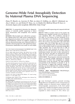
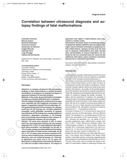
Trisomy 13 detection in the first trimester of pregnancy using a chromosome-selective
cell-free DNA analysis method
Ghalia ASHOOR1, Argyro SYNGELAKI1, Eric WANG2, Craig STRUBLE2, Arnold OLIPHANT2,
Ken SONG2, Kypros H. NICOLAIDES1,3
London, England
1. Harris Birthright Research Centre for Fetal Medicine, King’s College Hospital, London, UK.
2. Ariosa Diagnostics, Inc., San Jose, CA
3. Department of Fetal Medicine, University College London Hospital, London, UK.
Conflict of interest statement:
[Ariosa] are paid employees of Ariosa Diagnostics
Sources of Funding: The study was supported by a grant from The Fetal Medicine Foundation
(UK Charity No: 1037116). Analysis of samples was performed by Ariosa Diagnostics at 5945
Optical Court San Jose, CA 95138, USA.
Correspondence
Professor KH Nicolaides
Harris Birthright Research Centre for Fetal Medicine
King's College Hospital
Denmark Hill, London SE5 9RS
Telephone: (0044) 2032998256
Fax: (0044) 2077339534
E-mail: [email protected]
This article has been accepted for publication and undergone full peer review but has not been through the
copyediting, typesetting, pagination and proofreading process, which may lead to differences between this
version and the Version of Record. Please cite this article as doi: 10.1002/uog.12299.
Copyright © 2012 ISUOG. Published by John Wiley & Sons, Ltd.
Abstract
Objective: To assess the performance of chromosome-selective sequencing of maternal plasma
cell-free DNA (cfDNA) in non-invasive prenatal testing (NIPT) for trisomy 13.
Study design: Two phase case-control study on a single plasma sample per case. The first
phase was used to optimize the trisomy 13 algorithm which was then applied to a second
dataset to determine risk score for trisomy 13 by laboratory personnel who were blinded to fetal
karyotype
Results: In the first phase, trisomy 13 risk scores were given for 11 cases of trisomy 13 and 145
euploid cases at 11-13 weeks’ gestation. The test identified 7 (63.6%) cases of trisomy 13 with
no false positives. The trisomy 13 algorithm was subsequently modified and the trisomy 13 risk
score was >99% in all 11 cases of trisomy 13 and <0.01% in all 145 euploid cases. In the
second phase, the new algorithm was used to generate trisomy 13 risk scores for 10 cases of
trisomy 13 and 1,939 euploid cases. The trisomy 13 risk scores were >99% in 8 (80%, 95% CI
49.0%-94.3%) cases of trisomy 13. In the 1,939 euploid cases the risk score for trisomy 13 was
<0.01% in 1,937 (99.9%), 0.79% in 1, and >99% in 1. Therefore, at the predefined risk cut-off of
1% for classifying a sample as high versus low-risk, the false positive rate (FPR) was 0.05%
(95% CI 0.0%-0.3%).
Conclusions: Chromosome-selective sequencing of cfDNA can detect the majority of cases of
trisomy 13 at FPR of less than 0.1%.
Key words: Non-invasive prenatal testing; Trisomy 13; First trimester.
Copyright © 2012 ISUOG. Published by John Wiley & Sons, Ltd.
Introduction
Chromosome-selective sequencing of loci from chromosomes 21 and 18 in maternal plasma
cell-free DNA (cfDNA) has been successfully applied in non-invasive prenatal testing (NIPT) for
fetal trisomies 21 and 18 [1-5]. In pregnancies with a trisomic fetus, the incremental cfDNA
molecules derived from the extra fetal chromosome can be detected given the higher proportion
of molecules relative to a reference disomic chromosome.
Sparks et al., used a training set of euploid and trisomic pregnancies to perform selective
sequencing of cfDNA and develop a novel algorithm for estimation of individualized trisomy risk
[2]. They subsequently applied this approach for assessment of a blinded validation set and
correctly discriminated the 36 cases of trisomy 21 and 8 cases of trisomy 18 from the 123
euploid cases. Ashoor et al., performed a nested case-control study of cfDNA in plasma
obtained at 11-13 weeks before chorionic villous sampling (CVS) from 300 euploid pregnancies,
50 pregnancies with trisomy 21 and 50 pregnancies with trisomy 18 [3]. Chromosome-selective
sequencing correctly detected all cases of trisomy 21 and 49 (98%) of the cases of trisomy 18
with false positive rate (FPR) of 0% [3]. Norton et al., performed chromosome selective
sequencing on chromosomes 21 and 18 in a multicenter cohort of high-risk pregnancies at 1039 (mean 17) weeks’ gestation [4]. They correctly detected all 81 cases of trisomy 21 with FPR
of 0.03% (1/2888 normal cases) and detected 37 (97.4%) of the 38 cases of trisomy 18 with
FPR of 0.07% (2/2888). More recently, the chromosome-selective sequencing approach was
applied for NIPT in pregnant women undergoing routine screening for aneuploidies at 11-13
weeks’ gestation [5]. All 8 cases of trisomy 21 and 2 cases of trisomy 18 had a trisomy risk
score of >99%, whereas the risk score for trisomy 21 was <1% in all 1,939 euploid pregnancies
(FPR 0%) and the risk score for trisomy 18 was <1% in 1,937 (FPR 0.1%).
The objective of this study is to assess the performance of selective sequencing of cfDNA in
maternal plasma for the prenatal detection of fetal trisomy 13.
Copyright © 2012 ISUOG. Published by John Wiley & Sons, Ltd.
Materials and Methods
Study population
Two groups of women with singleton pregnancies were examined. In Group 1, all women had
fetal karyotyping carried out by CVS because screening by maternal age, serum free ß-hCG
and PAPP-A and ultrasound examination at 11-13 weeks’ gestation demonstrated that there was
a high risk for aneuploidies [6,7]. Maternal blood samples for research were obtained before
CVS and the plasma stored at -80oC. This group included 150 euploid and 15 trisomy 13 cases
and they were all recruited at King’s College Hospital, London, UK. Essentially, we searched our
database to identify cases of trisomy 13 with a minimum of 2 mL available stored plasma and
each trisomy 13 case was matched with 10 euploid controls for length of storage of their plasma
samples. Maternal blood was collected between April 2006 and February 2011.
Group 2 consisted of 1,992 euploid pregnancies and 10 confirmed trisomy 13 pregnancies. The
euploid pregnancies underwent routine first trimester combined screening (between October
2010 and January 2011) and subsequently delivered phenotypically normal neonates at King’s
College Hospital, London, UK. Maternal blood samples for research were collected at the time
of screening and the plasma stored at -80oC. The findings of NIPT for trisomies 21 and 18 in
these patients were previously reported [5]. The confirmed trisomy 13 cases were recruited from
US centers. Maternal blood samples for research were collected at 13-26 weeks’ gestation post
confirmation of trisomy as determined by CVS or amniocentesis and the plasma stored at 80oC.
All women who participated in the study provided written informed consent under approved
protocols by local ethics committees.
Sample collection and processing
Maternal venous blood was collected in ethylene diamine tetraacetic acid (EDTA) BD
vacutainer™ tubes (Becton Dickinson UK limited, Oxfordshire, UK) or Cell-free™ BCT tubes
(Streck, Omaha, NE). Plasma was isolated from each sample via a double centrifugation
protocol of 2,000 xg for 10 minutes, followed by 16,000 xg for 10 minutes, after a tube transfer
following the first spin. cfDNA was isolated from plasma using the Dynabeads® Viral NA DNA
Copyright © 2012 ISUOG. Published by John Wiley & Sons, Ltd.
purification kit (Dynal) protocol, with minor modifications as previously described [1,2].
Trisomy analysis
A single aliquot of plasma for each case that met minimum fetal fraction concentration of 4%
was analyzed by laboratory personnel at Ariosa Diagnostics who were blinded to trisomy status.
Fetal fraction was determined as previously described [2]. Samples were analyzed with the
DANSR assay and FORTE algorithm as previously described with the addition of 576 nonpolymorphic loci on chromosome 13 to the existing non-polymorphic loci on chromosomes 18
and 21 as part of the DANSR assay [1,2]. Briefly, this method uses ligation of locus-specific
oligonucleotides to produce a sequencing template only from selected genomic loci, thus
reducing the amount of DNA sequencing needed. The FORTE algorithm was used to estimate
the risk of aneuploidy for chromosome 13 in each sample. Proportion metrics for chromosomes
13, 18, and 21 are determined by computing the mean cfDNA counts of the loci for each
chromosome and then dividing the mean count of each chromosome by the sum of all three
means.The FORTE risk score is determined by calculating the odds ratio for trisomy based on
chromosomal cfDNA counts (proportion metrics), and fraction of fetal cfDNA in the sample, then
applying this as a likelihood ratio to the a priori trisomy risk based on the maternal age and
gestational age. A predefined cut-off value of 1 in 100 (1%) was designated as the threshold for
classifying a sample as High-risk versus Low-risk. Results were provided on the risk of trisomy
13
Group 1 samples were used in the first phase of the study. 384 loci on chromosome 13 were
selected based primarily on previously studied euploid pregnancies. Of the 165 samples that
were analyzed, 9 failed amplification and sequencing and no risk score could be provided. The
cases where a risk score was provided were then unblinded and these samples were used to
train and improve the trisomy 13 algorithm by selection of a new set of 384 loci from
chromosome 13, which were able to best discriminate trisomy 13 from euploid samples using
the FORTE algorithm.
In the second phase of the study, the new set of 384 loci from chromosome 13 was used to
analyze the samples of Group 2. Of the 2,002 samples analyzed, 53 of the euploid samples
failed amplification and sequencing and no risk score could be provided.
Copyright © 2012 ISUOG. Published by John Wiley & Sons, Ltd.
Statistical analyses
Descriptive data were presented in median and standard deviation (SD) for continuous variables
and in numbers and percentages for categorical variables (Table 1). Comparison between the
outcome groups was by Fisher exact test for categorical variables and Mann-Whitney-U test for
continuous variables.
The statistical software R version 2.15.1 was used for data analyses.
Results
First phase of the study: Group 1
In the first phase of the study, trisomy 13 risk scores were given for 156 of the 165 samples in
Group 1, including 145 of the 150 euploid and 11 of the 15 trisomy 13 cases. The characteristics
of this population of 156 pregnancies are summarized in Table 1.
The risk score for trisomy 13 was >1% in 7 (63.6%, 95% CI 35.4%-84.8%) of the 11 cases of
trisomy 13 and the score was <0.01% in all 145 euploid cases (FPR of 0.0%, 95% CI 0%-2.6%)
(Figure 1). After un-blinding and modification of the trisomy 13 algorithm, the trisomy 13 risk
score was >99% in all 11 cases of trisomy 13 and <0.01% in all 145 euploid cases.
Second phase of the study: Group 2
In the second phase of the study, trisomy 13 risk scores were given for 1,949 of the 2,002
samples in Group 2, including 1,939 of the 1,992 euploid and all 10 trisomy 13 cases. The
characteristics of these cases are summarized in Table 1.
In the 10 cases of trisomy 13, the estimated trisomy 13 risk score was >99% in 8 (80%) and
<0.01 in 2 (Figure 2). In the 1,939 euploid cases, the estimated trisomy 13 risk score was
<0.01% in 1,937 (99.9%), 0.79% in 1 and >99% in 1. Therefore, at the predefined risk cut-off of
1% for classifying a sample as High-risk versus Low-risk, the detection rate of trisomy 13 was
80% (95% CI 49.0%-94.3%) with FPR of 0.05% (95% CI 0%-0.03%).
Copyright © 2012 ISUOG. Published by John Wiley & Sons, Ltd.
Discussion
Principal findings of this study
This two phase case-control study shows that chromosome-selective sequencing of cfDNA in
maternal plasma can detect the majority of trisomy 13 pregnancies with FPR of 0.05%. The first
phase of the study was used to optimize the trisomy 13 algorithm which was then applied via a
blinded analysis to a second set of cases. In 8 of the 10 cases of trisomy 13, the estimated risk
for this aneuploidy was >99%, whereas in 99.9% of the euploid cases the risk score for trisomy
13 was <0.01%.
Comparison of the findings with previous studies in the literature
The detection rate of trisomy 13 by chromosome-selective sequencing of maternal plasma
cfDNA is lower than that for detection of trisomies 21 and 18. Previous studies utilizing DANSR
and FORTE, both in high-risk and routinely screened populations reported that the detection
rates of trisomy 21 and 18 were 100% and 97-98%, respectively, with false positive rates of
0.1% or less [2-5].
Three previous studies, utilizing non-selective massively parallel shotgun sequencing (MPSS),
have examined NIPT for trisomy 13 from analysis of cfDNA in maternal plasma. Chen et al.,
examined 25 trisomy 13 pregnancies and reported that with the use of a previously
standardized z-score method developed for trisomy 21, they detected 9 (36%) of the cases of
trisomy 13 at FPR of 7.6% [8]. After adjustments, including removal of repeat-masking and
correction for the GC content bias in the sequencing data, the detection rate increased to 100%
and the FPR decreased to 1.1%. Palomaki et al., used a similar approach and detected 11 of 12
(91.7%) cases of trisomy 13 at FPR of 0.9% [9]. Bianchi et al., examined 16 cases of trisomy 13
and with a z-score cut-off of 2.5 the detection rate and FPR were 81.2% (13/16) and 0%,
respectively, whereas at a z-score cut-off of 4.0 the detection rate was 68.8% (11/16) at FPR of
0% [10,11].
One possible explanation for the observed lower detection rate of trisomy 13 with NIPT,
Copyright © 2012 ISUOG. Published by John Wiley & Sons, Ltd.
compared to trisomy 21, is that in pregnancies with trisomy 13 fetuses there is placenta
confined mosaicism with a high proportion of cells being disomic, whereas in trisomy 21 all
placental cells are invariably trisomic [12]. As the primary source of cfDNA in the maternal
circulation is thought to be the placenta [13], a mosaic placenta that is predominantly disomic
would lead to a false negative result when using NIPT for trisomy13 detection. Likewise, a
mosaic placenta that is predominantly trisomic in which the fetus is euploid could lead to a false
positive result.
Current approach to prenatal screening for trisomy 13
At 11-13 weeks’ gestation, the relative prevalence of trisomies 18 and 13 to trisomy 21 are 1:3
and 1:7, respectively [14-16]. However, in trisomies 18 and 13 the rate of spontaneous abortion
or fetal death between 12 weeks of gestation and 40 weeks is about 80% and therefore the
relative prevalence of these trisomies to trisomy 21 at birth are 1:12 and 1:28, respectively.
Effective first-trimester screening for trisomy 21 is provided by a combination of maternal age,
fetal NT thickness and maternal serum-free ß-hCG and PAPP-A with a detection rate of more
than 90% for a false positive rate (FPR) of 5% [6]. A beneficial consequence of screening for
trisomy 21 is the early diagnosis of about 75% of cases of trisomies 18 and 13, which are the
second and third most common chromosomal abnormalities. All three trisomies are associated
with increased maternal age, increased fetal NT and decreased maternal serum PAPP-A, but in
trisomy 21 serum free ß-hCG is increased whereas in trisomies 18 and 13 this is decreased [6,
7,17-19]. When specific algorithms for trisomies 18 and 13 in addition to the one for trisomy 21
are also used about 90% of fetuses with trisomy 21 and 95% of those with trisomies 13 and 18
can be detected for an overall increase in FPR of only 0.1% [7]. This incremental FPR has
generally been considered acceptable despite the low prevalence of trisomies 13 and 18.
Trisomy 13 is often associated with abnormalities that can be readily identified by ultrasound,
not only in the second but also in the first-trimester [20.21]. A study of 181 fetuses with trisomy
13 reported that at the 11-13 weeks scan 92 (50.2%) had holoprosencephaly, exomphalos,
and/or megacystis and in 129 (71.3%) the heart rate was above the 95th percentile [20]. Another
study of holoprosencephaly, exomphalos and megacystis detected at 11-13 weeks reported that
these defects are associated with a high incidence of chromosomal abnormalities, mainly
trisomies 18 and 13, found in about 65%, 55% and 30% of cases, respectively [21].
Copyright © 2012 ISUOG. Published by John Wiley & Sons, Ltd.
Implications for practice
The performance of NIPT in screening for both trisomy 21 and trisomy 18 is far superior to that
of currently available screening methods with a substantial increase in detection rate and
decrease in FPR [5,6]. A study of NIPT with a chromosome-selective sequencing approach in
pregnant women undergoing routine screening for aneuploidies at 11-13 weeks’ gestation
reported that the estimated trisomy risk score was >99% in all cases of trisomy 21 and trisomy
18 and <1% in 99.9% of the euploid cases [5].
This study has shown that NIPT is also useful in screening for trisomy 13. Although the total
number of cases of trisomy 13 examined is too small for accurate assessment of detection rate,
the FPR was only 0.05%. Consequently, if the NIPT result is positive for trisomy 13 there is a
1600-fold (80/0.05) increase in risk for this trisomy and therefore such patients should be offered
the option of invasive diagnostic testing. If the NIPT result is negative for trisomy 13 there is a 5fold (99.95/20) decrease in the a-priori risk. In NIPT there is a delay of 1-2 weeks between
sampling and obtaining results. The blood sample could be collected at 9-10 weeks so that the
results would be available by 12 weeks, which is the best time to carry out the first-trimester
ultrasound examination [6]. If the 12 weeks scan demonstrates holoprosencephaly, exomphalos
or megacystis, where the risk for aneuploidies is very high, the 5-fold reduction in risk following
a negative NIPT test is unlikely to reassure the parents and they should still be offered invasive
testing. Since the prevalence of these defects is less than 0.1% the effect on the overall
proportion of pregnancies requiring an invasive test would be minimal.
The sensitivity and specificity of NIPT for trisomies 21, 18 and 13 is not 100% and therefore
NIPT should not be considered a diagnostic test to replace invasive testing in high-risk
pregnancies. It is a new high performance screening test that identifies a high-risk group
requiring further investigation by invasive testing. Similarly, the introduction of NIPT in universal
screening for trisomies 21, 18 and 13 will complement rather than replace the 11-13 weeks
scan. The latter is not only useful in screening for aneuploidies but it is also a diagnostic test for
many major fetal defects, some of which, such as holoprosencephaly, require further
investigation by invasive testing, and additionally, in combination with biochemical and other
biophysical markers, the scan can provide effective early screening for pregnancy
complications, including preeclampsia and preterm birth [22-25].
Copyright © 2012 ISUOG. Published by John Wiley & Sons, Ltd.
Conclusions
Trisomy 13 is associated with a high rate of fetal death and the prevalence in live births is about
30 times lower than that of trisomy 21. Additionally, unlike individuals with trisomy 21 that
survive for more than 60 years, individuals with trisomy 13 rarely survive beyond the first few
months of life. Chromosome-selective sequencing of cfDNA can detect the majority of cases of
trisomy 13 with FPR of less than 0.1%, but the detection rate is lower than that reported for
trisomies 21 and 18.
Copyright © 2012 ISUOG. Published by John Wiley & Sons, Ltd.
Table 1. Characteristics of the cases and controls
Phase 1
Phase 2
Euploid
Trisomy 13
Euploid
Trisomy 13
n = 145
n = 11
n=1,939
n=10
Maternal age in yrs, median* (SD)
37.2 (6.0)
32.8 (4.2)
31.8 (5.6)
37.5* (5.3)
Gestational age in weeks* (SD)
13.3 (0.8)
12.0 (0.6)
12.6 (0.56)
20.9* (3.8)
128 (88.3)
9 (81.8)
1370 (70.7)
8 (80.0)
African, n (%)
7 (4.8)
2 (18.2)
387 (20.0)
2 (20.0)
Asian, n (%)
6 (5.6)
-
131 (6.8)
-
Mixed, n (%)
2 (1.4)
-
51 (2.6)
-
11.5
7.6
10.0
14.0
(4.6-25.5)
(6-11.8)
(4.1-31)
(6.1-24)
Racial origin
Caucasian, n (%)
Fetal fraction in %, median* (range)
Comparisons between trisomy 13 and euploid pregnancies are by Mann Whitney-U test for
continuous variables and by Fisher exact test for categorical variables
* p-value <0.05 between trisomy 13 and euploid cases in each of the phases
Copyright © 2012 ISUOG. Published by John Wiley & Sons, Ltd.
Figure 1. Phase 1- Estimated risk score for trisomy 13 in trisomy 13 (red circles, n=11) and
euploid (black circles, n=145) pregnancies plotted in relation to the fetal fraction.
Figure 2. Estimated risk score for trisomy 13 in trisomy 13 (red circles, n=10) and euploid (black
circles, n=1,939) pregnancies plotted in relation to the fetal fraction.
Copyright © 2012 ISUOG. Published by John Wiley & Sons, Ltd.
References
1. Sparks AB, Wang ET, Struble CA, Barrett W, Stokowski R, McBride C, Zahn J, Lee K, Shen
N, Doshi J, Sun M, Garrison J, Sandler J, Hollemon D, Pattee P,Tomita-Mitchell A, Mitchell
M, Stuelpnagel J, Song K, Oliphant A: Selective analysis of cell-free DNA in maternal blood
for evaluation of fetal trisomy. Prenat Diagn 2012;6:1-7.
2. Sparks AB, Struble CA, Wang ET, Song K, Oliphant A: Noninvasive prenatal detection and
selective analysis of cell-free DNA obtained from maternal blood: evaluation for trisomy 21
and trisomy 18. Am J Obstet Gynecol 2012;206:319.e1-9.
3. Ashoor G, Syngelaki A, Wagner M, Birdir C, Nicolaides KH: Chromosome-selective
sequencing of maternal plasma cell-free DNA for first-trimester detection of trisomy 21 and
trisomy 18. Am J Obstet Gynecol 2012;206:322.e1-5.
4. Norton ME, Brar H, Weiss J, et al. Non-invasive chromosomal evaluation (NICE) study:
results of a multicenter prospective cohort study for detection of fetal trisomy 21 and trisomy
18. Am J Obstet Gynecol. 2012 Jun 1. [Epub ahead of print]
5. Nicolaides KH, Syngelaki A, Birdir C, Touzet G, Ashoor G: Non-invasive prenatal testing for
fetal trisomies in a routinely screened first-trimester population. Am J Obstet Gynecol 2012;
[in press]
6. Nicolaides KH: Screening for fetal aneuploidies at 11 to 13 weeks. Prenat Diagn 2011;31:715.
7. Kagan KO, Wright D, Valencia C, Maiz N, Nicolaides KH: Screening for trisomies 21, 18 and
13 by maternal age, fetal nuchal translucency, fetal heart rate, free beta-hCG and
pregnancy-associated plasma protein-A. Hum Reprod 2008;23:1968-75.
8. Chen EZ, Chiu RW, Sun H, Akolekar R, Chan KC, Leung TY, Jiang P, Zheng YW, Lun FM,
Chan LY, Jin Y, Go AT, Lau ET, To WW, Leung WC, Tang RY, Au-Yeung SK, Lam H, Kung
YY, Zhang X, van Vugt JM, Minekawa R, Tang MH, Wang J, Oudejans CB, Lau TK,
Copyright © 2012 ISUOG. Published by John Wiley & Sons, Ltd.
Nicolaides KH, Lo YM: Noninvasive prenatal diagnosis of fetal trisomy 18 and trisomy 13 by
maternal plasma DNA sequencing. PLoS One 2011;6:e21791.
9. Palomaki GE, Deciu C, Kloza EM, Lambert-Messerlian GM, Haddow JE, Neveux LM, Ehrich
M, van den Boom D, Bombard AT, Grody WW, Nelson SF, Canick JA: DNA sequencing of
maternal plasma reliably identifies trisomy 18 and trisomy 13 as well as Down syndrome: an
international collaborative study. Genet Med 2012;14:296-305.
10. Bianchi DW, Platt LD, Goldberg JD, Abuhamad AZ, Sehnert AJ, Rava RP: Genome-wide
fetal aneuploidy detection by maternal plasma DNA sequencing. Obstet Gynecol
2012;119:890-901.
11. Benn P, Cuckle H, Pergament E: Genome-wide fetal aneuploidy detection by maternal
plasma DNA sequencing. Obstet Gynecol 2012;119:1270.
12. Kalousek DK, Barrett IJ, McGillivray BC: Placental mosaicism and intrauterine survival of
trisomies 13 and 18. Am J Hum Genet 1989;44:338-43.
13. Alberry M, Maddocks D, Jones M, Abdel Hadi M, Abdel-Fattah S, Avent N, Soothill PW. Free
fetal DNA in maternal plasma in anembryonic pregnancies: confirmation that the origin is the
trophoblast. Prenat Diagn 2007;27:415-8.
14. Snijders RJM, Holzgreve W, Cuckle H, Nicolaides KH: Maternal age-specific risks for
trisomies at 9–14 weeks’ gestation. Prenat Diagn 1994;14:543-552.
15. Snijders RJM, Sebire NJ, Cuckle H, Nicolaides KH: Maternal age and gestational agespecific risks for chromosomal defects. Fetal Diagn Ther 1995;10:356-367.
16. Snijders RJM, Sundberg K, Holzgreve W, Henry G, Nicolaides KH: Maternal age and
gestation-specific risk for trisomy 21. Ultrasound Obstet Gynecol 1999;13:167-170.
17. Wright D, Kagan KO, Molina FS, Gazzoni A, Nicolaides KH: A mixture model of nuchal
translucency thickness in screening for chromosomal defects. Ultrasound Obstet Gynecol
2008;31:376-383.
Copyright © 2012 ISUOG. Published by John Wiley & Sons, Ltd.
18. Snijders RJ, Noble P, Sebire N, Souka A, Nicolaides KH: UK multicentre project on
assessment of risk of trisomy 21 by maternal age and fetal nuchal-translucency thickness at
10–14 weeks of gestation. Lancet 1998;352:343-346.
19. Spencer K, Ong C, Skentou H, Liao AW, Nicolaides KH: Screening for trisomy 13 by fetal
nuchal translucency and maternal serum free beta hCG and PAPP-A at 10–14 weeks of
gestation. Prenat Diagn 2000;20:411-416.
20. Papageorghiou AT, Avgidou K, Spencer K, Nix B, Nicolaides KH: Sonographic screening for
trisomy 13 at 11 to 13(+6) weeks of gestation. Am J Obstet Gynecol 2006;194:397-401.
21. Kagan KO, Staboulidou I, Syngelaki A, Cruz J, Nicolaides KH: The 11-13-week scan:
diagnosis and outcome of holoprosencephaly, exomphalos and megacystis. Ultrasound
Obstet Gynecol 2010;36:10-4.
22. Nicolaides KH: Turning the pyramid of prenatal care. Fetal Diagn Ther 2011;29:183-96.
23. Syngelaki A, Chelemen T, Dagklis T, Allan L, Nicolaides KH: Challenges in the diagnosis of
fetal non-chromosomal abnormalities at 11-13 weeks. Prenat Diagn 2011;31:90-102.
24. Greco E, Gupta R, Syngelaki A, Poon LC, Nicolaides KH: First-trimester screening for
spontaneous preterm delivery with maternal characteristics and cervical length. Fetal Diagn
Ther 2012;31:154-61.
25. Akolekar R, Syngelaki A, Poon L. Wright D, Nicolaides KH: Competing risks model in early
screening for preeclampsia by biophysical and biochemical markers. Fetal Diagn Ther 2012;
DOI: 10.1159/000341264.
Copyright © 2012 ISUOG. Published by John Wiley & Sons, Ltd.
100
Trisomy 13 risk (%)
10
1.0
0.1
0.01
4
9
14
19
Fetal fraction (%)
Figure 1
24
100
Trisomy 13 risk (%)
10
1.0
0.1
0.01
4
9
14
19
24
Fetal fraction (%)
Figure 2
29
34
Scaricare