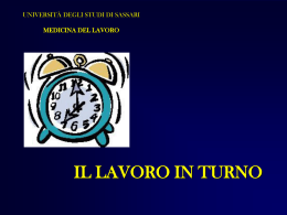
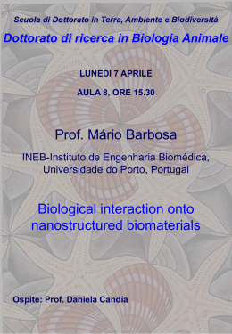
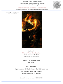
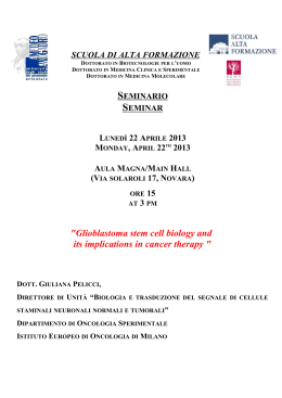
Università degli Studi di Sassari Scuola di Dottorato in Storia, Letterature e Culture del Mediterraneo XXVI ciclo Utilization of Epidemiological, Archaeological, and Genomic Data for Assessing the Health of Historic Communities Direttore Tutor Prof. M. Milanese Candidata Nikki Ann Kelvin A.A. 2014/2015 i Table of Contents Table of Contents .......................................................................................................................................... i Acknowledgements ..................................................................................................................................... iv Chapter One Introduction ............................................................................................................................ 1 History of Castelsardo ............................................................................................................................... 1 History of the Cattedrale Sant’Antonio Abate .......................................................................................... 4 Bioarchaeology and ancient DNA in a crypt of Cattedrale Sant’Antonio Abate ..................................... 13 Thesis hypothesis and objectives ............................................................................................................ 18 Chapter Two Infectious Disease, Epidemics and Analysis of Death Records in Northern Sardinia from the 16th to the 19th Centuries .................................................................................................................. 20 A brief history of infectious disease in Europe from the 14th to the 19th century ................................. 20 Analysis of population, baptisms, and death records in Casterllaragonese/Castelsardo, Sassari, and Nulvi (later 16th and 19th century) .......................................................................................................... 23 High mortality rates for children 1750-1836 ......................................................................................... 55 Chapter Three Methodology I Bioarchaeological Removal of Specimens for an Excavation (Safety and Control of Contaminants) .......................................................................................................................... 62 Introduction ........................................................................................................................................... 62 Active Research ...................................................................................................................................... 64 Personal protective equipment and sample acquisition ....................................................................... 71 Personal protective equipment ............................................................................................................... 71 Site assessment to identify required protective equipment ................................................................. 74 Personal protective equipment used for the excavation of the crypt of Sant’Isidoro .......................... 75 Tyvek protective equipment (coveralls) ................................................................................................ 78 Respirators and face masks .................................................................................................................... 83 Sterile disposable gloves ........................................................................................................................ 90 Protective eyewear ................................................................................................................................ 96 Hair covers .............................................................................................................................................. 97 Footwear covers ................................................................................................................................... 102 Precautions and personal safety in different types of environments ................................................. 103 Sample acquisition, storage, cataloguing, and processing .................................................................. 106 Sample acquisition ............................................................................................................................... 106 Good practices for sterile sample acquisition ...................................................................................... 109 Sample collection protocols ................................................................................................................. 110 Sample storage and preservation ........................................................................................................ 113 Sample cataloguing .............................................................................................................................. 114 Chapter Four Laboratory Methods DNA Extraction from Bones ........................................................... 117 Decontamination of Workspace .......................................................................................................... 117 Materials .............................................................................................................................................. 117 Reagent set up ...................................................................................................................................... 118 Extraction of aDNA from bone samples................................................................................................ 119 Nikki Ann Kelvin, Utilization of Epidemiological, Archaeological, and Genomic Methods for Assessing the Health of Historic Communities. Tesi di Dottorato in Scuola di Dottorato in Storia, Letterature e Culture del Mediterraneo, Università degli Studi di Sassari ii DNA release........................................................................................................................................... 120 DNA purification.................................................................................................................................... 121 DNA library construction....................................................................................................................... 123 Polymerase chain reaction .................................................................................................................... 123 Histology ............................................................................................................................................... 124 Chapter Five Bioarchaeological material recovery and analysis; A case study from the Crypt of San Isadoro, San Antionio Abate, Castelsardo .............................................................................................. 126 Summary of archaeological findings ..................................................................................................... 126 The mummified individuals................................................................................................................... 144 Mother-child family burials................................................................................................................... 155 Individuals 32 and 32A ................................................................................................................. 155 Possible Family Burial .................................................................................................................. 160 Possible mother and child ............................................................................................................ 165 Basket burial ................................................................................................................................ 168 Genetic analysis of human remains for mitochondrial haplotype (genotype) and possible pathogens .............................................................................................................................................................. 168 What is mitochondrial DNA?........................................................................................................ 171 DNA purification........................................................................................................................... 171 Library construction, sequencing and analysis of mitochondrial DNA......................................... 174 Analysis of aDNA sequences ........................................................................................................ 174 Chapter Six Discussion The beginning and end of burials in the crypt of Saint Isodoro........................ 189 Mother-infant-child burials ................................................................................................................... 192 The making of mummies, the process of mummification in the crypt of Sant’Isodoro ....................... 193 Conclusion ............................................................................................................................................. 197 References ................................................................................................................................................ 199 Nikki Ann Kelvin, Utilization of Epidemiological, Archaeological, and Genomic Methods for Assessing the Health of Historic Communities. Tesi di Dottorato in Scuola di Dottorato in Storia, Letterature e Culture del Mediterraneo, Università degli Studi di Sassari iii Dedicated to my Mother Nikki Ann Kelvin, Utilization of Epidemiological, Archaeological, and Genomic Methods for Assessing the Health of Historic Communities. Tesi di Dottorato in Scuola di Dottorato in Storia, Letterature e Culture del Mediterraneo, Università degli Studi di Sassari iv Acknowledgements Words seem so inadequate to express the gratitude I feel for all the people who helped me complete a journey that began over 30 years ago, but they are all I have. I am deeply grateful to my supervisor, Professor Marco Milanese, for agreeing to take me on as a student. His guidance and support are much appreciated. There are no words to express my gratitude to archaeologists Franco G. R. Campus, Antonietta Maria Demurtas, and Luca Sanna. I am quite confident that they didn’t know what they were getting into when they agreed to let me participate in what they thought was going to be a quick, routine excavation – until I brought in all my Tyvek suits, masks, gloves, and rules about how to retrieve and store samples without contaminating them with modern DNA. Their cooperation was phenomenal, and I am extremely grateful for their patience and teaching. I am very grateful to Professor Salvatore Rubino for suggesting that I should finish my degree and encouraging me to do it at the University of Sassari. This journey would not have begun if he had not lit the spark under the fire. I am also indebted to his colleagues, the members of the Department of Biomedical Science and Clinical Medicine, especially Professor Vittorio Mazzarello, Daniella Chessa, Patrizia Marongiu, and Manuela Murgia, as well as all the other microbiologists, radiologists, entomologists who helped investigate the samples from the crypt. The same indebtedness goes to all the laboratory members at the University Health Network in Toronto, Canada, and the team at the International Institute of Infection and Immunity at the University of Shantou, Shantou, China. Special thanks go to Jeff Coombs, Alyson Kelvin, David Banner, Stephen Wang, Alberto Leon, Luoling Xu, and Mavis Pan – thank you all so much. Nikki Ann Kelvin, Utilization of Epidemiological, Archaeological, and Genomic Methods for Assessing the Health of Historic Communities. Tesi di Dottorato in Scuola di Dottorato in Storia, Letterature e Culture del Mediterraneo, Università degli Studi di Sassari v I am very appreciative of the assistance I received from everyone at the Archivio Storico di Tempio Ampurias, particularly Don Francesco Tamponi and Fabio Ardau, for their help with translations and pointing out interesting sources. Thanks to Laura, Antonio, and Giuseppe for their moral support. I am also grateful to Don Usai for allowing me the privilege of researching the library inside the Cattedrale Sant’Antonio Abate. I am grateful to the Li Ka Shing Foundation, Shantou, China, for financial assistance. I would like to express my thanks to all the wonderful professors and students at the University of Sassari, who welcomed me into their midst and made this one of the most memorable experiences of my life. I am honoured to have been able to participate in your excavations. I am indebted to Giustina Casu and Michele Guirguis for helping me through the system. More than anything, I am grateful to my wonderful family for all their love, support, and encouragement, and for putting up with my absences and craziness. I love you all very much. Nikki Ann Kelvin, Utilization of Epidemiological, Archaeological, and Genomic Methods for Assessing the Health of Historic Communities. Tesi di Dottorato in Scuola di Dottorato in Storia, Letterature e Culture del Mediterraneo, Università degli Studi di Sassari CHAPTER ONE Introduction Where then? Spain or Sardinia. Spain or Sardinia. Sardinia, which is like nowhere. Sardinia, which has no history, no date, no race, no offering. Let it be Sardinia. They say neither Romans nor Phoenicians, Greeks nor Arabs ever subdued Sardinia. It lies outside; outside the circuit of civilisation. Like the Basque lands. Sure enough, it is Italian now, with its railways and its motor-omnibuses. But there is an uncaptured Sardinia still. - D. H. Lawrence, Sea and Sardinia (1921) History of Castelsardo D. H. Lawrence’s Sea and Sardinia is said to be one of his finest pieces of writing, as its descriptive language captures the beauty and essence of the island in spite of the writer’s negative attitude throughout the book toward his journey. This same irony is seen in Lawrence’s cheeky proclamation that Sardinia “has no history”, as the statement that follows gives his readers a glimpse into the island’s rich and culturally diverse past, which as Lawrence’s brief history lesson suggests, is distinctly tied to its physical location. Sardinia’s position bounded by the Mediterranean and Tyrrhenian Seas has inexorably contributed to its history (Figure 1.1). Lying about 8 kilometres due south of the island of Corsica, it is roughly 200 kilometres west of mainland Italy across the Tyrrhenian Sea and 200 kilometres north of Tunisia across the Mediterranean (Encyclopaedia Brittanica (Online) 2013; Dyson and Rowland 2007: 2). This location made it a natural stopping place for early settlers, traders, and conquerors coming from all regions of the Mediterranean. Nikki Ann Kelvin, Utilization of Epidemiological, Archaeological, and Genomic Methods for Assessing the Health of Historic Communities. Tesi di Dottorato in Scuola di Dottorato in Storia, Letterature e Culture del Mediterraneo, Università degli Studi di Sassari 2 Figure 1.1 Map showing location of Sardinia. Source: http://www.sardegnadigitallibrary.it/index.php?xsl=615 &s=17&v=9&c=4461&id=13471 Nikki Ann Kelvin, Utilization of Epidemiological, Archaeological, and Genomic Methods for Assessing the Health of Historic Communities. Tesi di Dottorato in Scuola di Dottorato in Storia, Letterature e Culture del Mediterraneo, Università degli Studi di Sassari 3 According to Stephen L. Dyson and Robert J. Rowland, Jr. (2007: 17-20), Sardinia’s earliest inhabitants likely arrived via land bridges or short sea journeys in the middle Palaeolithic during glaciation periods when low sea levels and temporary land bridges made the island more easily accessible. The oldest artifacts from this time, made from flint and quartz typical of Clactonian culture, were found in the Anglona region in the north part of the island and date to about 150,000 years ago (Vona 1997: 71; Dyson and Rowland 2007: 17-20). That these early settlers and their descendants flourished on the island over the millennia is seen in the evidence they left behind, from the Mesolithic tools and fossils found at Grotta su Coloru and Corbeddu, to the ceramics, religious icons and Domus de Janas of the Ozieri in the pre-nuraghic (4000-3200 BCE), and later to the thousands of nuraghi that were constructed about four thousand years ago (Dyson and Rowland 2007: 19-54; Melis 2002: 1331-1332; Spoor 1999: 297-302; Vona 1997: 72-73). As an island, Sardinia had port cities that were active in commerce with major trading partners of the day, even in ancient times (Dyson and Rowland 2007: 2, 24-25; Melis: 13311332; Vona 1997: 72), but this accessibility for trade also left the inhabitants open to attack from other seafaring civilizations, and consequently, Sardinia was repeatedly occupied by various invaders for more than two thousand years. Francesco Cesare Casùla (2000; 2012b) states that from the nuragic period up to the Middle Ages, the most notable conquerors of Sardinia included the Phoenicians (1000 - 509 BCE), the Carthaginians (509 - 238 BCE), the Romans (238 - 456 CE), the Vandals (456 - 534 CE), and the Byzantines (534 - ca. 900 CE). In about 900, the four indigenous kingdoms of Sardinia – Gallura, Torres, Arborea, and Calari – were created. Although they each had their own governments with kings, coats of arms, parliaments, and laws, they also had their own alliances with outside influences, particularly Calari with Genoa, and Nikki Ann Kelvin, Utilization of Epidemiological, Archaeological, and Genomic Methods for Assessing the Health of Historic Communities. Tesi di Dottorato in Scuola di Dottorato in Storia, Letterature e Culture del Mediterraneo, Università degli Studi di Sassari 4 Torres and Arborea with Pisa (Casùla 2000; 20b). All these periods in Sardinian history were marked by urban development, increased trade, and the cultural influences of each of the occupying peoples; however, the early history of Sardinia is outside the scope of this thesis. The basis for my thesis project has its roots in the time that the city of Castelsardo (Figure 1.2) began to take shape in the Middle Ages. Although there is evidence that early Roman settlements existed in the area around Castelsardo (Brigaglia et al. 2014: 56-59; Pittau 2007; Zucca 2007), there is little doubt that the origins of the city proper are tied to the building of the castle during the Doria period in the Middle Ages when the village was called Castelgenovese; however, the exact date is disputed, ranging from 1102 (Cano 2009: 360-365) to about 1260-1275, with scholarly consensus today leaning heavily toward the latter period (Castellaccio 2007: 285-286; Soddu 2007: 237-238; Milanese 2010a: 17). While there are no written records to verify when the building of the castle was initiated (Milanese 2010a: 17), the earliest recorded documents referring to Castelgenovese are dated between 1272 and 1274 (Castellaccio 2007: 286; Soddu 2007: 239-240; Milanese 2010a: 17). The city was acquired by the Kingdom of Aragon in 1448, and the name was changed to Castellaragonese about 1520 (Porcu Gaias 2007: 684; Milanese 2010a: 17). History of the Cattedrale Sant’Antonio Abate During this period, a Romanesque church, Sant’Antonio Abate (Figure 1.3), was built close to the sea when the city became the seat of the diocese by the order of Pope Julius II Porcu Gaias: 686). A historical plaque outside today’s cathedral (Figure 1.4) proclaims that the old Nikki Ann Kelvin, Utilization of Epidemiological, Archaeological, and Genomic Methods for Assessing the Health of Historic Communities. Tesi di Dottorato in Scuola di Dottorato in Storia, Letterature e Culture del Mediterraneo, Università degli Studi di Sassari 5 Figure 1.2 Castelsardo. The star on the map showsCastelsardo the location of the city on the northern coast of Sardinia. Map: http://i.infoplease.com/images/mitaly.gif Nikki Ann Kelvin, Utilization of Epidemiological, Archaeological, and Genomic Methods for Assessing the Health of Historic Communities. Tesi di Dottorato in Scuola di Dottorato in Storia, Letterature e Culture del Mediterraneo, Università degli Studi di Sassari 6 Figure 1.3 Cattedrale Sant’Antonio Abate. Figure 1.3. Cattedrale Sant’Antonio Abate. Nikki Ann Kelvin, Utilization of Epidemiological, Archaeological, and Genomic Methods for Assessing the Health of Historic Communities. Tesi di Dottorato in Scuola di Dottorato in Storia, Letterature e Culture del Mediterraneo, Università degli Studi di Sassari 7 Figure commemorating the consecration of theof church in 1502.in 1502. Figure1.4. 1.4 Historical Historicalplaque plaque commemorating the consecration the church The arrow highlights the date of the consecration. Nikki Ann Kelvin, Utilization of Epidemiological, Archaeological, and Genomic Methods for Assessing the Health of Historic Communities. Tesi di Dottorato in Scuola di Dottorato in Storia, Letterature e Culture del Mediterraneo, Università degli Studi di Sassari 8 church “[. . .] was consecrated on 26th [sic] November 1503.” The church was granted the status of cathedral when it was expanded during a restoration ordered by Bishop Giovanni Sanna that lasted from 1597 to 1606 (Porcu Gaias 2007: 686). The cathedral has undergone several more restorations since then, most notably in the 18th century when the location of the entrance was moved from the west side of the church to the south and a 700-pipe organ (Figure 1.5) made by Leonardo Spensatello (Moretti 1997: 98; Ardau 2013: 35) was installed above the original door. A plaque in the cathedral commemorates the completion of the expansion in 1727. Architecturally, the Cattedrale Sant'Antonio Abate is a blend of Catalan Gothic and Renaissance elements. The church follows the Latin cross design consisting of a single nave with a transept, barrel vaults, and side chapels (Figure 1.6). Interestingly, the bell tower predates the church and is not attached to the cathedral, but is part of the city wall, located about five metres from the cathedral door. According to a notation in the Delibere Capitolari del Capitolo della Cattedrale di Ampurias 1606-1630 (dsc_0002/0003/0004.jpg), Bishop Juan Sanna personally funded the building of the tower at the end of 1500 (Ardau 2013: 34). The bell tower has a small majolica dome and, in addition to the bell, it also has a lantern which was used as a signal for ships at sea (Porcu Gaias 2007: 686-687). A museum for the Diocese of Ampurias, which was constructed out of an old cistern, contains relics of saints, well-preserved music books, and paintings by the famous but anonymous “Master of Castelsardo”, including Saint Michael the Archangel and Enthroned Madonna and Child with Angel Musicians (Figure 1.7). The hypogea on the north and the east sides of the church were used as tombs. Various hypotheses have been postulated to explain the origins of the use of crypts as places of burial. One suggests that crypt burials possibly began in the early Christian period, when churches were built over pagan places of worship, such as Roman temples and mithrea. Nikki Ann Kelvin, Utilization of Epidemiological, Archaeological, and Genomic Methods for Assessing the Health of Historic Communities. Tesi di Dottorato in Scuola di Dottorato in Storia, Letterature e Culture del Mediterraneo, Università degli Studi di Sassari 9 Figure 1.5 organ Figure 1.5 The The700-pipe 700-pipeorgan organbuilt builtby700-pipe by700-pipe th organ built by Leonardo Spensatello the 18th built by Leonardo Spensatello in the 18in century. century. Figure 1.6. Interior Interiorview viewofofCattedrale Cattedrale Sant’Antonio Figure 1.6. Sant’Antonio Abate. Nikki Ann Kelvin, Utilization of Epidemiological, Archaeological, and Genomic Methods for Assessing the Health of Historic Communities. Tesi di Dottorato in Scuola di Dottorato in Storia, Letterature e Culture del Mediterraneo, Università degli Studi di Sassari 10 Nikki Ann Kelvin, Utilization of Epidemiological, Archaeological, and Genomic Methods for Assessing the Health of Historic Communities. Tesi di Dottorato in Scuola di Dottorato in Storia, Letterature e Culture del Mediterraneo, Università degli Studi di Sassari 11 The secret cult of Mithras, which was based on the Persian god, emerged in Rome in the latter part of the first century CE (Volken 2004; Griffith 2000); its followers built underground sanctuaries, or mithraea (Griffith 2000), in which to hold their ceremonies. These chambers provided ready-made crypts for Christian basilicas when the spaces were repurposed. Archaeological evidence of mithrea under churches and basilicas is found throughout Europe, with as many as 17 known mithraea beneath piazzas, ancient senators’ houses, and Christian churches in Rome, including St. Peter’s Basilica and the Basilica of San Clemente (Griffith 2000), and under several churches in Ostia (Torres 2008: 72-74). Another hypothesis contends that followers of the new Christian faith, who had converted from either Judaism or the pagan Roman religion and were therefore already familiar with the Jewish and Roman customs of underground burials, simply carried on with the same traditional practices, using their family tombs, and inviting other members of the faith to use them as well, to keep their dead safe and to bring members of the Christian community together in one place. Some sources, including the Institute of Salesiano San Callisto, which manages the Christian catacombs in Rome, suggests that this is how the catacombs began (Istituto Salesiano San Callisto; Torres; 146; Waal). When the catacombs fell into disuse, the bones of martyrs and saints, and other relics were moved into hypogea in churches for safe-keeping; these chambers were usually under the altar or the apse of the church. Gregory T. Armstrong (1974; 9-16) and Milton L. Torres (2008: 1; 235-239) note that in Pope Constantine’s time, it became custom to construct basilica ad corpus, that is, basilicas with mausoleums built under the apse, to contain bodies of the dead and religious relics, again with the purpose of bringing the Christian community together, and also to spiritually protect the dead. Nikki Ann Kelvin, Utilization of Epidemiological, Archaeological, and Genomic Methods for Assessing the Health of Historic Communities. Tesi di Dottorato in Scuola di Dottorato in Storia, Letterature e Culture del Mediterraneo, Università degli Studi di Sassari 12 Regardless of the origins of crypts, their significance lies in their location inside the church, under the consecrated altars, and therefore under the protection of the Church and God. The consecration of religious places is an ancient rite, also observed by pagan religions predating Christianity, so it follows that Christians would adopt the familiar custom to consecrate their grounds, especially to purify those spaces that had been previously used by pagan religious groups. Noting Christian historian Eusebius’s account of public consecrations of churches after Constantine granted freedom to Christians, Augustin Joseph Schulte states in the Catholic Encyclopedia that “The consecration of churches dates probably from Apostolic times and is, in a sense, a continuation of the Jewish rite instituted by Solomon” (http://www.newadvent.org/cathen/04276a.htm). In Book X, Chapter III of his treatise, Ecclesiastical History, Eusebius, Bishop of Cæsarea, wrote: After this was seen the sight which had been desired and prayed for by us all; feasts of dedication in the cities and consecrations of the newly built houses of prayer took place, bishops assembled, foreigners came together from abroad, mutual love was exhibited between people and people, the members of Christ's body were united in complete harmony. […] And there was one energy of the Divine Spirit pervading all the members, and one soul in all, and the same eagerness of faith, and one hymn from all in praise of the Deity. Yea, and perfect services were conducted by the prelates, the sacred rites being solemnized, and the majestic institutions of the Church observed, here with the singing of psalms and with the reading of the words committed to us by God, and there with the performance of divine and mystic services; and the mysterious symbols of the Saviour's passion were dispensed. At the same time people of every age, both male and female, with all the power of the mind gave honor unto God, the author of their benefits, in prayers and thanksgiving, with a joyful mind and soul. And every one of the bishops present, each to the best of his ability, delivered panegyric orations, adding luster to the assembly. (Eusebius ~340: Church History X.1.iii. Retrieved from http://www.documentacatholicaomnia.eu/03d/02650339,_Eusebius_Caesariensis,_Church_History,_EN.pdf) file:///D|/Documenta%20Chatolica%20Omnia/99%20-%20Provvi...Library/001%20Da%20Fare/01/EusebiusChurchHistory9-3.htm2006-06-03 11:30:30) Nikki Ann Kelvin, Utilization of Epidemiological, Archaeological, and Genomic Methods for Assessing the Health of Historic Communities. Tesi di Dottorato in Scuola di Dottorato in Storia, Letterature e Culture del Mediterraneo, Università degli Studi di Sassari 13 The consecration of the church of Sant’Antonio Abate by the bishop in 1503 would have held just as much importance for the people of Castellaragonese as the consecrations of the churches did for the early Christians who had had been persecuted until Constantine issued the Edict of Milan, which allowed Christians to worship freely (Eusebius ~340: Church History IX.xii). For the people of Castellaragonese, the church brought the community together in a new place of worship, and its crypts offered a sanctified final resting place under the protection of God, ensuring their place in heaven in the afterlife. Bioarchaeology and ancient DNA in a crypt of Cattedrale Sant’Antonio Abate From an archaeological and scientific point of view, the use of the church’s crypts for burials concentrates several centuries of archaeological, bioarchaeological, and genomic data in one place, within an enclosed structure. During the period in which the cathedral’s crypts were used for burials, three different regimes ruled the city, which had already undergone a recent change in rule when Castelgenovese became Castellargonese. The Aragonese had been in power for only about 55 years before the church was consecrated in 1503, and their rule lasted about 200 years until the Kingdom of Sardinia was granted to Victor Amadeus II of Savoy, who became King of Sardinia in 1720 (Frey and Frey 1995: 471-472). In 1730, Amadeus abdicated to his son, Carlo Emanuele III, who renamed the city Castelsardo in 1767 (Porcu Gaias 2007: 688). This historical perspective underscores the importance of the data that can be obtained through an archaeological investigation of the crypts in the Cattedrale Sant’Antonio Abate. Evidence of the city’s changing cultural, political, and religious influences and the impact of the influx of people owing to its role as a major port are recorded in every level of the burials in the crypts throughout the years they were used. Ironically, the port that brought wealth through commerce Nikki Ann Kelvin, Utilization of Epidemiological, Archaeological, and Genomic Methods for Assessing the Health of Historic Communities. Tesi di Dottorato in Scuola di Dottorato in Storia, Letterature e Culture del Mediterraneo, Università degli Studi di Sassari 14 and trade to the city also opened the gate to infectious diseases arriving from other parts of Europe. In 2011, a preliminary survey of the Cattedrale Sant’Antonio Abate in Castelsardo was performed in preparation of excavating its tombs to expand the church’s growing museum. Upon opening one of the crypts, archaeologists discovered several individuals who appeared to have been naturally mummified by the ambient temperature and humidity in the room (Figures 1.8A and B). The crypt, which is associated with the chapel of Sant'Isidoro l'agricoltore (Figure 1.9), was used for burials from the 17th century to the 19th century. During this period, several intense outbreaks and epidemics of infectious diseases occurred in Sardinia, including Yersinia pestis, smallpox, malaria, measles, influenza, and typhus (Manconi 1994; Tognotti 2013); therefore, a proposal was placed before the comune of Castelsardo and the Diocese of Ampurias to excavate the contents of this crypt in the context of a bioarchaeological and genomic analysis of the individuals and biological matter, with a view toward gaining a better perspective on the epidemiology of disease and the health of the citizens in the past. There is no one definition for the word “bioarchaeology.” Its meaning has changed greatly since the term was first coined by Grahame Clarke in 1972 when he used it to refer to the collection of faunal remains from an archaeological site (Buikstra and Beck 2006: xvii). Later in the 1970s, Jane Buikstra proposed a revised view that involved a multidisciplinary approach that included not only the investigation of human remains, but also anthropological studies (Buikstra and Beck 2006: xviii). Today, European and North American archaeologists approach the topic from different perspectives. The Collins Dictionary illustrates the difference between the two approaches in its simplified definition of bioarchaeology: “The study of biological remains from Nikki Ann Kelvin, Utilization of Epidemiological, Archaeological, and Genomic Methods for Assessing the Health of Historic Communities. Tesi di Dottorato in Scuola di Dottorato in Storia, Letterature e Culture del Mediterraneo, Università degli Studi di Sassari 15 Figure 1.8A. The two mummies as seen through the aperture in the exterior wall of the crypt Figure 1.8AThe two mummies as seen through the aperture in the exterior wall of the crypt. Figure 1.8B Close-up of the two mummies lying on a wooden plank in the crypt. Figure 1.8B. Close-up of the two mummies lying on a wooden plank in the crypt Nikki Ann Kelvin, Utilization of Epidemiological, Archaeological, and Genomic Methods for Assessing the Health of Historic Communities. Tesi di Dottorato in Scuola di Dottorato in Storia, Letterature e Culture del Mediterraneo, Università degli Studi di Sassari 16 Figure 1.9 The chapel of Sant'Isidoro l'agricoltore. Figure 1.9Photograph: The chapelF.ofArdau Sant'Isidoro l'agricoltore. Photograph: F. Ardau Nikki Ann Kelvin, Utilization of Epidemiological, Archaeological, and Genomic Methods for Assessing the Health of Historic Communities. Tesi di Dottorato in Scuola di Dottorato in Storia, Letterature e Culture del Mediterraneo, Università degli Studi di Sassari 17 archaeological sites (UK); The study of human remains, particularly bones, from archaeological sites (US)” (http://www.collinsdictionary.com/submission/2622/bioarchaeology). Other sources offer complex descriptions involving the many disciplines which scientists today consider make up the field of bioarchaeology, including archaeology, anthropology, biology, ecology, genetics, and microbiology; a Google search performed for the term “bioarchaeology definition” produced over 17,000 results. For the purposes of this thesis, the definition offered by MedineNet.com is the most appropriate, as it assigns the term a specifically functional meaning related to submitting archaeological materials to various biological techniques, similar to the methodologies applied in this study: “Bioarchaeology: The use of a range of biological techniques on archaeological material in order to learn more about past populations. In bioarchaeology, one might isolate and amplify DNA from very old bones such as from the frozen body of the 9,000-year-old Ice Man who was found in the Italian Alps.” (http://www.medterms.com/script/main/art.asp?articlekey=33158). Although this thesis focuses on the analysis of human remains for aDNA analysis, other ongoing studies with collaborators have also analysed insects and soil from the crypt (Paglietti et al. 2014; Murgia et al. 2013). The term “ancient DNA (aDNA)” is commonly used to refer to DNA found in ancient biological specimens. In this context, “ancient” can be applied to specimens dating from 50 years to several thousands of years old. The successful isolation, characterization and sequencing of ancient Neanderthal and woolly mammoth genomes illustrates the value and power of nextgeneration sequencing techniques and the analysis of genomes of extremely ancient specimens (Miller et al. 2008; Johnson et al. 1985; Ovchinnikov et al. 2000). Archaeologists can use ancient DNA analysis of historic populations to determine the genetic relatedness and the sex of individuals, as well as to investigate cultural practices such as marriage and migration (Kaestle Nikki Ann Kelvin, Utilization of Epidemiological, Archaeological, and Genomic Methods for Assessing the Health of Historic Communities. Tesi di Dottorato in Scuola di Dottorato in Storia, Letterature e Culture del Mediterraneo, Università degli Studi di Sassari 18 and Horsburgh 2002). Furthermore, aDNA analysis of skeletal tissue, dental pulp, and occasionally soft tissue can be used to identify and characterize infectious diseases that may have been the causative agent in the death of an individual. Thesis hypothesis and objectives The crypt in Castelsardo provided a unique opportunity to gain specific knowledge about the way of life and health of the people living in Castelsardo from the 17th century to the 19th century. Its sealed environment offered the perfect opportunity to employ cutting-edge microbiological and genomic methods and techniques to gather, record, and interpret data in ways that very few investigators get to explore. Knowing the dates that the crypt had been used, having access to local historical records, and being able to employ both archaeological and biological methods to collect data from such a contained space made the crypt the perfect site to determine how the use of microbiological techniques can work in conjunction with archaeology and epidemiology to contribute to our understanding of a past culture. My hypothesis is that genomic information, archaeological data, and historical and epidemiological records can be used to recreate a model of human health and disease in Castelsardo, Sardinia, during the 17th to the 19th centuries. The objectives for this project were as follows: 1. Obtain historical records and identify epidemiological periods of excessive death due to infectious disease. Nikki Ann Kelvin, Utilization of Epidemiological, Archaeological, and Genomic Methods for Assessing the Health of Historic Communities. Tesi di Dottorato in Scuola di Dottorato in Storia, Letterature e Culture del Mediterraneo, Università degli Studi di Sassari 19 2. Develop sterile collection methodologies to retrieve bioarchaeological samples (e.g., teeth, bones, hair) during the archaeological excavation of the crypt of Sant’Isodoro l'agricoltore in the Cattredale Sant’Antonio Abate. 3. Extract, purify, and sequence ancient DNA to identify potential pathogens and genetic history. 4. Integrate the archaeological, epidemiological, and genomic data to recreate a model of human health and disease in during the 17th to the 19th centuries. The ultimate goal is to demonstrate how the use of a combination of archaeological, epidemiological, and genomic data can provide a comprehensive picture of the health status of a historic population. Nikki Ann Kelvin, Utilization of Epidemiological, Archaeological, and Genomic Methods for Assessing the Health of Historic Communities. Tesi di Dottorato in Scuola di Dottorato in Storia, Letterature e Culture del Mediterraneo, Università degli Studi di Sassari CHAPTER TWO Infectious Disease, Epidemics and Analysis of Death Records in Northern Sardinia from the 16th to the 19th Centuries A brief history of infectious disease in Europe from the 14th through the 19th century According to the World Health Organization, infectious disease is the second leading cause of human mortality and morbidity throughout the modern world today (WHO: http://www.who.int/mediacentre/factsheets/fs310/en/). Throughout the medieval and modern historical periods, outbreaks, epidemics, and pandemics caused large numbers of deaths across all social levels (Manconi 1994). In Sardinia, as in most of Europe, all members of society – the impoverished, the merchants, the wealthy, and even royalty – were susceptible to infectious disease (Hollingsworth and Hollingsworth 1971; Kelly 2006). Eleonora of Arborea, for example, died of the plague in 1403, contributing to the conquest of Sardinia by the Aragonese (Casùla 2012; Gazano 1777). The very young, the elderly, and the poor were by far the largest groups to succumb to infectious diseases, with disproportionate numbers of the poor dying during plague epidemics (Manconi 1994; Kelly 2006; Moote and Moote 2004; McNeill 1998; Sherman 2007). In Europe, the waves of plague are the most widely known of the infectious diseases to circulate during the medieval and post medieval periods. Mortality rates were estimated Nikki Ann Kelvin, Utilization of Epidemiological, Archaeological, and Genomic Methods for Assessing the Health of Historic Communities. Tesi di Dottorato in Scuola di Dottorato in Storia, Letterature e Culture del Mediterraneo, Università degli Studi di Sassari 21 to exceed 50% . (Hollingsworth and Hollingsworth 1971; Kelly 2006; Moote and Moote 2004; Dewitte 2014). The high numbers of dead during plague epidemics spread fear and panic and caused major shifts in social structure and demographics (Herlihy 1997; Johnson 2006; Moote and Moote 2004). Plague was, however, not the only notable infectious disease; many other pandemic, epidemic, and endemic infectious diseases were prevalent during these same periods (Carrell 2004; Cosse 1843; Bruno 1977; Azuni 1802; Buntgen et al. 2012; Corridore 1902; Bascome 1851; Drancourt and Raoult 2011; Fenner et al. 1988; Gibbons 2013; Kannisto et al. 1999, Maatouk and Moutran 2014; Manconi 1994; Manno 1826; Pinna 1898; Seitz 1877; Tognotti 2000; Tognotti 2013; Zink et al. 2002). Historical records point out that smallpox (Variola), typhoid fever, typhus, malaria, measles, syphilis, tuberculosis, scarlet fever (scarlatina), influenza, diphtheria and chickenpox also caused many deaths and much suffering in Europe from the 14th to the 19th century (see Table 2.1; Carrell 2004; Cosse 1843; Azuni 1802; Buntgen et al. 2012; Corridore 1902; Drancourt and Raoult 2011; Fenner 1988a, 1988b; Gibbons 2013; Kannisto et al. 1999; Maatouk and Moutran 2014; Manconi 1994; Manno 1826; Pinna 1898; Seitz 1877; Tognotti 2000; Tognotti 2013; Zink et al. 2002; Haeser 1862; Farris 2013). The causative agents of these diseases were not fully characterized until the late 19th and 20th centuries (Butler, 2014; Crawford 2007; Magner 2009; Gaynes 2011; Hsu 2013; Oldstone 2009; Sherman 2013). The understanding of contagious diseases continuously evolved throughout the 16th century to the 19th century, reflecting medical and scientific discoveries and, more importantly, the need to control the spread of infectious diseases from one region to another (Barnes 2006; Butler, 2014; Nikki Ann Kelvin, Utilization of Epidemiological, Archaeological, and Genomic Methods for Assessing the Health of Historic Communities. Tesi di Dottorato in Scuola di Dottorato in Storia, Letterature e Culture del Mediterraneo, Università degli Studi di Sassari 22 Table 2.1. Epidemic and endemic diseases prevalent in Europe during the 14th to the Late 19th centuries Disease Pathogen Epidemic or Endemic Plague Yersina pestis Epidemic episodes Smallpox Variola (major & minor) Endemic with occasional epidemics Measles Paramyxovirus Endemic Chickenpox Varicella zoster Endemic Tuberculosis Mycobacterium tuberculosis Endemic Scarlet Fever Streptococcus pyrogenes Endemic Malaria P. falciparum Endemic with periodic epidemics Diphtheria Corynebacteium diphtheriae Epidemic Endemic Syphilis Treponema pallidum Endemic Typhoid Fever Salmonella enterica Epidemic Typhus Rickettsia sp. Epidemic Carrell 2004; Cosse 1843; Azuni 1802; Buntgen et al. 2012; Corridore 1902; Drancourt and Raoult 2011; Fenner 1988a, 1988b; Gibbons 2013; Kannisto et al. 1999; Maatouk and Moutran 2014; Manconi 1994; Manno 1826; Pinna 1898; Seitz 1877; Tognotti 2000; Tognotti 2013; Zink et al. 2002; Haeser 1862; Farris 2013 Nikki Ann Kelvin, Utilization of Epidemiological, Archaeological, and Genomic Methods for Assessing the Health of Historic Communities. Tesi di Dottorato in Scuola di Dottorato in Storia, Letterature e Culture del Mediterraneo, Università degli Studi di Sassari 23 Crawford 2007; Magner 2009; Gaynes 2011; Hsu 2013; Oldstone 2009; Sherman 2013; Tognotti 2000; Tognotti 2013; Stevens Crawshaw 2012). Sardinia experienced many epidemics caused by the circulating pathogens in Europe from the mediaeval period to the late 19th century (Table 2.2)(Tognotti 2000; Tognotti 2013; Manconi 1994; Gazano 1777; Pinna 1898; Farris 2014; Cosse 1843). As an island, Sardinia was susceptible to the importation of infectious agents through commerce and trade. The vast majority of the epidemics in Sardinia documented from the mediaeval period to the late 1800s were caused by commercial or naval vessels arriving from distant ports that were in the midst of ongoing outbreaks and epidemics (Tognotti 2000; Tognotti 2013; Manconi 1994; Gazano 1777; Pinna 1898; Farris 2014; Cosse 1843). Naval vessels landed in Castellaragonese (present day Castelsardo) in 1528 and brought with them the plague, making the city the epicenter of a plague outbreak (Gazano 1777). The disease spread from Castellaragonese to Sassari and then on to Alghero and the Anglona region (Gazano 1777; Manconi 1994). This pattern of transmission of disease through port entry was repeated many times in Sardinia’s history. Several examples will be shown later in this chapter. Analysis of population, baptisms, and death records in Castellaragonese/Castelsardo, Sassari, and Nulvi (late 16th to 19th century) To identify periods of excessive mortality in northern Sardinia, I examined the Quinque Libri of the Cattedrale Sant’Antonio Abate, Castellaragonese/Castelsardo; the Nikki Ann Kelvin, Utilization of Epidemiological, Archaeological, and Genomic Methods for Assessing the Health of Historic Communities. Tesi di Dottorato in Scuola di Dottorato in Storia, Letterature e Culture del Mediterraneo, Università degli Studi di Sassari 24 Tognotti 2000; Tognotti 2013; Manconi 1994; Gazano 1777; Pinna 1898; Farris 2014; Cosse 1843 Nikki Ann Kelvin, Utilization of Epidemiological, Archaeological, and Genomic Methods for Assessing the Health of Historic Communities. Tesi di Dottorato in Scuola di Dottorato in Storia, Letterature e Culture del Mediterraneo, Università degli Studi di Sassari 25 transcribed Quinque Libri of the parishes of San Donato and San Nicola, Sassari; and the Quinque Libri of the city of Nulvi. I concentrated on the period from the15th century to the mid-19th century and tabulated the number of deaths for each year. My hypothesis was that periods of excessively high mortalities for a given year or years may reflect infectious disease outbreaks, epidemics or pandemics. The motive for reconstructing periods of outbreaks and epidemic infectious diseases is that this type of information could be used to develop specific field methodologies for excavation of the crypt beneath the chapel of Sant’Isodoro located in the Cattedrale Sant’Antonio Abate, Castelsardo (Chapter 3). The information could also be used to design specialized laboratory reagents to detect specific infectious agents of the period (Chapter 4). The number of recorded deaths in the Defunti and Polivolenti volumes of the Quinque Libri (Archivio Storico della Diocesi di Tempio Ampurias) of Sant’Antonio Abate covering the period from the presumed initial operation of the crypt (late 1500s) to the end of its use (mid-1800s) are shown in Figures 2.1A, B and C. As noted in the previous chapter, customary funeral practices for this period were to bury the dead on sacred land, often – and preferentially – in crypts located beneath the church floor. The cessation of crypt burials is not easy to identify because each church and parish may have ended this practice at different times. In general, however, it is thought the practice ended in the mid-19th century. A more detailed analysis is presented in the Discussion. Yearly deaths were tabulated for Castelsardo/Castellaragonese from 1581 to 1860. In a similar way, yearly deaths were tabulated for Sassari from 1609 to 1836 (Figures 2.2A, B, and C) and for Nulvi from 1628 to 1855 (Figures 2.3A, B, and C). To put the Nikki Ann Kelvin, Utilization of Epidemiological, Archaeological, and Genomic Methods for Assessing the Health of Historic Communities. Tesi di Dottorato in Scuola di Dottorato in Storia, Letterature e Culture del Mediterraneo, Università degli Studi di Sassari 26 th th Figure 2.1 Deaths per year based from late 16 to the mid-19 century. (A) 1581-1607; (B) 1662-1735; (C) 1773-1860. Data was derived from the Quinque Libri of the parish of Cattedrale Sant’Antonio Abate, Castelsardo. Data is missing for 1598-1601, 1607-1662, and 1735-1773. Nikki Ann Kelvin, Utilization of Epidemiological, Archaeological, and Genomic Methods for Assessing the Health of Historic Communities. Tesi di Dottorato in Scuola di Dottorato in Storia, Letterature e Culture del Mediterraneo, Università degli Studi di Sassari 27 Figure 2.2. Deaths per year for Sassari from (A) 16061699; (B) 1700-1799; and (C) 1800-1836. Nikki Ann Kelvin, Utilization of Epidemiological, Archaeological, and Genomic Methods for Assessing the Health of Historic Communities. Tesi di Dottorato in Scuola di Dottorato in Storia, Letterature e Culture del Mediterraneo, Università degli Studi di Sassari 28 Nulvi: Deaths per year 1628-1699 A 600 500 Figure 2.3. Deaths per year in Nulvi (A) 1628 to 1699; (B) 1700-1799; (C 1800-1855 Deaths per year 400 300 200 100 1688 1691 1694 1697 1788 1792 1796 1685 1784 1682 1679 1676 1673 1670 1667 1664 1661 1658 1655 1652 1649 1646 1643 1640 1637 1634 1631 1628 0 Year B 100 50 1780 1776 1772 1768 1764 1760 1756 1752 1748 1744 1740 1736 1732 1728 1724 1720 1716 1712 1708 1704 0 1700 Deaths per year Nulvi: Deaths per year 1700-1799 150 Year Nulvi: Deaths per year 1800-1855 C 300 200 150 100 50 0 1800 1802 1804 1806 1808 1810 1812 1814 1816 1818 1820 1822 1824 1826 1828 1830 1832 1834 1836 1838 1840 1842 1844 1846 1848 1850 1852 1854 Deaths per year 250 Year Figure 2.3. Deaths per year in Nulvi for (A) 1628 to 1699; (B) 1700-1799; (C) 1800-1855. Data is missing for late 1652 to 1661. Nikki Ann Kelvin, Utilization of Epidemiological, Archaeological, and Genomic Methods for Assessing the Health of Historic Communities. Tesi di Dottorato in Scuola di Dottorato in Storia, Letterature e Culture del Mediterraneo, Università degli Studi di Sassari 29 data regarding the yearly deaths into perspective with the number of inhabitants in Castelsardo/Castellaragonese, Sassari, and Nulvi, the populations for each city were obtained from Pinna (1898) and Corridore (1902), and are graphed in Figures 2.4A, B, and C. The population of Castellaragonese in the late 16th century was estimated at around 1,100 people (Corridore 1902). Additional information from the Quinque Libri of Sant’Antonio Abate from the late 16th century supports this number of inhabitants. About 800 inhabitants were registered with the church as confessors (Archivio Storico di Diocesi di Tempio Ampurias, Fondo della Parrocchia di Castelsardo, Quinque Libri 1581-1609), and adding an estimated 300 to 400 children to this number (children usually account for 30-40% of a community), the total population of Castelsardo/Castellagonese would have been 1,000 to 1,200 individuals. Over the next two and a half centuries, the population of Castellaragonese/Castelsardo gradually rose to just under 2,000 (Figure 2.4A). Sassari is the largest urban center in northwestern Sardinia and is somewhat centrally located between the port cities of Castelsardo, Porto Torres, and Alghero. The population was estimated to be in the area of 10,000 during the late 16th century and increased to about 20,000 inhabitants by the mid-19th century (Figure 2.4B). The five- to ten-fold difference in population compared to Castelsardo/Castellagonese can provide information that may confirm epidemics in Castelsardo, or may reveal regional differences between the two cities. Nulvi, on the other hand, is a smaller city with a population of around 2,000 in the 17th century, increasing to 3,500 in the mid-19th century (Figure 2.4 C). It is located between Castelsardo and Sassari and has a Quinque Libri that contains good historical records. Nikki Ann Kelvin, Utilization of Epidemiological, Archaeological, and Genomic Methods for Assessing the Health of Historic Communities. Tesi di Dottorato in Scuola di Dottorato in Storia, Letterature e Culture del Mediterraneo, Università degli Studi di Sassari 30 Figure 2.4. Population numbers for (A) Castellaragonese/ Castelsardo 1591-1901; (B) Sacer/Sassari 1678-1901; and (C) Nului/Nulvi 1678-1901. Data was based on Census or estimates of people in households (Pinna 1898 and Corridore 1902). Nikki Ann Kelvin, Utilization of Epidemiological, Archaeological, and Genomic Methods for Assessing the Health of Historic Communities. Tesi di Dottorato in Scuola di Dottorato in Storia, Letterature e Culture del Mediterraneo, Università degli Studi di Sassari 31 The analysis of the number of deaths per year for Castellaragonese/Castelsardo, Sassari, and Nulvi is very revealing. An increase in the number of deaths can be seen for the years 1592, 1602, 1681, 1716-1718, the early 1780s, 1829, and 1855 for Castellaragonese/Castelsardo, while Sassari has peaks at 1618, 1638, 1652, 1681, 1716, 1729, 1754, 1758, 1783, 1812, 1816, and 1829 (Figures 2.2A, B, and C). There is also the cholera outbreak of 1855 (Tognotti 2000). Nulvi had peaks of mortality in 1638, 1652, 1681, 1709, 1728-1729, 1763, 1768, 1781-1783, 1789, 1812, 1829, and 1855 (Figures 2.3A, B, and C). Table 2.3 summarizes the years that have common peaks of mortality with the three cities. In Castellaragonese, the peak years of 1592 and 1602 were qualitatively different regarding who died in each of these years. All age groups had increased numbers of deaths in 1592, including adults and children, while in 1602 the deaths were composed of mostly (80%) young children (Zucca 1997). Neither Sassari nor Nulvi, has data for either of these years. During the 17th century, four prominent peaks stand out in Sassari’s records, namely 1618, 1638, 1652 and 1681. The years of 1618 and 1638 have large numbers of deaths for children. Currently, the cause of these deaths is not known, although the large numbers of children in these years suggest it could be one of the endemic diseases such as smallpox, or perhaps the emergence of a novel infectious agent such as diphtheria (Seitz 1877). In the late 16th century and early 17th centuries, diphtheria emerged in Spain and Naples (Seitz 1877; Loeffler 1908). From Naples the disease spread to Sicily and Sardinia around 1618 (Seitz 1877; Loeffler 1908). The daily epidemic charts from the Nikki Ann Kelvin, Utilization of Epidemiological, Archaeological, and Genomic Methods for Assessing the Health of Historic Communities. Tesi di Dottorato in Scuola di Dottorato in Storia, Letterature e Culture del Mediterraneo, Università degli Studi di Sassari 32 Table 2.3. Similarity in peak years of death in Castelsardo, Sassari, and Nulvi Castelsardo Sassari Nulvi Notes Data not available 1638 1638 Unknown Restricted movement into Castelsardo 1652 1652 Plague (Pestis) 1681 1681 1681 Famine and infectious disease Not a peak 1709 1709 Unknown 1716-1718 1716 1716 Unknown 1783 1783 1781-1783 Unknown Not a peak 1812 1812 Typhus? 1829 1829 1829 Smallpox (Variola) 1855 1855 1855 Cholera Nikki Ann Kelvin, Utilization of Epidemiological, Archaeological, and Genomic Methods for Assessing the Health of Historic Communities. Tesi di Dottorato in Scuola di Dottorato in Storia, Letterature e Culture del Mediterraneo, Università degli Studi di Sassari 33 data of the San Nicola Quinque Libri indicate that the deaths in 1618 and 1638 followed a curve indicative of an outbreak of infectious disease (data not shown). Diphtheria is a disease caused by the bacteria Corynebacteium diphtheria and is spread from person to person through droplets (e.g., coughs and sneezes). Infection with this bacteria when it contains the tox gene (from a bacteriophage, which is a virus that infects bacteria), leads to an upper respiratory infection in which a membrane coats the throat, making breathing very difficult (Seitz 1877; Loeffler 1908). In children this can lead to suffocation and death because their larynges are much narrower than those of adults. In the 17th century, diphtheria was known as garrotillo, which is Spanish for “strangler”. The impact this disease had on communities at the time is evident in that it is the topic of a famous painting by Goya, which shows a child who has diphtheria being examined (Figure 2.5). From the 17th century onward, diphtheria became an endemic disease in Europe and America, causing significant mortality and morbidity among children (Seitz 1877). Analysis of the yearly mortality figures for Nulvi in the 17th century (Figures 2.3A, B and C) shows a picture similar to that of Sassari for the same time. Peaks for 1638, 1652, and 1681 are easily recognized (Figure 2.2A). The year of 1652 is wellknown in northern Sardinia as a year of the great pestilence or plague. This was, in fact, the first year of a period of a series of epidemics from 1652 to 1658 as the plague spread throughout the island. Plague was a problem in Sardinia from the 14th century to the 17th century, with documented outbreaks and epidemics occurring every 25 to 50 years during that period (Manconi 1994; Pinna 1898; Corridore 1902; Gazano 1777). It should be pointed out that the symptoms of people infected with Yersinia pestis, the causative agent Nikki Ann Kelvin, Utilization of Epidemiological, Archaeological, and Genomic Methods for Assessing the Health of Historic Communities. Tesi di Dottorato in Scuola di Dottorato in Storia, Letterature e Culture del Mediterraneo, Università degli Studi di Sassari 34 Figure 2.5. Garotillo by Francisco De Goya (1819). Source: S. K. Vora, "Death of Seurat." (2005) In Emerging Infectious Diseases 11 (1):162. Nikki Ann Kelvin, Utilization of Epidemiological, Archaeological, and Genomic Methods for Assessing the Health of Historic Communities. Tesi di Dottorato in Scuola di Dottorato in Storia, Letterature e Culture del Mediterraneo, Università degli Studi di Sassari 35 of the plague, are not readily apparent, and it is possible that some of the epidemics or outbreaks attributed to the plague were caused by other infectious agents. Even though molecular evidence from recent studies supports Y. pestis for playing a central role in causing a number of plague epidemics, pandemics, and outbreaks throughout Europe (Tran et al. 2011a; Tran et al. 2011b; Tran et al. 2011c; Tsiamis 2011a; Tsiamis 2011b; Tsiamis 2014; Vergnaud et al. 2007; Bos et al. 2011; Devault et al. 2014), the of role of Y. pestis as the causative agent of individual epidemics remains controversial. For example, it is argued that the plagues of Iceland do not fit a pattern indicative of Y. pestis (Karlsson 1996), so molecular investigation of the causative agent of Sardinian plagues will be an important objective for bioarchaeology. Many of the plague epidemics and outbreaks in Sardinia can be traced to a port city where an infected ship landed. This was the case for the previously mentioned plague epidemic of 1528, which originated in the port of Castellaragonese, and the 1582 epidemic which began in the port city of Alghero (Gazano 1777; Manconi 1994; Bianucci et al. 2013; Milanese 2010b). The 1652 epidemic in northern Sardinia was no different from previous epidemics: it began with the docking of the ship Veguer in the port of Alghero in April 1652 (Manconi 1994). The ship had traveled from the Tarragona region in Catalonia, where an epidemic of the bubonic plague had raged. The first documented death from plague in Alghero was in April and the disease spread to Sassari in May, becoming a major epidemic in that city by July (Ardu 2008a; Ardu 2008b; Manconi 1994). From Sassari the disease spread to neighboring communities, including the community of Nulvi with its 2,000 inhabitants, and the community of Sedini. Nikki Ann Kelvin, Utilization of Epidemiological, Archaeological, and Genomic Methods for Assessing the Health of Historic Communities. Tesi di Dottorato in Scuola di Dottorato in Storia, Letterature e Culture del Mediterraneo, Università degli Studi di Sassari 36 The first death in Nulvi occurred in August and by mid-September of 1652 the epidemic was in full force (Quinque Libri Nulvi). On September 17, in the middle of the epidemic, in the early evening, the priest recording the deaths of the day witnessed a total eclipse of the moon and recorded his observations in the Quinque Libri (Figure 2.6): Oggi al 17 del presente Settembre alle sei ore di sera e tre quarti si è eclissata la luna ed è durata detta eclisse fino alle otto di sera e un quarto. [Today, the 17th of September at six o'clock in the evening and three quarters, the moon eclipsed and the eclipse lasted until eight and a quarter in the evening.] (Archivio Storico della Diocesi di Tempio Ampurias, Fondo del Capitolo di Nulvi, Defunti 1645-1652, File_122.) The drama of the celestial event was probably ominous to the priest, as comet sightings and lunar and solar eclipses were often thought of as portents of bad – if not disastrous – times ahead. The Quinque Libri from Sedini (Figure 2.7) also document cases of plague, noting “morte de male contagiosum” (“death caused by contagious disease”). Plague also spread south to the Oristano area, and eventually caused an extensive epidemic in the Cagliari region (Manconi 1994). The epidemic lasted from 1652 to 1656 and it is estimated that 25% of the population of Sardinia died in the series of epidemics in these years. The numbers of dead are only estimates, as accurate recording of deaths could not be performed. Even the Quinque Libri for both Sassari and Nulvi recorded deaths due to the plague early in the epidemic, but after the number of deaths began to mount, the recordings abruptly ended. The epidemic began in May in Sassari, and was followed by an exponential increase in the number of deaths in June and early July (Ardu Nikki Ann Kelvin, Utilization of Epidemiological, Archaeological, and Genomic Methods for Assessing the Health of Historic Communities. Tesi di Dottorato in Scuola di Dottorato in Storia, Letterature e Culture del Mediterraneo, Università degli Studi di Sassari 37 Figure 2.6. Nulvi 1652 pestis epidemic. Note the “C” for contagiousum, to indicate death from a contagious disease. Halfway down the page is an account of the total eclipse of the moon on the night of September 17. Source: Archivio Storico della Diocesi di Tempio Ampurias, Fondo della Parracchia di Nulvi, Quinque Libri, Defunti 1645-1652, file_122.jpg. Photograph: UBCE Tempio Ampurias. Nikki Ann Kelvin, Utilization of Epidemiological, Archaeological, and Genomic Methods for Assessing the Health of Historic Communities. Tesi di Dottorato in Scuola di Dottorato in Storia, Letterature e Culture del Mediterraneo, Università degli Studi di Sassari 38 Figure 2.7. Sedini 1652 Y. pestis epidemic. Note the number of entries with “morte de male contagiousum” as the cause of death. All entries on page 58 have the most visible notation. Source: Archivio Storico della Diocesi di Tempio Ampurias, Fondo della Parrocchia di Sedini, Polivolenti, Defunti 1600-1662, File_049. Photograph: UBCE Tempio Ampurias. Nikki Ann Kelvin, Utilization of Epidemiological, Archaeological, and Genomic Methods for Assessing the Health of Historic Communities. Tesi di Dottorato in Scuola di Dottorato in Storia, Letterature e Culture del Mediterraneo, Università degli Studi di Sassari 39 2008a; Ardu 2008b; Manconi1994); then on 13 July, the recordings of plague deaths stopped (Quinque Libri Sassari; Ardu 2008a; Ardu 2008b). The epidemic likely continued for some length of time but recordings of deaths did not take place. The extent to which the 1652 epidemic spread appears to have encroached on several cities and communities. Manconi (1994) presents evidence that the Gallura region as well as Castellaragonese may have been spared from the epidemics of 1652-1656, or at least suffered fewer mortalities than Sassari and other regions in Sardinia. He points out that the city of Castellaragonese (Castelsardo), having reliable information of a devastating epidemic in Sassari in the summer of 1652, instituted strict measures that prevented travellers from infected areas, including Sassari, from entering Castellaragonese (Manconi 1994). The policy of restricted movement in the plagueravaged region created economic havoc and exacerbated rural and urban health conditions by limiting much needed food and supplies (Manconi 1994). Likely the disappearance of villages and urban centers following the 1652 to 1656 plague epidemic was as much a consequence of the loss of life as well as diminished economic conditions (Manconi 1994). Did the 1652 epidemic to extend to Castellaragonese? The answer is presently unknown, but archaeological and bioarchaeological studies of burial sites during the mid17th century located in Castelsardo may provide additional information on this question. One of the most striking spikes in the number of deaths for Castelsardo/Castellaragonese, Sassari, and Nulvi was recorded in the year 1681 (Figures 2.1A, 2A, and 3A). Several historical documents indicate this year was the culmination of Nikki Ann Kelvin, Utilization of Epidemiological, Archaeological, and Genomic Methods for Assessing the Health of Historic Communities. Tesi di Dottorato in Scuola di Dottorato in Storia, Letterature e Culture del Mediterraneo, Università degli Studi di Sassari 40 environmental conditions causing a Sardinian-wide famine (Pinna 1898, Corridore 1902, Manconi 1982). In the Delibere Capitolari del Capitolo di Castelsardo 1677-1682 (Files 098 – 099.jpg) (Figure 2.8 and Figure 2.9), it is pointed out that the great number of deaths was associated with many people going without food. One of the priests wrote: Inoltre, avendosi discussi e trattato dell’assistenza continuata che ogni giorno fanno a tutte le ore nella chiesa e fuori di quella i Curati, ovvero i Beneficiati Antioco Bernardi, Michele Cavallero e Nicola De Nou in questo tempo di tanta mortalità (mortandad) che per la fame (hambre) generale di questa città e regno, in modo che compiere puntualmente l’amministrazione dei sacramenti non possono nella propria settimana e turno assistere al coro, e quindi guadagnare le distribuzioni del proprio beneficio. Ha voluto il detto e illustre Capitolo avere compassione di quelli, nonostante sarebbe compito dell’ordinario assicurar loro il salario o emolumenti necessari per il proprio sostentamento e mai è toccato alla mensa capitolare dare ai curati alcuna cosa che è pertinente la cura delle anime ai signori Vescovi… [Also, discussed and noted now, that after every day they, the Curati, i.e., Beneficiati Antiochus Bernardi, Michael Cavallero and Nicola De Nou, continued to assist at all hours both in and out of the church, in this time of so much death (mortandad) from general hunger (hambre) in this city and kingdom, to make timely administration of the sacraments, so that they cannot assist in their own weekly turn to watch the choir and then make distributions for its benefit. He asked the illustrious Chapter to have compassion on those, despite the ordinary task, to ensure their wages or salary to support themselves, and never touched the savings to give to the curates anything that is relevant to the care of souls to the lord Bishops ...] (Archivio Storico della Diocesi di Tempio Ampurias, Fondo del Capitolo di Castelsardo, Delibere Capitolari 1677-1682. Files_098 – 099 (10 febbraio 1681) It should be noted that, during times of famine, people who go long periods while undernourished are highly susceptible to infectious disease, and they usually die of infectious disease and a failing immune system (Ó Gráda 1999). Interesting text in the Nikki Ann Kelvin, Utilization of Epidemiological, Archaeological, and Genomic Methods for Assessing the Health of Historic Communities. Tesi di Dottorato in Scuola di Dottorato in Storia, Letterature e Culture del Mediterraneo, Università degli Studi di Sassari 41 Capitolo di Castelsardo shown in Figure 2.9 link “fame” with “peste.” Another entry which notes that other illnesses were brought on by the famine states: … A favore dei detti Curati e dei loro successori, in previsione che si presentino altre cause simili o maggiori delle riferite di fame, di peste o di qualsiasi altra causa universale o particolare, poiché in qualsiasi tempo e compito dei Prelati (Vescovi) e Ordinari suddetti, di sostenere i Curati di questa cattedrale e città e che così nel cessare per la misericordia di Dio la mortalità della fame di questi pochi mesi, non siano ritenuti detti Beneficiati Curati… …For the benefit of those Curates and their successors, in anticipation of being presented with other similar cases or more of the related famine, plague, or any other universal or particular cause, since at any time it is the duty of prelates (bishops) to support the Curates of this cathedral and city, and so in the end by the mercy of God for the death of the hunger for these few months, the Beneficati Curates are not considered… (Archivio Storico della Diocesi di Tempio Ampurias, Capitolo di Castelsardo, Delibere Capitolari 1677-1682. Files_098 – 099. 10 Febbraio 1681) The 248 deaths tallied in 1681 was an exceedingly high number for Castelsardo/Castellaragonese. If the population of Castelsardo/Castellaragonese was 1,300 to 1,400 (Figure 2.4), the number of deaths for this year would represent 18% to 20% of the total population of the city. The year of 1681 was a catastrophe for the entire island of Sardinia, with similar devastation and death in many areas, including Sassari and Nulvi, two neighboring cities. Even though several peaks of high mortality are seen for the early 1700s, exact information on infectious diseases causing outbreaks or epidemics is lacking. Febrile illnesses circulated in the 1718-1720 period, but their cause is difficult to ascertain. Nikki Ann Kelvin, Utilization of Epidemiological, Archaeological, and Genomic Methods for Assessing the Health of Historic Communities. Tesi di Dottorato in Scuola di Dottorato in Storia, Letterature e Culture del Mediterraneo, Università degli Studi di Sassari 42 Figure 2.8. Pages from the Delibere Capitolare of Castelsardo in which the writer discussses the famine. Source: Archivio Storico della Diocesi di Tempio Ampurias, Fondo della Parrocchia di Castelsardo, Delibere Capitolari 16671682, Files_098-099. Photograph: UBCE Tempio Ampurias. Nikki Ann Kelvin, Utilization of Epidemiological, Archaeological, and Genomic Methods for Assessing the Health of Historic Communities. Tesi di Dottorato in Scuola di Dottorato in Storia, Letterature e Culture del Mediterraneo, Università degli Studi di Sassari 43 Figure 2.9. Pages from the Delibere Capitolare of Castelsardo in which the writer discussses the famine. Source: Archivio Storico della Diocesi di Tempio Ampurias, Fondo della Parrocchia di Castelsardo, Delibere Capitolari 1667-1682, Files_098-099. Photograph: UBCE Tempio Ampurias. Nikki Ann Kelvin, Utilization of Epidemiological, Archaeological, and Genomic Methods for Assessing the Health of Historic Communities. Tesi di Dottorato in Scuola di Dottorato in Storia, Letterature e Culture del Mediterraneo, Università degli Studi di Sassari 44 The early part of the 1780s was also a period of increased mortality for Castelsardo/Castellaragonese, Sassari, and Nulvi (Figures 2.1B; 2.2B; and 2.3B). Again, the specific cause is also unknown, but in 1783 volcanic eruptions in Iceland and Sicily have been proposed as causing climatic changes in Europe (Hansell and Oppenheimer 2004; Horwell and Baxter 2006). Furthermore, several European countries noticed significant numbers of deaths associated with pulmonary disease, a possible direct or indirect effect of volcanic activity (Pinna 1898, Corredore 1902). Other explanations, however, are equally likely, with influenza as one possibility that would fit the symptoms of recognized illnesses. A general increase in the numbers of deaths per year was seen from around 1805 to 1818 in Castelsardo, Sassari, and Nulvi (Figures 2.1C, 2.2C, and 2.3C). These years were associated with the circulation of typhus, typhoid fever, smallpox, and other endemic diseases (Farris 2013; Tognotti 2000; Tognotti 2013; Pinna 1898). In the year 1829, an epidemic of smallpox caused 127 deaths in Castelsardo (Figure 2.1C), as well as significant deaths in Sassari (Figure 2.2C), Nulvi (Figure 2.3C), and most of the other population centres in Sardinia. The outbreak in Castelsardo began in September of that year (Figure 2.10A), with the first death recorded late in the month. The Quinque Libri of Castelsardo has a special “V” (for Variola) notation beside each fatality to identify deaths due to smallpox during the course of the outbreak (Figure 2.11). In Nulvi, a cross (“+”) was used to denote deaths due to smallpox (Figure 2.12). The outbreak peaked at around 15 November 1829, and by the end of December the outbreak had run its course. The outbreak began in July and peaked in August for Sassari (Figure 2.10B), while it began Nikki Ann Kelvin, Utilization of Epidemiological, Archaeological, and Genomic Methods for Assessing the Health of Historic Communities. Tesi di Dottorato in Scuola di Dottorato in Storia, Letterature e Culture del Mediterraneo, Università degli Studi di Sassari 45 Figure 2.10. Number of deaths in each month during smallpox outbreaks in (A) Castelsardo (yellow bars identify smallpox deaths; blue bars represent total deaths); (B) Sassari; and (C) Nulvi (green bars identify smallpox deaths; blue bars represent total deaths). Nikki Ann Kelvin, Utilization of Epidemiological, Archaeological, and Genomic Methods for Assessing the Health of Historic Communities. Tesi di Dottorato in Scuola di Dottorato in Storia, Letterature e Culture del Mediterraneo, Università degli Studi di Sassari 46 Figure 2.11. Quinque Libri of the Parrocchia di Castelsardo, Cattedrale Sant’Antonio Abate, showing deaths marked with a “V” for Variola (smallpox) 1829. Source: Archivio Storico della Diocesi di Tempio Ampurias, Fondo della Parrocchia di Castelsardo, Quinque Libri, Defunti 1818-1859, File_075. Photograph: UBCE Tempio Ampurias. Nikki Ann Kelvin, Utilization of Epidemiological, Archaeological, and Genomic Methods for Assessing the Health of Historic Communities. Tesi di Dottorato in Scuola di Dottorato in Storia, Letterature e Culture del Mediterraneo, Università degli Studi di Sassari 47 Figure 2.12. Nulvi. First smallpox death in 1829. (Antonio Pinnus Campus, page 11.) Note the cross “+” demoting each death from smallpox. Source: Archivio Storico della Diocesi di Tempio Ampurias, Fondo della Parrocchia di Nulvi, Quinque Libri, Defunti 1828-1837, File_008. Photograph: UBCE Tempio Ampurias. Nikki Ann Kelvin, Utilization of Epidemiological, Archaeological, and Genomic Methods for Assessing the Health of Historic Communities. Tesi di Dottorato in Scuola di Dottorato in Storia, Letterature e Culture del Mediterraneo, Università degli Studi di Sassari 48 in August for Nulvi and peaked in October (Figure 2.10C). These dates suggest that the epidemic moved eastward from Sassari to Nulvi and then on to Castelsardo. Examination of the ages of the deceased shows that the outbreak killed only children and pre-teenagers in Castelsardo (Figure 2.13A). Almost all of the children were younger than the age of seven (Figure 2.13A); however, a handful of children were 10 to 12 years old. Given that the baptismal rate for the 1820s was approximately 105 per year (Figure 2.14), the total number of children baptized younger than age seven in 1829 would have been around 735. Based on the yearly baptismal rates and death rates for children, there were probably about 400 to 500 children living in Castelsardo in 1829. The 119 deaths means that the city lost between 20 and 25% of its children in the 1829 outbreak. The ages of the victims who died during the smallpox epidemic in 1829 in Sassari are similar to what was seen in Castelsardo, with almost exclusively children below the age of 10 (Figure 2.13B) dying from the disease. The ages of those who died in Nulvi during the 1829 smallpox epidemic were also mostly children below the age of 10, with a number of teens and two individuals older than 20 years (Figure 13C). A closer examination of the families who lost children to smallpox in 1829 shows that 10 families lost more than one child (Table 2.4). The secondary attack rate is the measurement of the number of uninfected people who become infected from the first case (index case) that brought a disease into the community (Rothman 2012: 239-241). The attack rate of smallpox was about 7% in the 19th century, but the attack rate jumped to over 50% when uninfected people lived with an infected patient (Meltzer et al. 2001). Nikki Ann Kelvin, Utilization of Epidemiological, Archaeological, and Genomic Methods for Assessing the Health of Historic Communities. Tesi di Dottorato in Scuola di Dottorato in Storia, Letterature e Culture del Mediterraneo, Università degli Studi di Sassari 49 Figure 2.13. Age distribution of deaths during the 1829 smallpox epidemic for (A) Castelsardo: <1 refers to less than one year of age; whole numbers refer to the entire year of age. (B) Sassari: <1, refers to less than one year of age; <2 refers to less than two years of age but greater than one year of age and so on. (C) Nulvi: <1 refers to less than one year of age; whole numbers refer to the entire year. Nikki Ann Kelvin, Utilization of Epidemiological, Archaeological, and Genomic Methods for Assessing the Health of Historic Communities. Tesi di Dottorato in Scuola di Dottorato in Storia, Letterature e Culture del Mediterraneo, Università degli Studi di Sassari 50 Nikki Ann Kelvin, Utilization of Epidemiological, Archaeological, and Genomic Methods for Assessing the Health of Historic Communities. Tesi di Dottorato in Scuola di Dottorato in Storia, Letterature e Culture del Mediterraneo, Università degli Studi di Sassari 51 That means that if a family member was infected with smallpox, anyone living in the same household with the patient had a one in two chance of also becoming infected with smallpox. The 1829 Sardinian smallpox epidemic was extremely lethal, leaving a devastated community and many questions as to why this epidemic had such high mortality rates. The large numbers of deaths for 1829 due to smallpox infection will be discussed in the last chapter of this thesis. In Nulvi, nine families lost more than one child to smallpox in 1829, with one family losing a set of twins and another family losing three children (Table 2.5). In early 1855, the city of Livorno was in the midst of a large outbreak of cholera. Ships with contaminated goods and people left the city and sailed to Porto Torres, Sardinia, where through exchange of goods with local workers cholera was spread to the Sardinian villages of Torralba and Florinas (Tognotti 2000). The outbreak spread to Porto Torres and then on to Sassari in late July. From Sassari the epidemic then moved to many of the villages and small towns close to Sassari, including Castelsardo and Nulvi (Figures 2.15A and B); examples of original entries in the death records are shown in Figure 2.16. In Castelsardo, the first victim died in mid-August (Figure 2.15A) and over the next two months, 290 people in Castelsardo would die of this disease based on my analysis of the Quinque Libri of Castelsardo for 1855. With a population of 2,000 inhabitants in the 1850s, Castelsardo would have experienced a mortality rate of 14.5%. This is probably an underestimate of the mortality figures because many people likely fled the city and died elsewhere. Interestingly, the library located in the Cattedrale Sant’Antonio Abate, Castelsardo, had a pamphlet from the 1850s showing how to treat patients for cholera. Nikki Ann Kelvin, Utilization of Epidemiological, Archaeological, and Genomic Methods for Assessing the Health of Historic Communities. Tesi di Dottorato in Scuola di Dottorato in Storia, Letterature e Culture del Mediterraneo, Università degli Studi di Sassari 52 Table 2.5. Nulvi families who lost more than one child during the 1829 smallpox epidemic* Age Name Parents 2 Corsu Murgia Helena 1 Philippi Cossu and Joanna Mungia 8 Cossu Murgia Michael + 1 Philippi Cossu and Joanna Murgia 3 Piras Posadium Clara Maria + 2 Quirici Piras and Maria Antonia Posadinu Pirras Posadinu Matheus + 2 Quirici Piras and Maria Antonia Posadinu 4 Moro Meloni Bachisius + 3 Antonii Moro and Maria Dominica Meloni 6 months Moro Meloni Joanna + 3 Antonii Moro and Maria Dominica Meloni 6 months Moro Meloni Maria Josepha + 3 Antonii Moro and Maria Dominica Meloni 6 Campus Buscarino Maria Francisca + 4 Francisci Campus Meli and Antoniae Buscarino 10 Campus Meli Buscarino Antonius + 4 Francisci Campus Meli and Antoniae Buscarino 6 Sanna Usai Antonina + 5 Antonii Michaelis Sanna and Francisca Usai 2 months Sanna Usai Antonius + 5 Antonii Michaelis Sanna and Francisca Usai 16 Pileri Tedde Antonius + 8 Gavini Pileri and Victoria Tedde 20 Pileri Tedde Maria + 8 Gavini Pileri and Victoria Tedde 20 Dieschuru Maria Ignatia 9 Josephi Dieschuru and Joanna Musu 25 Dieschuru Salvator 9 Josephi Dieschuru and Joanna Musu 4 Buscarino Carta Joanna Angela + 10 Joannis Buscarino and Gavina Carta 10 Joannis Buscarino and Gavina Carta 7 months 2 or 3 months Buscarino Salvator *Unless stated otherwise, the ages given are in years Nikki Ann Kelvin, Utilization of Epidemiological, Archaeological, and Genomic Methods for Assessing the Health of Historic Communities. Tesi di Dottorato in Scuola di Dottorato in Storia, Letterature e Culture del Mediterraneo, Università degli Studi di Sassari 53 Figure 2.14. Yearly baptisms in Castelsardo 1820-1855 Nikki Ann Kelvin, Utilization of Epidemiological, Archaeological, and Genomic Methods for Assessing the Health of Historic Communities. Tesi di Dottorato in Scuola di Dottorato in Storia, Letterature e Culture del Mediterraneo, Università degli Studi di Sassari 54 There is an intriguing note in the Quinque Libri from Nulvi, at the beginning of the cholera outbreak. The passage identifies the index case, Domenico Manca Pintus, who acquired the disease from his ill son, who had lived and died from the disease in Sassari. Although it is long, it is worth citing here, as it describes how the priest recognized how easily the disease spread from a single person to the community, how devastating it was to the entire community, and how one type of infection can initiate the development of other illnesses in people with challenged immune systems. The transcribed and translated passage reads: Il giorno 5 del mese di Agosto dell’anno 1855 si lamenta il primo caso di morte per il pestifero morbo Cholera che sta portando alla morte tutto il popolo e sta causando dolore e ha fatto fluttuare fra la morte e la vita fino al giorno 10 Dicembre nel quale arrivò l’ultimo caso di Cholera. Sono deceduti in 237 così come si può vedere in questo libro alla pagina seguente 157 fino alla 200 con la quale questo libro finisce. La peste è ciò che sotto il nome Cholera di Dio ha devastato tutta la Sardegna, dalla città di Sassari fino al popolo di Nulvi, portata dal padre Domenico Manca Pintus che andando a trovare il figlio che abitava a Sassari, vide perire il detto figlio preso dal morbo e morto a Sassari, e portò (il corpo) a Nulvi. Suo padre Domenico fu il primo toccato dal Cholera e morto, e di lui 3 figli furono colpiti dal Cholera e gli altri suddetti 237 a causa della peste furono infettati e per questa terribile causa seppelliti. Dio avverte che la peste è così terribile da decimare tutte le nostre famiglie dai cari e amati, così come si vede dal nostro popolo e dalla Sardegna nostra. Da questa ira terribile, il morbo del Cholera, siamo grati al degno Dio ottimo e massimo, per essere stati assolti. Così oggi, come frutto per indicare la vita che rinasce per mantenere questa pestifera memoria, il Decano e tutti i Canonici pregano per questi (defunti) perché abbiano una eterna felicità. E da notare per maggiore futura chiarezza che il MORBO DELLA PESTE COLERA perdurò fino al 22 del mese di Settembre del detto anno 1855, dove alcuni furono presi da altri mali TIFO e VAIOLO che oltre al principale morbo CHOLERA che fu la loro origine, ci furono in numero di Nikki Ann Kelvin, Utilization of Epidemiological, Archaeological, and Genomic Methods for Assessing the Health of Historic Communities. Tesi di Dottorato in Scuola di Dottorato in Storia, Letterature e Culture del Mediterraneo, Università degli Studi di Sassari 55 237 morti che a causa del morbo Cholera e del Tifo e Vaiolo perirono a partire dal 5 Agosto fino al 10 Dicembre 1855. [The 5th day of the month of August of the year 1855 they [people in general] complain about the first case of death by the pestilential disease Cholera, which is leading to the death of all the people and has caused pain and [people] fluctuating between death and life, until the day December 10, in which came the last case of Cholera. 237 people died, as you can see in this book, from the next page, 157, to 200, with which this book ends. The name of the plague that God has ravaged the whole island with, from the city of Sassari up to the people of Nulvi, is Cholera. Manca Pintus, brought by his father Domenico who went to find his son, who lived in Sassari, and saw that his son was taken by the disease and died in Sassari, brought (the body) to Nulvi. His father Domenico was the first hit by Cholera and died, and his three sons were affected by Cholera and the other above-mentioned 237 because of the plague which infected and buried everyone from this terrible cause. God warns that the plague was so terrible that it decimated all our families and dearly beloved, as you can see from our people and from our Sardinia. From this terrible wrath, the disease of Cholera, we are grateful to the worthy and most excellent God, to be acquitted. So today, as a result to indicate the living is to keep this pestiferous memory, the Dean and Canons all pray for them (the dead) because they have eternal happiness. It should be noted that for greater clarity in the future that the DISEASE of the PLAGUE OF CHOLERA lasted until 22 of September of that year, 1855, where some were taken from other evils, TIFO [typhus] and POX, that in addition to the primary disease CHOLERA that was their origin, there were in number 237 deaths due to the disease Cholera and typhus and Smallpox who perished from August 5 to December 10, 1855.] (Archivio Storico della Diocesi di Tempio Ampurias, Fondo del Capitolo di Nulvi, Defunti 1848-1855_File 077. High mortality rates for children 1750-1836 One of the changes in the tabulation of deaths in the Quinque Libri of San Nicola in Sassari was that from 1750 onward children were included in the death statistics for Nikki Ann Kelvin, Utilization of Epidemiological, Archaeological, and Genomic Methods for Assessing the Health of Historic Communities. Tesi di Dottorato in Scuola di Dottorato in Storia, Letterature e Culture del Mediterraneo, Università degli Studi di Sassari 56 Figure 2.15. A: Cholera deaths per month during 1855 outbreak in Castelsardo; B: Cholera deaths per month during the 1855 outbreak in Nulvi. Nikki Ann Kelvin, Utilization of Epidemiological, Archaeological, and Genomic Methods for Assessing the Health of Historic Communities. Tesi di Dottorato in Scuola di Dottorato in Storia, Letterature e Culture del Mediterraneo, Università degli Studi di Sassari 57 Figure 2.16. Quinque Libri showing death entries and numbers for the cholera epidemic in Castelsardo 1855. Source: Archivio Storico della Diocesi di Tempio Ampurias, Fondo della Parrocchia di Castelsardo, Quinque Libri, Defunti 1818-1859, File_214. Photograph: UBCE Tempio Ampurias. Nikki Ann Kelvin, Utilization of Epidemiological, Archaeological, and Genomic Methods for Assessing the Health of Historic Communities. Tesi di Dottorato in Scuola di Dottorato in Storia, Letterature e Culture del Mediterraneo, Università degli Studi di Sassari 58 each year (Figures 2.3B and C). This, of course, resulted in an increase in the numbers of deaths per year compared to previous years, when children were not included in the enumeration. To gain a better understanding of mortality for children during the period from 1750 to 1836, I tabulated the total the number of deaths for each decade of life for the years from 1750 to 1836 (Figure 2.17A). The total number of deaths for the first decade of life (0 to 9 years of age) was just over 5,000 deaths. Each decade of life after nine years of age contains fewer than one thousand deaths and this rate remains fairly constant for each decade of life until the last two decades, 80 to 89 years and 90 to 99 years of age (Figure 2.17A). In terms of percentages, this means that over 45% of the total number of deaths in the period from 1750 to 1836 were children under the age of 10. The next seven decades of life account for 5% to 7% of the deaths (Figure 2.17 B). To understand the distribution of mortality among children under the age of 10, the age groups were recalculated for the first month of life, the next 11 months, and then each year until the age of ten (Figure 2.18 A and B). The highest single group for absolute number and highest percentage of deaths is that of children younger than one year of age. They represent 20% of all mortalities from 1750 to 1836. This analysis demonstrates that children, especially those under the age of three years, were in the highest risk group for death during this period. Furthermore, children younger than one year of age were the highest risk group for death among children. Infectious disease was the major cause of these early deaths. In summary, Sardinia, like other countries in Europe, witnessed multiple epidemics from the 16th to the 19th centuries. These included plague, typhus, typhoid Nikki Ann Kelvin, Utilization of Epidemiological, Archaeological, and Genomic Methods for Assessing the Health of Historic Communities. Tesi di Dottorato in Scuola di Dottorato in Storia, Letterature e Culture del Mediterraneo, Università degli Studi di Sassari 59 Figure 2.17. A: Deaths per age group 1750 to 1836; B: Percent deaths per age group 1750 to 1836. Ages are shown in years. Nikki Ann Kelvin, Utilization of Epidemiological, Archaeological, and Genomic Methods for Assessing the Health of Historic Communities. Tesi di Dottorato in Scuola di Dottorato in Storia, Letterature e Culture del Mediterraneo, Università degli Studi di Sassari 60 fever, smallpox, diphtheria, scarlet fever, malaria, cholera, and influenza. The Quinque Libri is a good source for identification of increased numbers of deaths. In this regard, several peak periods can be identified. The exact causes of many of the deaths during these peak periods remain unknown. The clustering of deaths in children and families is evident during many of the outbreaks and epidemics. In the next chapters I will discuss the methodologies utilized in unraveling the identification of the pathogens that may have caused these epidemics, as well as identification of family members buried together. Nikki Ann Kelvin, Utilization of Epidemiological, Archaeological, and Genomic Methods for Assessing the Health of Historic Communities. Tesi di Dottorato in Scuola di Dottorato in Storia, Letterature e Culture del Mediterraneo, Università degli Studi di Sassari 61 Figure 2.18. A: Deaths per age group 1750 to 1836. B: Percent deaths per age group 1750 to 1836. Note that children accounted for the vast number of deaths during this period. Nikki Ann Kelvin, Utilization of Epidemiological, Archaeological, and Genomic Methods for Assessing the Health of Historic Communities. Tesi di Dottorato in Scuola di Dottorato in Storia, Letterature e Culture del Mediterraneo, Università degli Studi di Sassari CHAPTER THREE Methodology I Bioarchaeological Removal of Specimens for an Excavation (Safety and Control of Contaminants) Introduction The crypt associated with the chapel of Sant'Isidoro l'agricoltore in the Cattedrale Sant’Antonio Abate was used for burials during the 17th and 19th centuries. During this period, several intense outbreaks and epidemics of infectious diseases occurred in Sardinia, including Yersinia pestis, smallpox, malaria, measles, influenza, and typhus. The aim of this study was to understand the health status and incidence of pathogenic infectious disease in the people of Castelsardo during the 17th to the 19th centuries. My hypothesis was that DNA sequencing, archaeological data, historical and epidemiological records, and the physical condition of human remains can work together to create a picture of human health and disease in historic communities. To test the hypothesis, this project had three objectives: • Obtain historical records (e.g. church records, diaries and other writings) • Obtain bioarcheological samples (e.g. teeth, bones, hair, cloth) from individuals in the crypt • Sequence and analyse DNA to identify ancient pathogens that may have caused the deaths of some of the individuals in the crypt and to identify any familial relationships among the individuals Nikki Ann Kelvin, Utilization of Epidemiological, Archaeological, and Genomic Methods for Assessing the Health of Historic Communities. Tesi di Dottorato in Scuola di Dottorato in Storia, Letterature e Culture del Mediterraneo, Università degli Studi di Sassari 63 The ultimate goal was to integrate the archaeological, epidemiological, and genomic data to show how the use of microbiological techniques can work in conjunction with archaeology and epidemiology to contribute to our understanding of a past culture and provide a comprehensive picture of the health status of the people of Castelsardo during the 17th to the19th centuries. To achieve these goals, this work was conducted in three stages, archive research, sample acquisition, and sample processing and testing, during 2011 to 2014. Part I of this chapter explains the methodology used to identify specific outbreak and epidemic years by analyzing the Quinque Libri of Castelsardo, Sassari, Sorso, Nulvi, Sedini and Porto Torres. By tabulating the number of baptisms and deaths in these cities during epidemics, I was able to identify the peak years and gain a better understanding of the outbreaks and epidemics during the time that the crypt was in use, and of how the diseases spread from one community to another. The sample acquisition aspect of my project involved developing and implementing specialized techniques specifically for the purpose of gathering biological samples in as sterile a manner as possible. Part II of this chapter describes those methodologies. A sterile collection method was necessary to obtain bioarchaeological samples for two reasons: first, even though no known no deaths or illnesses caused by ancient pathogens have been recorded among archaeologists (Handwerk, National Geogaphic Science, http://science.nationalgeographic.com/science/archaeology/curse-of-the-mummy/), crypts contain modern mould and bacteria that can cause serious lung and skin infections (Cox and Kneller 2000; Langley and Abbott; Crist 2001). Furthermore, as Baxter et al. point out, viable smallpox virus was found in scabs that had been stored in a laboratory for 13 years (1988). Prudence dictates caution when working with potentially dangerous pathogens; therefore, Nikki Ann Kelvin, Utilization of Epidemiological, Archaeological, and Genomic Methods for Assessing the Health of Historic Communities. Tesi di Dottorato in Scuola di Dottorato in Storia, Letterature e Culture del Mediterraneo, Università degli Studi di Sassari 64 archaeologists should wear personal protection equipment (PPE) while working in potentially hazardous environments to protect themselves from possible infection. Secondly, ancient DNA is in low abundance and is degraded over time (Allentoft); consequently, if a biological sample becomes contaminated with modern DNA, the stable nature and high abundance of modern DNA limits accurate detection of the ancient DNA. This is very problematic when using amplification protocols and techniques such as polymerase chain reaction (PCR). The strategies and the various types of personal protective equipment described here protect archaeologists from potential health and safety hazards caused by infectious diseases, and also decrease the possibility of contaminating ancient DNA (aDNA) in bioarchaeological samples with modern DNA during the excavation. The protocols followed to meet the third objective of identifying ancient pathogens and familial relationships are explained elsewhere, in Chapter 4, which describes the methodologies used to sequence, purify, and amplify DNA from bioarchaeological samples. Archive research During 2011 to the present, the archives in Sassari (L’Archivio Storico Diocesano di Sassari; L’Archivio di Stato di Sassari; and L’Archivio Storico di Comunale di Sassari) and in Tempio Pausania (L’Archivio Storico della Diocese di Tempio Ampurias) were studied to obtain information regarding the births, deaths, outbreaks, epidemics, and infectious diseases in Sassari and in Castelsardo. The books of the Defunti in the Quinque Libri for Sassari were invaluable for providing information regarding several epidemics which occurred in that city during the time that the crypt in Castelsardo was in use, as this data could be compared with that of Castelsardo to gain a better understanding of the epidemiology of past infectious diseases and how they were Nikki Ann Kelvin, Utilization of Epidemiological, Archaeological, and Genomic Methods for Assessing the Health of Historic Communities. Tesi di Dottorato in Scuola di Dottorato in Storia, Letterature e Culture del Mediterraneo, Università degli Studi di Sassari 65 spread from one community to another. To formulate a more complete picture of the epidemiology and the spread of diseases in northern Sardinia during the outbreaks, the data pertaining to deaths, baptisms, and testimonials in Nulvi and Sedini, were also obtained from the Quinque Libri and other sources in the Archivio Storico della Diocese di Tempio Ampurias. Over 25,000 entries were manually examined and numerically recorded. The methodology employed to examine the Quinque Libri from the cities mentioned above consisted mainly of physically counting the number of individuals for each category (i.e., births, baptisms, deaths, place of burial); recording the information in Excel spreadsheets; and formulating graphs to illustrate age, seasonal, and geographical trends in deaths. Individual entries were also read for specific content, as the priests recording the information sometimes strayed from the standard formula for the wording and added more information than usual. In most cases, the entries in the Defunti contained only the name, age, and date of death of the deceased; the names of the deceased person’s parents; where the person lived; whether confession or last rites were performed or a testimonial was given; and where the deceased was buried. For example, one typical entry from Castelsardo reads: Columbino Antonius. Nell’anno del Signore 1855, giorno 25, stesso mese. Figlio dei viventi Josephi e Maria Antonia Spezziga, dell’età di 24 anni circa in comunione con la Santa Madre Chiesa, avendo ricevuto i Sacramenti, il suo corpo giace in questa città a Santa Maria. Columbino Antonius. In the year of the Lord 1855, 25th day, the same month [as the previous entry]. Son of the living Joseph and Maria Antonia Spezziga, age 24 years or so, in communion with the Holy Mother Church, having received the sacraments, his body lies in this city in Santa Maria. (Archivio Storico della Diocesi di Tempio Ampurias, Fondo della Parrocchia di Castelsardo, Quinque Libri, Defunti, Registration 82. File 208.) Nikki Ann Kelvin, Utilization of Epidemiological, Archaeological, and Genomic Methods for Assessing the Health of Historic Communities. Tesi di Dottorato in Scuola di Dottorato in Storia, Letterature e Culture del Mediterraneo, Università degli Studi di Sassari 66 On occasion, however, the priests also wrote the cause of death or some interesting piece of information about the deceased in the entry; this additional information was also recorded in the Excel files. Other sources, including books and writings in chronicles such as the Delibere Capitolari (Castelsardo Resolutions) and Brevi notizie della Collegiata di Nulvi e Della Sua Fondazione (Brief News of the Nulvi Collegiate and Its Foundation) were read either in the original texts or on digitized .jpg files. Assistance was gratefully received from the director of the archives, Don Francesco Tamponi, and archivist Fabio Ardau. Analysis of the Quinque Libri from Sassari was facilitated by the fact that the books had been transcribed and printed, and I was able to obtain hard-copy volumes for my own personal use. The typed pages made counting and reading the names, ages, and dates for each of the entries fairly simple to do (Figures 3.1A and B). In Tempio, however, most of the books of the Quinque Libri and other resources have so far only been photographed and digitized as .jpg files containing either one leaf or two opposing leaves of each book per .jpg file; therefore, it was necessary to upload the files to a computer and scroll through several volumes of photographed antique pages to count the entries. Each digitized volume contained approximately 100 to 300 pages (files), with roughly ten to twenty entries on each page, depending on the handwriting of the priest who entered the information. Furthermore, since the books ranged in dates from the mid-1500s to the late 1800s, the legibility of the pages varied greatly. Apart from the fact that the pages were old and some pages were faded and torn (Figures 3.2A and B), the entries were written in Latin, Sard, Old Spanish, and in the case of the chronicles and books, Old Italian. It was not unusual to find some combination of these languages on the same page and often even in the same entry. For example, one of the first entries written in the Polivolenti for Castellaragonese for 1638 is written in Latin and Sard: Nikki Ann Kelvin, Utilization of Epidemiological, Archaeological, and Genomic Methods for Assessing the Health of Historic Communities. Tesi di Dottorato in Scuola di Dottorato in Storia, Letterature e Culture del Mediterraneo, Università degli Studi di Sassari 67 A 30 de Triulas de 1638 est morta Merensiana Canu at [ . . . ] totu sos sanctos sacramentos si est interrada in su cimoteriu quiale dela pnte villa. [On the 30th of July of 1638 Merensiana Canu died at [. . .] with all the sacraments and was buried in the cemetery of this town.] (Archivio Storico della Diocesi di Tempio Ampurias, Fondo della Parrocchia di Castelsardo, Quinque Libri, Defunti.) The Sard words “Triulas” (July), “interrada” (buried), and “cimoteriu” (cemetery) stand out in the otherwise Latin inscription. Moreover, the handwriting was sometimes difficult to decipher, varying from elegant, old-fashioned calligraphy to messy scrawls, depending on the priest and the circumstances. Interestingly, it was noted that the legibility of the priest’s writing usually deteriorated during stressful periods, such as during epidemics, when many deaths were recorded on single days. Figures 3.3A and B illustrate representative pages from the Quinque Libri from Castelsardo. The death statistics from the Quinque Libri were calculated in stages, broken down by city, parish, year, month, day, age, and in the case of Castelsardo, place of burial. The data for each of these variables were calculated separately: that is, for the first reading of the books, the total number of deaths for each parish in each city was calculated by counting and recording each entry for each year. Next, a second reading was performed in the same manner to determine the number of deaths per month for each year, which was followed by a third reading to determine the number of deaths per day for each year. It was important to count the entries to calculate each variable separately to avoid making mistakes that could be made by attempting to manipulate too much data at one time. Since children and the elderly are important indicators of the types of infections that might have caused the outbreaks and epidemics, this formula was repeated to establish the number of deaths by age for each year, month, and day. (Details of the role of Nikki Ann Kelvin, Utilization of Epidemiological, Archaeological, and Genomic Methods for Assessing the Health of Historic Communities. Tesi di Dottorato in Scuola di Dottorato in Storia, Letterature e Culture del Mediterraneo, Università degli Studi di Sassari 68 Figures 3.1A and B. Sassari Quinque Libri. The books have been transcribed and published. Nikki Ann Kelvin, Utilization of Epidemiological, Archaeological, and Genomic Methods for Assessing the Health of Historic Communities. Tesi di Dottorato in Scuola di Dottorato in Storia, Letterature e Culture del Mediterraneo, Università degli Studi di Sassari 69 Figures 3.2A and B. Pages from the Quinque Libri for Parrocchia di Castelsardo. The books are still in the process of being transcribed and published. A: Archivio Storico della Diocesi di Tempio Ampurias, Fondo della Parrocchia di Castelsardo, Quinque Libri, Defunti 1802-1818, File 001. B: Archivio Storico della Diocesi di Tempio Ampurias, Fondo della Parrocchia di Castelsardo, Quinque Libri, Defunti 1802-1818, File 037. Photograph: UBCE Tempio Ampurias. Nikki Ann Kelvin, Utilization of Epidemiological, Archaeological, and Genomic Methods for Assessing the Health of Historic Communities. Tesi di Dottorato in Scuola di Dottorato in Storia, Letterature e Culture del Mediterraneo, Università degli Studi di Sassari 70 A A: Archivio Storico della Diocesi di Tempio Ampurias, Fondo della Parrocchia di Castelsardo, Quinque Libri, Defunti 1662-1735 File 276. B: Archivio Storico della Diocesi di Tempio Ampurias, Fondo della Parrocchia di Castelsardo Quinque Libri, Defunti 1818-1859 File 153. B Figures 3.3A Quinque Libri.Libri. Legibility dependsdepends on the age of the Figures 3.3Aand andB.B.Castelsardo Castelsardo Quinque Legibility onand thecondition age book, as well as the priest’s handwriting. Foto UBCE Tempio Ampurias and condition of the book, as well as the priest’s handwriting. Photograph: UBCE Tempio Ampurias. Nikki Ann Kelvin, Utilization of Epidemiological, Archaeological, and Genomic Methods for Assessing the Health of Historic Communities. Tesi di Dottorato in Scuola di Dottorato in Storia, Letterature e Culture del Mediterraneo, Università degli Studi di Sassari 71 children as vectors in the spread of infectious diseases and the susceptibility of the elderly to some particular diseases are presented in the Discussion.) Finally, to define the years that the crypts in the cathedral were in use, the place of burial was noted for each individual in Castelsardo for the years 1830-1847. These dates were chosen because a preliminary examination of the entries in the Quinque Libri prior to 1830 showed that the vast majority of people were buried in the crypt, and that after 1850 all burials were in the cemetery. Personal protective equipment and sample acquisition Personal protective equipment Under normal circumstances, archaeologists face many dangers and health risks in the field. Apart from the obvious physical risks related to working on uneven, rocky, or slippery ground, often with heavy equipment or sharp tools and usually in extreme weather conditions, archaeologists may also face health challenges from poisonous plants, animals, reptiles, and insects. Infectious diseases such as malaria, West Nile fever and many other illnesses carried by ticks, fleas, mosquitoes, and parasites are only a few of the concerns archaeologists must consider when working in sandy, grassy, or tropical environments. Middens can harbour parasites, as well as rodent droppings that may be infected with Hantavirus. Excavations held in farmers’ fields where pesticides have been used, in sites near drainage or sewage systems, or in sites where hazardous chemicals were used in the past, can pose risks for cancers or lung diseases. Infections from bat guano, bird droppings, and mould present dangers in caves and in crypts. All these hazards are important reasons for archaeologists to follow basic health and safety practices using personal protective equipment (PPE) on every site, but working in crypts and burial grounds presents additional risks from a number of ancient bacterial and possibly viral Nikki Ann Kelvin, Utilization of Epidemiological, Archaeological, and Genomic Methods for Assessing the Health of Historic Communities. Tesi di Dottorato in Scuola di Dottorato in Storia, Letterature e Culture del Mediterraneo, Università degli Studi di Sassari 72 infections such as smallpox and Yersinia pestis that still may be present in human remains or soil (Baxter et al. 1988; Cox and Kneller 2000; Langley and Abbott 2000; Crist 2001). Much has already been written to help archaeologists recognize risks and prevent injury and illness while working at various types of sites. These publications range from general pamphlets and detailed field guides to scientific papers directed at specific diseases associated with archaeologists, as well as official government regulations and recommendations for working with biohazardous materials. For example, the section on “Basic Health and Safety” in the British Archaeological Jobs and Resources handbook contains fundamental personal safety recommendations such as keeping the excavation site clear; wearing hard hats, work gloves and steel-toe capped footwear; and knowing where the first-aid kit is located (British Archaeological Jobs and Resources 2005). Examples of medical research articles directed at specific diseases include those published by Bonifaz et al. in the Journal of the German Society of Dermatology (Bonifaz et al. 2010, Bonifaz et al. 2011) and Peterson et al. (2004) in Emerging Infectious Diseases on the risks of fungal infections of the lungs and skin; a paper by Werner et al. (1972) that describes a mycosis outbreak among archaeology students in Northern California (New England Journal of Medicine 286(10):507–512); and an article by Baxter and colleagues (1988) that describes the excavation of a church crypt that contained smallpox victims. “Health and safety issues in archaeology: Are archaeologists at risk?” by Ricky L. Langley, M. D. and Lawrence E. Abbott, Jr. is a concise yet thorough field guide that discusses many of the specific dangers that archaeologists face, some of which include working with heavy equipment and power tools, geographical and biological hazards, and health hazards in an archaeological laboratory environment. Citing American national and state labour laws as well as experts in infectious diseases, the authors provide suggestions for how to manage each of the dangers they Nikki Ann Kelvin, Utilization of Epidemiological, Archaeological, and Genomic Methods for Assessing the Health of Historic Communities. Tesi di Dottorato in Scuola di Dottorato in Storia, Letterature e Culture del Mediterraneo, Università degli Studi di Sassari 73 have listed. The paper also includes a table that describes many of the infections that archaeologists may acquire while working in various types of environments, the agents/vectors of these diseases, symptoms to be aware of, and whether protective vaccines are available. Many of the infectious agents listed by Langley and Abbott may be present in crypts and burial grounds, and could pose serious threats to archaeologists. In 2001, David A. Poirier and Kenneth L. Feder published Dangerous Places: Health, Safety, and Archaeology, a collection of 15 scholarly articles by archaeologists, each addressing a different potential health risk in the field, including Lyme disease, coccidioidomycosis, Hantavirus, smallpox, lead, and even unexploded ordnance devices. Several books and papers describe the importance of wearing personal protective equipment when working specifically in burial sites, particularly if the excavation includes opening caskets which may contain wet, partially decomposed remains that would contain high levels of different types of bacteria. In Crypt Archaeology: an approach, Margaret Cox and Paul Kneller (IFA paper No.3, 2000) provide an overview of procedures for crypt archaeology in England, which includes a discussion contributed by Kneller on health and safety that recommends the use of personal protective equipment, such as disposable coveralls and respirators. Although these and other publications are very informative regarding health and safety issues for archaeologists and also provide good suggestions for protective measures, little has been written to explain the purpose and correct use of the personal protective equipment in an archaeological context with a specific view toward sterile bioarchaeological sample collection for DNA analysis (Cox and Kneller 2000). The following pages describe the personal protective equipment used in the excavation of the crypt of Sant’Isidoro l’agricoltore in the Cattedrale Nikki Ann Kelvin, Utilization of Epidemiological, Archaeological, and Genomic Methods for Assessing the Health of Historic Communities. Tesi di Dottorato in Scuola di Dottorato in Storia, Letterature e Culture del Mediterraneo, Università degli Studi di Sassari 74 Sant’Antonio Abate. The role of the equipment in protecting both the archaeologist and the bioarchaeological samples is explained, and directions for the correct use of the equipment is also provided. Site assessment to identify required protective equipment To identify the potential health and safety risks on any archaeological excavation, a site assessment must be made. The site assessment should began with consulting the national, provincial/state, and regional regulations for health and safety on archaeological sites to ensure that all laws regarding personal protective equipment and safety are being observed. The regulations will vary depending on the country and jurisdiction and often may be included in general laws regarding workplace safety. The excavation at the Cattedrale Sant’Antonio Abate in Castelsardo was performed under the jurisdiction of the Archaeological Superintendent of Sassari and Nuoro, continuing the work that had previously been started in 2001. The site was under the direction of Giovanni Battista Gallus, architect, with Franco G. R. Campus the chief archaeologist in collaboration with Dr. Luca Sanna. Maria Antoinetta Demurtas was responsible for the production of graphic documentation. As in 2001, Franco Manca Osilo Construction supported all activities regarding good safety practices at the site. Once the regulations were ascertained, a site-specific assessment was conducted to determine what items of personal protective equipment would be required to excavate human remains in an environment that potentially contained victims of the Black Death and other infectious diseases. Personal protective equipment helps to protect archaeologists from health and safety hazards, including bacterial and viral pathogens that may be present in a site, and it also helps to prevent the contamination of biological samples by ensuring that modern DNA from the archaeologist is not transferred to bioarchaeological samples. Nikki Ann Kelvin, Utilization of Epidemiological, Archaeological, and Genomic Methods for Assessing the Health of Historic Communities. Tesi di Dottorato in Scuola di Dottorato in Storia, Letterature e Culture del Mediterraneo, Università degli Studi di Sassari 75 Each site should be assessed in advance for potential hazards and contaminants, and the bioarcheaological objectives of the excavation also should be established to determine what supplies are needed for the project. The following list of questions was composed before visiting the site to help determine what types of personal protective equipment would be necessary: What is the nature of the site (outdoor or enclosed environment)? What is the nature of the terrain (rocky, craggy, level?) What environmental conditions can be expected (hot and sunny, cold, humid, windy, etc.)? Are there dangers from wildlife (birds, bats, wild and domesticated animals)? Are there dangers from insects or parasites? Are there risks for infection from chemicals or gases? What are the risk factors for infection from mould or spores in the environment? What are the risks factors for infection from bacterial or viral diseases in the environment? What types of biological samples will be collected? What type of equipment will be used to excavate and collect samples? Personal protective equipment used for the excavation of the crypt of Sant’Isidoro Based on the answers to the above questions and an on-site analysis of the crypt of Sant’Isidoro, it was determined that the risk of acquiring respiratory complications due to dust particles, chemicals, allergens, or infectious disease was high (Figure 3.4). The crypts in the Cattedrale Sant’Antonio Abate in Castelsardo were in use from approximately 1500 to 1850. As shown previously in Chapter 2, several outbreaks and epidemics of infectious diseases occurred during this period and the data from the Quinque Libri indicate that many of the individuals who died from these diseases are likely buried in the crypt. Furthermore, as noted above in this Nikki Ann Kelvin, Utilization of Epidemiological, Archaeological, and Genomic Methods for Assessing the Health of Historic Communities. Tesi di Dottorato in Scuola di Dottorato in Storia, Letterature e Culture del Mediterraneo, Università degli Studi di Sassari 76 Figure 3.4. Health and safety risks in the crypt. Photograph: Luca Sanna and Franco G. R. Campus Nikki Ann Kelvin, Utilization of Epidemiological, Archaeological, and Genomic Methods for Assessing the Health of Historic Communities. Tesi di Dottorato in Scuola di Dottorato in Storia, Letterature e Culture del Mediterraneo, Università degli Studi di Sassari Animals, animal products Birds Flea bacterium Clostridium tetani bacterium Vibrio Anthrax Psittacosis, ornithosis Plague Tetanus Vibriosis Bats, birds Histoplasmosis Mosquitoes Rodents Variola virus Fever; headache; stiff neck Fatigue; fever; muscle pain Fever; muscular pain; rash; skin lesions Lump under skin; chest pain; cough Cough; pneumonia; skin infection Breathlessness; cough; meningitis; pneumonia Breathlessness; chest pain; cough; fever Fever; pneumonia Pneumonia Muscle spasms Cramps; diarrhea; fever; vomiting Skin infection; pneumonia Symptoms Mosquito bites Contact with rodent urine, saliva, or feces in soil No known cases from excavating human remains of victims, but precautions should be taken Transmitted by mosquitoes that have bitten infected dogs Spores released by digging in soil contaminated with bird and/or bat droppings Airborne spores released by digging in soil contaminated by bird droppings Spores in the damp environment Bird droppings Human remains of plague victims Puncture wounds Human remains of cholera victims Spores in soil or in artefacts such as leather shoes; human remains from anthrax victims Possible source in crypt Adapted from Langley, Ricky L., M.D. and Lawrence E. Abbott, Jr. (2000) Health and Safety Issues in Archaeology: Are Archaeologists at Risk? North Carolina Archaeology 49:23-42. Encephalitis Hantavirus Smallpox Viral Dirofilariasis/Roundworm Dogs Birds, soil Cryptococcosis Parasitic Mould Aspergillosis Fungal Vector Disease Bacterial Table 3.1. Potential pathogens in the crypt of Sant’Isodoro, Cattedrale Sant’Antonio Abate, Castelsardo 77 Nikki Ann Kelvin, Utilization of Epidemiological, Archaeological, and Genomic Methods for Assessing the Health of Historic Communities. Tesi di Dottorato in Scuola di Dottorato in Storia, Letterature e Culture del Mediterraneo, Università degli Studi di Sassari 78 chapter, a crypt environment also supports other environmental bacteria and moulds that can cause serious lung and skin infections when inhaled while excavating and cleaning a site. Table I lists several infectious diseases that could be contracted in crypt sites, their vectors, and the possible sources of infection in a crypt. Whether ancient pathogens such as smallpox or Yersinia pestis remain active after their DNA has degraded over time is a controversial topic (Baxter et al. 1988); however, given the presence of modern and possibly ancient pathogens in the crypt and the goal to acquire uncontaminated biological samples for DNA testing, the decision was made to follow standard laboratory procedures for working with infectious diseases and DNA. Personal protective equipment was used and a methodology for sterile sample collection and preservation was designed for the excavation of the crypt of Sant’Isodoro in the Cattedrale Sant’Antonio Abbate. The personal protective equipment that was recommended is listed below: Tyvek protective equipment (coveralls) Respirators/face masks Protective eyewear (goggles) Sterile disposable gloves Hair covers Foot covers Tyvek protective equipment (coveralls) Developed to protect emergency response, health-care, industrial, and laboratory personnel from biohazardous materials, Tyvek coveralls (Dupont, CITY, USA) are recommended to help protect archaeologists from pathogens that might be present in burial grounds or other sites where infectious agents or chemical hazards may be present (Figures 3.5A and B). The coveralls are made of high-density polyethylene developed by Dupont engineers. Nikki Ann Kelvin, Utilization of Epidemiological, Archaeological, and Genomic Methods for Assessing the Health of Historic Communities. Tesi di Dottorato in Scuola di Dottorato in Storia, Letterature e Culture del Mediterraneo, Università degli Studi di Sassari 79 Figures 3.5A and B. Tyvek suits. Nikki Ann Kelvin, Utilization of Epidemiological, Archaeological, and Genomic Methods for Assessing the Health of Historic Communities. Tesi di Dottorato in Scuola di Dottorato in Storia, Letterature e Culture del Mediterraneo, Università degli Studi di Sassari 80 This material provides a protective barrier against solids, liquids, and aerosols that may contain bacterial or viral pathogens, as well as dangerous mould spores. Various types of suits are available with different levels of protection to meet the safety requirements for the levels of biohazards that an archaeologist might encounter. Product data information sheets describing the protective properties and recommendations for use of the different types of suits are available on the Dupont website (http://www.dupont.com/products-and-services/personal-protectiveequipment/chemical-protective-garments/brands/tyvek-protective-apparel/products/tyvekcoveralls.html). Tyvek coveralls are very durable and, for an archaeologist’s purposes, most suits will last for the duration of an excavation; however, care should be taken to prevent contaminating them. Each member of the excavation team should have his or her own suit, and the coveralls should not be worn off-site. Biohazard level 3 Tyvek coveralls were chosen for use in the crypt for their durability and protective properties (Figure 3.5 A). This biohazard rating offers protection against small hazardous airborne particles, including dust, aerosols, and spores that may cause lung or skin infections. All members of the excavation team had their own Tyvek suits, and suits were stored onsite in the crew trailer. The Tyvek coverall is a full body suit with long sleeves, long pant legs, and a hood. The zippered opening facilitates putting on and taking off the suit, and seals it against contaminants. The sleeves and pant legs have elasticized openings to fit snugly around the wrists and ankles to prevent biohazardous materials from entering the suit. Additional protection was achieved by tucking the cuffs of disposable gloves inside the elastic of the sleeves, or alternatively, pulling the cuffs of the gloves over the sleeves (Figure 3.6) and taping the seams shut between the suit and gloves. Since air circulation in the crypt was poor, a disposable hair cover, such as that used Nikki Ann Kelvin, Utilization of Epidemiological, Archaeological, and Genomic Methods for Assessing the Health of Historic Communities. Tesi di Dottorato in Scuola di Dottorato in Storia, Letterature e Culture del Mediterraneo, Università degli Studi di Sassari 81 Figure 3.6. Double gloves with gloves tucked inside Tyvek suit. Nikki Ann Kelvin, Utilization of Epidemiological, Archaeological, and Genomic Methods for Assessing the Health of Historic Communities. Tesi di Dottorato in Scuola di Dottorato in Storia, Letterature e Culture del Mediterraneo, Università degli Studi di Sassari 82 Figures 3.7A, B, and C. A: Tyvek hood; B: surgical cap; C: plastic shower cap. Nikki Ann Kelvin, Utilization of Epidemiological, Archaeological, and Genomic Methods for Assessing the Health of Historic Communities. Tesi di Dottorato in Scuola di Dottorato in Storia, Letterature e Culture del Mediterraneo, Università degli Studi di Sassari 83 during surgical procedures or a plastic shower cap, was worn in lieu of the Tyvek hood when the environmental temperatures became very warm (Figures 3.7A, B, and C). A section of this chapter, “Precautions and Personal Safety in Different Types of Environments” (below) describes alternative protective methods that may be necessary to employ due to extreme environmental conditions during an excavation. In addition to protecting the archaeologist, Tyvek coveralls also help to prevent the contamination of bioarchaeological samples from modern DNA that may be transferred by the archaeologist. The hood and the zippered and elasticized openings prevent hair, skin cells, and natural fibres from clothing from falling onto biological samples. For extra protection against modern DNA contamination of bioarchaeological samples, disposable surgical caps or plastic shower caps were used in addition to (under) the hood of the Tyvek coverall when possible. Respirators and face masks Several studies have indicated that archaeologists are at risk for serious respiratory complications caused by inhaling and being exposed to dust and dirt particles, parasites, bacteria, and moulds while at work (Werner et al. 1972; Gilmore et al. 2005; Bonifaz et al. 2010; Bonifaz et al. 2011; Petersen et al. 2004); therefore, the use of respirators and face masks in addition to wearing a Tyvek coverall during excavations is recommended. Different classifications are used to categorize respirators and face masks based on the level of protection that they offer, which depends on the purpose of their use and the types of respiratory hazards expected to be encountered (Canadian Centre for Occupational Health and Safety: http://www.ccohs.ca/oshanswers/prevention/ppe/respslct.html). On an archaeological site, the location of the site and the type of work being done will indicate the type of contaminants that Nikki Ann Kelvin, Utilization of Epidemiological, Archaeological, and Genomic Methods for Assessing the Health of Historic Communities. Tesi di Dottorato in Scuola di Dottorato in Storia, Letterature e Culture del Mediterraneo, Università degli Studi di Sassari 84 can be anticipated. For example, using a pick-axe or shovel in a hot dry location will produce dust particles and mould spores; excavating a midden, a crypt, or a cave could release spores containing bacteria or parasites; and digging near a sewage facility could release chemical gases. Furthermore, because of the effects of air currents, performing such activities outdoors will have different levels of risk than what can be expected when performing them in an indoor environment such as a crypt or cave. In all cases, precautions should be taken whenever there is a possibility of inhaling airborne particles. Analysis of the site before digging will help determine which type of respirator or face mask would be best to use for the excavation. A supplied-air respirator (Figure 3.8) would be used in situations that require very high levels of protection, such as when lead, asbestos, or hazardous chemicals are known or suspected to be present on a site. As their name suggests, supplied-air respirators deliver clean air to the wearer from an air tank, and they may also involve the use of hoods, helmets, or masks that completely cover the head or face, depending on the contaminant. Most archaeological sites require only the use of air-purifying respirators. Instead of supplying air from a tank, an air-purifying respirator uses either filters or sorbent materials to purify the air that is breathed in from the environment by the wearer (Canadian Centre for Occupational Health and Safety: http://www.ccohs.ca/oshanswers/prevention/ppe/glasses.html). Various types of N95 air-purifying respirators are available, ranging from elaborate gear that have one or two large filters (Figure 3.9A), to simpler face-mask styles (Figures 3.9B, C, and D). It is very important, however, not to confuse the N95 mask with simple dust masks or surgical masks (Figures 3.10A and B). Although they look similar to some N95 respirator masks, disposable dust masks and surgical masks are made from materials that do not have the capacity Nikki Ann Kelvin, Utilization of Epidemiological, Archaeological, and Genomic Methods for Assessing the Health of Historic Communities. Tesi di Dottorato in Scuola di Dottorato in Storia, Letterature e Culture del Mediterraneo, Università degli Studi di Sassari 85 Figure 3.8. Supplied-air respirator with full face cover. Nikki Ann Kelvin, Utilization of Epidemiological, Archaeological, and Genomic Methods for Assessing the Health of Historic Communities. Tesi di Dottorato in Scuola di Dottorato in Storia, Letterature e Culture del Mediterraneo, Università degli Studi di Sassari 86 Figures 3.9A, B, C, and D. Different types of N95 respirator masks. A: with filters; B, C, and D: without filters. D. Photograph: 3M. http://www.3m.com/product/information/Respirator-8670F.html Nikki Ann Kelvin, Utilization of Epidemiological, Archaeological, and Genomic Methods for Assessing the Health of Historic Communities. Tesi di Dottorato in Scuola di Dottorato in Storia, Letterature e Culture del Mediterraneo, Università degli Studi di Sassari 87 X X Figures 3.10A and B. A: Dust mask; B: Surgical mask. These masks do not have the capacity to block very fine airborne particulates, spores, or pathogens and therefore should not be used as they do not provide adequate levels of protection against respiratory infection. A. Photograph: Stanley Personal Protection https://stanleypersonalprotection.com/products/Nuisance%20Disposable%20Dust% 20Mask/Nuisance_Disposable_Dust_Ma-large.jpg B. Photograph: Pyro-Energen http://www.pyroenergen.com/articles13/surgical-face-masks.htm Nikki Ann Kelvin, Utilization of Epidemiological, Archaeological, and Genomic Methods for Assessing the Health of Historic Communities. Tesi di Dottorato in Scuola di Dottorato in Storia, Letterature e Culture del Mediterraneo, Università degli Studi di Sassari 88 to block very fine airborne particulates, spores, or pathogens and therefore do not provide adequate levels of protection against respiratory infection. As demonstrated in a study by Larry E. Bowen, in which the efficacy of an N95 mask at blocking dust inhalation was compared with that of a surgical mask, a bandanna, and a dust mask, “The protective efficiencies were 33.3%, 11.3%, and 6.1% for the surgical, bandana, and dust masks, respectively. The N95 mask protective efficiency was 89.6%” (2010). To achieve the maximum benefit of the respirator and to protect biological samples, the face mask must be worn and handled correctly. The respirator should be put on before donning gloves to avoid contaminating sterile gloves. The mask must fit tightly around the cheeks and chin, over the nose, and below the eyes. If condensation appears on the wearer’s glasses or protective eyewear, then air is escaping from the mask, which means that it is not fitted properly, and conversely, pathogens can also get inside the mask and into the lungs. The style of N95 respirator used in the crypt can be adjusted by pinching the soft metal bar over the nosepiece so that it conforms to the shape of the wearer’s nose (Figure 3.11). If it is necessary to adjust the mask while working, this should be done away from the site to prevent contaminating any biological samples, and gloves should be removed prior to adjusting the mask to prevent transferring contaminants to the face. New gloves and a new mask should be donned before returning to the worksite. Risk of respiratory complications from inhaling dirt and dust comprised of decomposed biological matter and mould was high in the crypt; for this reason, N95 respirators were worn during the excavation. The styles of N95 mask shown in Figures 9B, C, and D and Figure 3.11 above were the preferred choice because of the amount of protection and comfort they offer to Nikki Ann Kelvin, Utilization of Epidemiological, Archaeological, and Genomic Methods for Assessing the Health of Historic Communities. Tesi di Dottorato in Scuola di Dottorato in Storia, Letterature e Culture del Mediterraneo, Università degli Studi di Sassari 89 Figure 3.11. Proper fit for N95 masks: pinch the metal strip over the bridge of the nose. Nikki Ann Kelvin, Utilization of Epidemiological, Archaeological, and Genomic Methods for Assessing the Health of Historic Communities. Tesi di Dottorato in Scuola di Dottorato in Storia, Letterature e Culture del Mediterraneo, Università degli Studi di Sassari 90 the wearer. Their low cost compared with the cost of other styles was another factor considered when choosing this style of mask: since three to five people were wearing masks every day, and changing the masks several times a day, a cost-efficient style was necessary to stay within budget. In addition, these styles provided better protection for the biological samples than the style shown in Figure 3.12, which was found to leak condensation of the wearer’s breath through the ventilator, which could then drip onto the samples and contaminate them with modern DNA. It is extremely important to remember that while using an N95 mask, the wearer is also inhaling his or her own carbon dioxide after taking in oxygen through the mask. Therefore, the mask should never be worn for extended lengths of time. Taking a ten- to fifteen-minute break every hour off-site to breathe fresh air and drink water is recommended. If dizziness or nausea is experienced, the wearer should remove the mask and leave the site immediately to breathe fresh air and drink water. All workers in the crypt of Sant’Isidoro were required to take a five- to tenminute fresh-air break every hour. Sterile disposable gloves Industrial work gloves are normally worn during excavations to protect the hands from injury when using shovels, picks or heavy equipment, and when clearing rocks or other matter from the site. Apart from being large and difficult to wear when doing work that requires fine motor skills, they are also often made of leather or cotton or other biological materials which would contaminate biological samples; therefore, industrial work gloves made from synthetic materials are recommended on archaeological sites where bioarchaeological samples are being collected for DNA analysis. Many different styles are available from several manufacturers, and Nikki Ann Kelvin, Utilization of Epidemiological, Archaeological, and Genomic Methods for Assessing the Health of Historic Communities. Tesi di Dottorato in Scuola di Dottorato in Storia, Letterature e Culture del Mediterraneo, Università degli Studi di Sassari 91 Figure 3.12. It is not advisable to use this type of N95 mask. Although it provides good protection for the archaeologist against airborne particulates, it can compromise the integrity of bioarchaeological samples, as condensation leaks from the respirator filter. Nikki Ann Kelvin, Utilization of Epidemiological, Archaeological, and Genomic Methods for Assessing the Health of Historic Communities. Tesi di Dottorato in Scuola di Dottorato in Storia, Letterature e Culture del Mediterraneo, Università degli Studi di Sassari 92 A B C D E X X Figures 3.13A, B, and C. A: Latex gloves; B: hold gloves only at the cuff; C and D: do not touch the outer hand and finger parts of the glove; E taping gloves Figures 3.13A, B, and C. A: Latex gloves; B: hold gloves only at the cuff; C and D: do not touch the outer hand and finger parts of the glove; do not use bare hands to push gloves into place around the fingers; E: taping gloves. Nikki Ann Kelvin, Utilization of Epidemiological, Archaeological, and Genomic Methods for Assessing the Health of Historic Communities. Tesi di Dottorato in Scuola di Dottorato in Storia, Letterature e Culture del Mediterraneo, Università degli Studi di Sassari 93 the decision on which type to use can be based on the type of work to be done and the wearer’s preference for fit, comfort, and style. Although work gloves should be worn whenever hard labour, such as digging or picking, is performed, disposable medical-style gloves that fit the hand tightly are more suitable than industrial work gloves for detailed work that demands the use of fine motor skills when retrieving biological samples. Disposable gloves (Figures 3.13A) may be made from latex, vinyl, nitrile, or polyethylene, and although they all appear to be similar, their differences are important when deciding which type to use. Some disposable gloves are intended to protect only the wearer from outside contaminants, and therefore have not been sterilized, while other gloves that have been designed to protect the wearer and another person, or a biological sample, have been sterilized. It is important to use sterile gloves when collecting bioarcheaological samples for DNA analysis to decrease any risk of contaminating samples. Another important consideration when choosing disposable gloves is that many people are allergic to latex, and some people may develop an allergy to latex gloves from wearing them for a long time, such as over the course of several weeks or months during an excavation. In addition, some styles have a powder lining, which may provide extra comfort for some wearers but cause irritation for others. It is best to have a variety of types and sizes of gloves at the site to accommodate the needs and preferences of all the workers. If gloves with powder are used, the powder should be DNA-free. Tradex International, a supplier of disposable gloves and other protective wear under the Ambitex brand (Cleveland, Ohio, USA), hosts the “Ambitex University” website that provides “disposable glove training from the industry experts” (http://www.gloveuniversity.com/selection/types.php). In addition to explaining the differences among the different types of gloves, the site also offers advice on which gloves are most suitable Nikki Ann Kelvin, Utilization of Epidemiological, Archaeological, and Genomic Methods for Assessing the Health of Historic Communities. Tesi di Dottorato in Scuola di Dottorato in Storia, Letterature e Culture del Mediterraneo, Università degli Studi di Sassari 94 for various types of work. I would caution, however, that the video on the site that demonstrates how to put on and take off gloves does not practice sterile methods. In the video, the demonstrator touches the outer hand and finger parts of the glove, which transfers DNA to the surface of the gloves, which would consequently contaminate bioarchaeological samples. It is important to put on and take off the gloves properly to prevent contaminating them with personal DNA. The correct procedures for putting on and taking off gloves, as well as the protocols for changing gloves are described below. These practices were strictly followed while working in the crypt of Sant’Isisdoro. To put on the gloves: To remove gloves from the box, grasp the cuff of the glove with fingertips (Figure 13B), taking care to avoid touching any other parts of the glove (Figures 13C and D). If the glove is stuck together at the cuff, do not blow into it to open it, since doing so will contaminate the glove. Instead, insert fingers into the cuff to loosen the sides of the gloves from the inside. To facilitate donning the gloves without touching their outer surfaces, choose a glove that is one size larger than what you would normally wear. Once the opening is wide enough, slide the hand gently into the glove, taking care not to touch the outside of the glove below the rolled cuff. Do not use the other hand to move the glove into place around the fingers, as this will transfer DNA from the hand to the glove, which will in turn contaminate the biological samples. Instead, repeat the above procedure to put on the second glove and then adjust the fit if necessary. For extra security, double-glove (i.e., wear a second set of gloves over the first), following the same procedure described above for donning the gloves. To protect the wrists and to prevent accidental contamination of samples from exposed skin or hair on the wrists, tuck the cuffs of the gloves either inside or Nikki Ann Kelvin, Utilization of Epidemiological, Archaeological, and Genomic Methods for Assessing the Health of Historic Communities. Tesi di Dottorato in Scuola di Dottorato in Storia, Letterature e Culture del Mediterraneo, Università degli Studi di Sassari 95 over the wrist openings of the Tyvek suit. For extra protection, the seams between the gloves and the suit should also be taped shut (Figure 3.13E). Conversely, taking off the gloves properly will help protect the archaeologist from touching pathogens such as mould in the soil that might have been transferred from the earth or from the samples to the gloves. To remove the gloves: Grasp the cuff and pull the glove over the hand, turning the glove inside out to avoid touching any contaminants that might be on the outside of the glove. Hold the used glove inside the hand that is still gloved, and repeat the above step, pulling the second glove inside out and over the first. Take care when removing gloves in very warm environments as perspiration will accumulate in the gloves. Step away from the site before removing gloves to prevent perspiration from dripping onto the site. Place the used gloves in an appropriate biological waste container for disposal. To ensure that bioarchaeological samples were not compromised by modern human DNA during the excavation and that the archaeologists were protected against pathogens, a selection of disposable gloves made from latex, vinyl, and nitrile in all available sizes was supplied to the archaeologists and support workers in the crypt. Disposable gloves were worn by all members of the excavation team, and the correct procedures for putting on and taking off the gloves were followed. As well as adhering to the correct procedures for putting on and taking off gloves described above, the following protocols were adopted to protect the archaeological team from pathogens and the samples from contamination: Nikki Ann Kelvin, Utilization of Epidemiological, Archaeological, and Genomic Methods for Assessing the Health of Historic Communities. Tesi di Dottorato in Scuola di Dottorato in Storia, Letterature e Culture del Mediterraneo, Università degli Studi di Sassari 96 Put the gloves on last, after the Tyvek suit, mask, protective eyewear, hair coverings, and footwear have been donned. Change gloves immediately after touching eyewear, masks, hair, or skin. Change gloves after sneezing or coughing into them, and remove gloves before and after using facial tissues. Change gloves before and after using any unsterilized tools, including measuring or digging tools, pens, pencils, markers, sample collection containers, plaques, and cameras. Change gloves after handling any samples, both biological samples and artefacts. Use new gloves for each separate identified individual when removing bones. Change gloves at regular intervals as a precautionary measure. Use new gloves whenever re-entering the site. Be vigilant: constantly be aware of the environment and personal activities such as touching clothes, face, or samples. Protective eyewear Protective eyewear (Figure 3.14) may be necessary on an archaeological site depending on the environment and the work being done, such as when digging with a shovel or using a pick-axe might cause stone or wood chips to fly, or when wind may blow sand, dust or pollen into the eyes. In a crypt environment, tight-fitting goggles or a face shield should be used if the excavation involves partially decomposed wet matter or airborne particles that could cause an infection in mucosal tissues in the eyes (Cox and Kneller 2000). In the Sant’Isidoro crypt in Castelsardo, it was at times necessary to loosen earth with shovels or pick-axes. Furthermore, the dry earth was comprised of decomposed biological matter that could cause infections. For these reasons, protective eye wear was necessary when working with shovels and pick-axes. With the exception of crying tears that could drip onto the site, the Nikki Ann Kelvin, Utilization of Epidemiological, Archaeological, and Genomic Methods for Assessing the Health of Historic Communities. Tesi di Dottorato in Scuola di Dottorato in Storia, Letterature e Culture del Mediterraneo, Università degli Studi di Sassari 97 eyes pose little risk for contaminating biological samples with DNA, so the use of goggles was not necessary to safeguard samples. Constantly rearranging safety glasses for comfort while working poses a hazard by distracting the wearer and possibly transferring pathogens to the eyes, skin, or lungs. Furthermore, touching the glasses increases the risk of transferring DNA from the face to biological samples. Therefore, for the safety of the wearer and to ensure that biological samples are not compromised, it is important that safety goggles fit correctly. The Canadian Centre for Occupational Health and Safety (http://www.ccohs.ca/oshanswers/prevention/ppe/glasses.html) recommends the following: Ensure [that] safety glasses fit properly. Eye size, bridge size, and temple length all vary. Safety glasses should be individually assigned and fitted. Wear safety glasses so that the temples fit comfortably over the ears. The frame should be as close to the face as possible and adequately supported by the bridge of the nose. Avoid touching the glasses or the face while wearing gloves. If it is necessary to adjust glasses, remove gloves first and put on new gloves before resuming work. Hair covers According to WebMD.com, it is not unusual for a person to lose up to 100 hairs per day during normal activities (http://www.webmd.com/skin-problems-and-treatments/hairloss/tc/hair-loss-topic-overview). For this reason, it is important that hair covers are worn during excavations in which biological materials are being collected for DNA analysis. A single hair falling on a bone or other sample can cause modern DNA to contaminate the ancient DNA so that only the modern DNA will be detected on analysis of the sample. The hood on a Tyvek suit, Nikki Ann Kelvin, Utilization of Epidemiological, Archaeological, and Genomic Methods for Assessing the Health of Historic Communities. Tesi di Dottorato in Scuola di Dottorato in Storia, Letterature e Culture del Mediterraneo, Università degli Studi di Sassari 98 Protective eyewear Figure 3.14. Protective eyewear. Source: First Responder Network, All Safe Industries, Inc. Source: First Responder Network http://www.firstrespondernetwork.com/store/p/7607-Safety-Splash-Goggle-334AF-ClearAnti-Fog-Lens-10-Case.aspx http://www.firstrespondernetwork.com/store/p/7607-SafetySplash-Goggle-334AF-Clear-Anti-Fog-Lens-10-Case.aspx Figure 3.14. Protective eyewear Nikki Ann Kelvin, Utilization of Epidemiological, Archaeological, and Genomic Methods for Assessing the Health of Historic Communities. Tesi di Dottorato in Scuola di Dottorato in Storia, Letterature e Culture del Mediterraneo, Università degli Studi di Sassari 99 surgical caps, and shower caps (Figures 3.7A, B, and C above) are all acceptable hair covers, providing all hair stays enclosed within the cover. Individuals with long hair should also use elastic bands, headbands, and/or hairpins under the hair covers to keep hair in place. For extra protection against sample contamination, a surgical cap or shower cap may be worn under the hood of the Tyvek suit. Bald men should wear head covers to prevent skin cells and perspiration from falling onto samples. Two separate excavations were conducted in the crypt, one in the late fall/early winter, and another in the spring; consequently, the ambient temperature within the crypt varied and the type of head cover worn was at times dependent on the environmental conditions. In cooler weather, archaeologists wore either the Tyvek hood alone (Figure 3.15) or the Tyvek hood with a surgical cap, while either a surgical cap or a shower cap alone was worn on hotter days to prevent the wearer from overheating. In addition, safety helmets were worn over the head covers whenever digging with shovels or pick-axes was performed. The following protocols regarding hair covers were adopted for the excavation of the crypt of Sant’Isidoro: Put on hair covers before donning gloves. Ask a co-worker to check that all hairs are in place under the cover. Avoid touching the hair or the face with gloved hands. If hair falls out of place or if the hair cover needs to be adjusted, step away from the site and remove gloves to make the necessary adjustments. Put on new gloves before resuming work. Nikki Ann Kelvin, Utilization of Epidemiological, Archaeological, and Genomic Methods for Assessing the Health of Historic Communities. Tesi di Dottorato in Scuola di Dottorato in Storia, Letterature e Culture del Mediterraneo, Università degli Studi di Sassari 100 Figure 3.15. Tyvek hood hair cover. Nikki Ann Kelvin, Utilization of Epidemiological, Archaeological, and Genomic Methods for Assessing the Health of Historic Communities. Tesi di Dottorato in Scuola di Dottorato in Storia, Letterature e Culture del Mediterraneo, Università degli Studi di Sassari 101 Figure 3.16. Footwear covers Figure 3.16. Footwear covers. Nikki Ann Kelvin, Utilization of Epidemiological, Archaeological, and Genomic Methods for Assessing the Health of Historic Communities. Tesi di Dottorato in Scuola di Dottorato in Storia, Letterature e Culture del Mediterraneo, Università degli Studi di Sassari 102 A section of this chapter, “Precautions and Personal Safety in Different Types of Environments” (below) describes alternative protective methods that may be necessary to employ due to extreme environmental conditions during an excavation. Footwear covers Disposable footwear covers are worn over shoes by hospital personal in intensive care units and operating rooms, and by scientists in laboratories conducting work with DNA, to reduce the risk of transferring contaminants to sterile environments (Figure 3.16). Although these footwear covers may also be worn by archaeologists over the steel-toed work boots that are recommended or even required by law in some jurisdictions on archaeological sites, the covers are not always practical for many excavations. Since the footwear covers are flimsy and insecure, they can present a risk of causing falls on ground that is rocky, uneven, or composed of sand or gravel that may be slippery or shift. The safety of the archaeologist should always be the most important consideration on an excavation site, so footwear must be tailored to the environment and wearing the covers should be optional, depending on the environment. However, certain measures, such as the following suggestions, can still be taken to decrease the risk of contaminating a site that is being excavated for biological materials if foot covers are not being utilized. Work boots made from synthetic, inert materials such as rubber, latex, or plastic should be worn instead of leather boots, which contain animal DNA. If possible, new boots that are dedicated to the project, worn only on the site, and decontaminated every day, should be purchased at the outset of the excavation. Build a metal (not wood, which is biological) platform or scaffolding to work from. Nikki Ann Kelvin, Utilization of Epidemiological, Archaeological, and Genomic Methods for Assessing the Health of Historic Communities. Tesi di Dottorato in Scuola di Dottorato in Storia, Letterature e Culture del Mediterraneo, Università degli Studi di Sassari 103 Design a work plan that allows the work to flow, beginning from a neutral area and then proceeding so that subsequent work is performed from previously cleared areas. Due to the rocky, uneven, and loose earth in the crypt in Castelsardo, sterile shoe coverings were not recommended, and conventional work boots and shoes were worn. Most work was performed from areas that had already been cleared to avoid stepping on biological samples. I wore new rubber rain boots dedicated to the site, which were stored in the crew trailer. Precautions and personal safety in different types of environments While the goal of obtaining sterile, uncontaminated bioarchaeological samples may be central to the purpose of a particular excavation, the most important consideration should always be the safety of the archaeologists. As noted above, the site assessment is the first step in deciding which items of personal protective equipment will be necessary for excavating biological materials, and the information gleaned from the assessment should be used to make rational decisions regarding safety. Under ideal circumstances, the equipment and methods listed above should be used to protect the integrity of the samples when collecting bioarchaeological material; however, in some situations, alternative methods can – and should – be used to protect the safety of the archaeologists. For example, it would be dangerous to wear a Tyvek suit, head covering, gloves, and mask in an open field in extreme heat under the sun, or in an enclosed area with poor air circulation over an extended period, or even for a short time in very hot weather. Below are suggestions on how to modify sample collection protocols to accommodate various environments. Nikki Ann Kelvin, Utilization of Epidemiological, Archaeological, and Genomic Methods for Assessing the Health of Historic Communities. Tesi di Dottorato in Scuola di Dottorato in Storia, Letterature e Culture del Mediterraneo, Università degli Studi di Sassari 104 In extreme heat: Wear regular clothes until the first signs of bones, teeth, or other biological materials are uncovered, and then put on the protective equipment. Wear the Tyvek suit and other protective equipment for a limited time, such as 30- to 40-minute intervals, and take frequent breaks. Work under an umbrella or a portable canopy while wearing protective equipment. Wear a disposable head covering, such as those used during surgical procedures, instead of the Tyvek hood. Substitute the Tyvek suit for a lighter, new, disposable coverall, providing it is made of synthetic material. Wear Tyvek sleeves (Figure 3.17) instead of the complete Tyvek suit when working in a small area. Take frequent breaks to remove face masks and gloves, and to drink water. Take care when removing face masks and gloves as perspiration will accumulate in them. It is not unusual when working outdoors in hot weather for disposable gloves to fill with perspiration, which can drip out and contaminate the site. Step away from the site before removing masks and gloves to prevent perspiration from dripping onto the site. Change gloves and masks frequently at regular intervals. In extreme cold: Tyvek coveralls make excellent barriers against the cold and wind, and fit comfortably over sweaters and jackets. When considering other items of warm clothing, the following points are important to remember: Avoid clothing made out of wool and other natural materials. Fibres may fall onto biological samples and contaminate them. Large/extra-large disposable sterile gloves can be worn over synthetic gloves. Nikki Ann Kelvin, Utilization of Epidemiological, Archaeological, and Genomic Methods for Assessing the Health of Historic Communities. Tesi di Dottorato in Scuola di Dottorato in Storia, Letterature e Culture del Mediterraneo, Università degli Studi di Sassari 105 Figure 3.17. Wear Tyvek sleeves when the environmental conditions are too hot to wear a full suit. Nikki Ann Kelvin, Utilization of Epidemiological, Archaeological, and Genomic Methods for Assessing the Health of Historic Communities. Tesi di Dottorato in Scuola di Dottorato in Storia, Letterature e Culture del Mediterraneo, Università degli Studi di Sassari 106 Boots made out of synthetic materials are preferable over leather boots or boots made from other natural materials, especially in northern territory excavations where animal remains are being collected. It is important to remember that these modifications may compromise your safety or the integrity of the bioarchaeological samples, or both. Therefore, increased vigilance and behaviour modification are necessary to protect the samples from contamination and the archaeologist from potential pathogens. Using the personal protective equipment and following the protocols described above, it was possible to achieve a balance between personal safety and preservation of the site and the biological samples while working in the crypt in Castelsardo. Sample acquisition, storage, cataloguing, and processing One of the main objectives of this research was to obtain bioarchaeological samples for DNA analysis. While it may seem that many specialized tools would be required for this purpose, in truth, very little equipment outside an archaeologist’s everyday tools were needed. The more important consideration was developing a protocol and following good practices to avoid contaminating samples while retrieving them. The following section lists the tools and describes the sterile collection methods used to obtain the samples from the crypt of Sant’Isidoro in the Cattedrale Sant’Antonio Abate. Sample acquisition Other than the biohazard personal protective equipment described above, very few specialized tools were required in the field to collect sterile archeaobiological samples for DNA Nikki Ann Kelvin, Utilization of Epidemiological, Archaeological, and Genomic Methods for Assessing the Health of Historic Communities. Tesi di Dottorato in Scuola di Dottorato in Storia, Letterature e Culture del Mediterraneo, Università degli Studi di Sassari 107 analysis. In addition to standard archaeological equipment (i.e., trowels, brushes, whisk brooms, probes, picks, measuring scales, tape measures, and calipers), the only specialized tools used during the excavation were the surgical instruments listed below (Figure 3.18) to obtain bone, skin, muscle, and tissue samples from some of the individuals in the crypt: Scalpels Surgical scissors Surgical forceps and disposable forceps Liquid isopropyl alcohol and sterile alcohol wipes Virox or Chlorox (5%) for cleaning instruments and tools Sterile storage containers For the purposes of sterile sample collection, the more important considerations were what the tools were made of and how they were handled to prevent contamination of the biological samples. It was essential that all equipment should be made of synthetic material free of nucleic acids, lipids and proteins. For example, the archaeologists’ staples for cleaning sites, whisk brooms and paint brushes, should be made from plastic and nylon instead of natural fibres such as plant materials and animal hair, which contain DNA. Despite the fact that animal and plant DNA can be filtered out of the human DNA during the DNA isolation process, possible future studies involving plant and animal DNA on some of the biological finds should not be discounted, so it is important to prevent contamination of all samples by modern DNA that could be transmitted by natural materials. Glass material should also not be used because DNA adheres to glass. As noted above, the more important consideration regarding the tools was how they were treated; therefore, protocols for handling tools to limit DNA contamination were developed. The Nikki Ann Kelvin, Utilization of Epidemiological, Archaeological, and Genomic Methods for Assessing the Health of Historic Communities. Tesi di Dottorato in Scuola di Dottorato in Storia, Letterature e Culture del Mediterraneo, Università degli Studi di Sassari 108 Figure 3.18. Disposable scalpels and forceps used in the crypt. Nikki Ann Kelvin, Utilization of Epidemiological, Archaeological, and Genomic Methods for Assessing the Health of Historic Communities. Tesi di Dottorato in Scuola di Dottorato in Storia, Letterature e Culture del Mediterraneo, Università degli Studi di Sassari 109 following section describes the methods that were used to handle the tools to limit DNA contamination of bioarchaeological samples. Good practices for sterile sample acquisition When handling all tools and instruments, it is important to never touch them with bare hands, since doing so will transfer DNA to the tool; disposable gloves should be used, and gloves should be changed following the protocols described above. The tools should also be held only by their handles and not the tool face that will touch the samples. Trowels should be cleaned with isopropyl alcohol or Virox wash or Chlorox bleach (5%) prior to excavating specific individuals. Additionally, trowels should be cleaned before starting work on a new individual. Care must be taken not to scrape the outer surface of bones with trowels or other tools to preserve the integrity of the inner marrow, which has the highest concentrations of DNA. New paint brushes with synthetic bristles should be purchased for cleaning the site, and a new brush should be used to expose and clean the area around each separate individual. Likewise, new whisk brooms should be used for cleaning around each individual. Metal tools such as probes, picks, calipers and surgical instruments should be cleaned with Virox or Chlorox (5%) or isopropyl alcohol prior to use. If possible, tools should be sterilized in an autoclave. Disposable instruments such as scalpels and forceps should be purchased so that a new instrument can be used to collect each biological sample. New scalpels and individually pre-packaged plastic forceps were used to collect samples from each individual in the crypt (Figure 3.18 above). Nikki Ann Kelvin, Utilization of Epidemiological, Archaeological, and Genomic Methods for Assessing the Health of Historic Communities. Tesi di Dottorato in Scuola di Dottorato in Storia, Letterature e Culture del Mediterraneo, Università degli Studi di Sassari 110 When collecting samples, it is important to resist the natural urge to clean them by brushing or rubbing off the dirt, as doing so will increase the risk of contamination. If something happens that might cause a sample to become contaminated accidentally, the details of the incident, with the sample name and number, should be recorded in the daily log notes. These notes can be cross-checked later in case an unusual result is observed in the laboratory work. Sample collection protocols The crypt of Sant’Isidoro in the Cattedrale Sant’Antonio Abate was unique in that it contained two mummified individuals and several partially mummified limbs. Additionally, many teeth, swatches of hair and fabric, and other bioarchaeological samples were found. To protect the integrity of the bioarchaeological samples for future DNA analysis, they were collected and stored in sterile containers immediately at the site. The following protocols were developed to help prevent contamination of samples during the collection process: Protective clothing as described above was worn. Random bones were moved with a trowel or a gloved hand, or retrieved by picking them up using the inside of sterile bag (Figure 3.19). Random teeth and hair samples and small, unbroken bones were collected using a new, prepackaged sterile forceps for each sample and deposited into a sterile container, or scooped into a sterile container as shown in Figure 3.20. Small (3 cm – 5 cm) samples of skin, muscles, tendons and tissues were cut using sterile surgical scissors or a new scalpel for each sample, and immediately deposited into a sterile container. At the same time that each sample was collected, it was labelled according to standard archaeological protocol, indicating the location of the site and the year (Castelsardo 2011; CSD11; Castelsardo 2013; CSD 2013), the ground level from which the sample was retrieved (numbered sequentially according to the level of Nikki Ann Kelvin, Utilization of Epidemiological, Archaeological, and Genomic Methods for Assessing the Health of Historic Communities. Tesi di Dottorato in Scuola di Dottorato in Storia, Letterature e Culture del Mediterraneo, Università degli Studi di Sassari 111 Figure 3.19. Move bones with trowel. Nikki Ann Kelvin, Utilization of Epidemiological, Archaeological, and Genomic Methods for Assessing the Health of Historic Communities. Tesi di Dottorato in Scuola di Dottorato in Storia, Letterature e Culture del Mediterraneo, Università degli Studi di Sassari 112 Figure 3.20. Scooping biological sample into tube. Nikki Ann Kelvin, Utilization of Epidemiological, Archaeological, and Genomic Methods for Assessing the Health of Historic Communities. Tesi di Dottorato in Scuola di Dottorato in Storia, Letterature e Culture del Mediterraneo, Università degli Studi di Sassari 113 excavation); the name and number of the sample; and the date that it was collected. All samples related to identifiable individuals (teeth, bone, cloth, earth, etc.) were also assigned the specific individual’s number. Gloves were changed before and after labelling and/or photographing the samples. Sample storage and preservation A variety of different types of containers were used to store the bioarchaeological samples. Standard laboratory Falcon tubes and specimen cups (Figure 3.21A) were used for small samples such as skin, muscle, tendons, tissue, teeth and hair. Larger samples were placed in new, unused food storage containers (i.e., plastic bags and plastic food boxes purchased directly from the distributor to ensure sterility). Fragile samples, such as mandibles and children’s bones, were also placed in sterile plastic boxes (Figures 21B and C). Large bones were picked up by gloved hands, using one set of gloves for each separate individual, and placed in new, large, clear plastic bags. When possible, large bones were retrieved by wrapping the inside of the bag over the sample, while holding the bag on the outside. The two mummified individuals were wrapped in new, sterile industrial plastic to be transported to the laboratory (Figure 22). Selected tissue and muscle samples were stored either in alcohol or at ˗80°C to preserve the integrity of the tissues for future biological investigation. Nikki Ann Kelvin, Utilization of Epidemiological, Archaeological, and Genomic Methods for Assessing the Health of Historic Communities. Tesi di Dottorato in Scuola di Dottorato in Storia, Letterature e Culture del Mediterraneo, Università degli Studi di Sassari 114 Figure 3.21. Use Falcon tubes, specimen cups, and sterile food bags and boxes to collect biological samples. Nikki Ann Kelvin, Utilization of Epidemiological, Archaeological, and Genomic Methods for Assessing the Health of Historic Communities. Tesi di Dottorato in Scuola di Dottorato in Storia, Letterature e Culture del Mediterraneo, Università degli Studi di Sassari 115 Figure 3.22. The mummies were protected in sterile industrial plastic. Nikki Ann Kelvin, Utilization of Epidemiological, Archaeological, and Genomic Methods for Assessing the Health of Historic Communities. Tesi di Dottorato in Scuola di Dottorato in Storia, Letterature e Culture del Mediterraneo, Università degli Studi di Sassari 116 Sample cataloguing Prior to being moved to storage the storage facility and laboratories, all tubes, plastic bags, and containers holding samples, as well as all Individuals, were recorded in a Microsoft Excel database (Redmond, Washington, USA) and transferred to numbered storage bins. Nikki Ann Kelvin, Utilization of Epidemiological, Archaeological, and Genomic Methods for Assessing the Health of Historic Communities. Tesi di Dottorato in Scuola di Dottorato in Storia, Letterature e Culture del Mediterraneo, Università degli Studi di Sassari CHAPTER FOUR Laboratory Methods DNA extraction from bones The following ancient DNA (aDNA) extraction from bone protocol is based on the methods of Rohland and Hofreiter. Decontamination of workspace Before initiating the extraction of DNA from bones, anti-contamination procedures and control procedures should be implemented. Specifically, scientific instruments and worktop benches must be anti-contaminated, such as the bone drilling equipment before and after aDNA extraction, PCR master mix preparation and tube loading, and PCR amplicon analyzers. Positive PCR controls are never performed when working with aDNA to avoid contaminating samples. All reagents and equipment (if possible) are bleached and UV irradiated (1.0 J/cm2, 254nm for 45 minutes) before use. Molecular biology grade reagents should be used and all reagents should be aliquoted into small volumes. Designated pipets and tips with filters should be used. Blank samples should be used for each extraction as a control for determining possible crosscontamination between samples and from the environment to the samples. A blank reaction is performed in parallel with each extraction, and several negative PCRs control are made. Use only pipet tips with filters. Materials Materials needed for aDNA extraction include: UV crosslinker; drilling equipment; centrifuges; ShakerIncubator 37°C and water bath 55°C;15ml/50ml sterile; polypropylene tubes; Nikki Ann Kelvin, Utilization of Epidemiological, Archaeological, and Genomic Methods for Assessing the Health of Historic Communities. Tesi di Dottorato in Scuola di Dottorato in Storia, Letterature e Culture del Mediterraneo, Università degli Studi di Sassari 118 tube holders of different sizes; pipets of different calibers; tips with filters of different sizes; aluminum foil; pH indicator strips; disposable plastic curvets; sensitive scales; small spatula;vortex; proteinase K (recombinant), PCR grade (Thermo Scientific #EO0491); Ethylenediaminetetraacetic acid disodium salt dihydrate (EDTA)(Wisent); #809-163-CL; water (HPLC grade)(Fisher Chemical, Fisher Scientific, #W5-1); Absolute alcohol; NaCl solution (5M, Wisent, 809-083-CL); Tris (Sigma), Silicon dioxide (Sigma); 30%HCl, GuSCN (Fisher Scientific); LoBind Safe-Lock Tube 1.5mL, DNA, PCR Clean, #13-6987-91 (Fisher Scientific). Reagent setup General considerations Prepare all buffers, solutions and suspensions with HPLC-grade water and use only disposable equipment to weigh chemicals and prepare buffers. To minimize the risk of contamination, never put leftover chemicals back into the storage container. UV irradiation could also be applied to UV-insensitive reagents, buffers, reaction tubes and surfaces to reduce the risk of contamination. Reagent setup is given for one sample. Do not forget to include sufficient buffer volume for at least one negative control during each extraction to check for crosscontamination and reagent contamination; a minimum of one extraction control is recommended. For more than seven samples, two or more extraction controls should be included. The extraction solution is composed of 0.45 M EDTA (0.5M EDTA (Wisent, 809-163CL)) with 0.25 mg/ml proteinase K (Proteinase K (to get 0.25 mg/ml; Thermo Scientific, EO0491)) at pH 8.0. Ten ml of extraction solution is needed for 500 mg of sample. Always prepare fresh extraction solution. Nikki Ann Kelvin, Utilization of Epidemiological, Archaeological, and Genomic Methods for Assessing the Health of Historic Communities. Tesi di Dottorato in Scuola di Dottorato in Storia, Letterature e Culture del Mediterraneo, Università degli Studi di Sassari 119 Binding buffer (41 ml) was prepared as follows: 40.7 ml H2O HPLC (Fisher Scientific, W5-1); 205 μl NaCl solution (5M, Wisent, 809-083-CL); 248 mg Tris (Sigma); 24 g GuSCN (Fisher Scientific). Binding buffer should be made fresh, although it can be stored for up to three weeks at room temperature (20-23°C). Washing buffer was prepared as follows: 3.6525 grams NaCl (Sigma), 0.6057 grams Tris (Sigma), 0.18612 grams EDTA (Wisent, 809-163-CL), and fill to 250 ml using H2O HPLC (Fisher Scientific, W5-1). pH the solution to 8.0. Add 1 volume of buffer to 1 volume of ethanol on the day of extraction. To prepare silica suspension for aDNA binding, weigh 4.8 grams of silicon dioxide and dissolve into 40 ml of H2O HPLC (Fisher Scientific, W5-1). Allow the solution to settle for 1 hour and then transfer 39 ml of the supernatant to a new Eppendorf tube and allow to sediment. Following a 4-hour incubation period, remove 35 ml of the supernatant and add 48 ul of 30% weight over volume (w/v) HCl to the pellet. Once the final suspension is produced, aliquot 850 ul portions into new Eppendorf tubes and store at room temperature in the dark. To prepare TE buffer (50 μl/sample) combine 4 μl of 0.5M EDTA with 2 ml H20 HPLCgrade and 2 mg Tris (Sigma). Extraction of aDNA from bone samples Bone preparation and powdering Prior to bone preparation, take inventory photos of bones, including identification and sizing with a ruler. If not all bones are used, make a record reflecting what has been left. Do not dispose of the original transport bag. Once the bones have been inventoried, wash the exterior of the bones with a brush and HPLC grade water. Scrape approximately 1 mm of the bone surface Nikki Ann Kelvin, Utilization of Epidemiological, Archaeological, and Genomic Methods for Assessing the Health of Historic Communities. Tesi di Dottorato in Scuola di Dottorato in Storia, Letterature e Culture del Mediterraneo, Università degli Studi di Sassari 120 away with a scalpel blade. Expose the bone to UV light in the crosslinker for 30 minutes on each side. Prepare equipment including instruments, drill machine, drill, pliers, plastic cuvettes, protective glasses, hygienic hats, gloves, spatulas, liquid nitrogen (or dry ice with alcohol), plastic box and plastic bags, tubes for samples, tube holders, and marker. Clean all working surfaces and instruments with water, 0.5% bleach, alcohol to neutralize bleach, and keep all instruments under UV (in hood or crosslinker). Perform drilling inside the plastic box (the box should be around 30 cm x 50 cm, to prevent dust contamination). Line the box with disposable plastic bags for easy cleaning. Use protective glasses, lab coat, hygienic hat and gloves. As the drilling is performed, collect the powder into a plastic curvet and transfer the sample into a labeled plastic tube. Once the drilling is completed, cool the drill to avoid overheating by using liquid nitrogen. Between the processing of each sample, clean and decontaminate the workplace surfaces and instruments by use of UV light and changing the plastic protection bag. Retain the remaining bone shells from drilling to be used for carbon dating. DNA release To extract the DNA from the bone sample, the ground bone sample must first be prepared into an aqueous solution. Add 10 ml extraction solution to each 500-mg sample powder. Also include a blank extraction (10 ml extraction solution without a sample). The blank should be treated identically to the experimental samples throughout the entire procedure. The purpose of the blank is to monitor for contamination of the reagents/environment and/or crosscontamination during the procedure. Positive controls should be avoided. If a positive control (of the same kind of material) is included, it is recommended that a different species is used to Nikki Ann Kelvin, Utilization of Epidemiological, Archaeological, and Genomic Methods for Assessing the Health of Historic Communities. Tesi di Dottorato in Scuola di Dottorato in Storia, Letterature e Culture del Mediterraneo, Università degli Studi di Sassari 121 control for cross-contamination by testing both the blank extraction and all extracts for DNA from the positive control. Use a separate pipette tip for each sample to avoid cross-contamination as each sample is processed. Seal the capped tubes with Parafilm and incubate with gentle agitation (e.g., slow rotation) at 37ºC for 24 hours in the dark. Next day, to improve DNA yields, incubate with agitation for an additional 1 hour at 56ºC. DNA purification Once the DNA has been released into an aqueous solution, silica beads are used to precipitate the DNA out of solution. Centrifuge the samples for 2 minutes at 5,000xg in an appropriate centrifuge for the tube size. Keep the remaining sample material; you may wish to retain it for a second round of extraction, especially if working with rare samples. Transfer the supernatant into 40 ml of binding buffer in a 50 ml conical tube. Add 100 μl silica suspension to the supernatant and adjust the pH to 4.0 by adding 300 μl of 30% w/v HCl. First add only 200 μl of 30% w/v HCl, mix gently and measure the pH by pipetting (this minimizes the chance of introducing contamination) a few microliters to indicator paper. If the pH is higher than 4.0, add more HCl in 25μl aliquots until a pH reading of 4.0 is reached. Vortex the silica suspension prior to pipetting, as the particles settle down quickly. The amount of HCl you need to add may vary from sample to sample, as the pH of the extraction solution depends on the amount and type of sample and the extent of decalcification (EDTA complexes calcium ions, thereby releasing hydrogen ions, and therefore influences the pH). Do not add too much HCl to the solution, as low pH will destroy DNA. NOTE: HCl is hazardous to the skin and eyes due to its extreme acidity; be sure to wear protective clothes and gloves. Nikki Ann Kelvin, Utilization of Epidemiological, Archaeological, and Genomic Methods for Assessing the Health of Historic Communities. Tesi di Dottorato in Scuola di Dottorato in Storia, Letterature e Culture del Mediterraneo, Università degli Studi di Sassari 122 Seal the tubes and wrap with Parafilm. Incubate on a rocking incubator shaker for 3 hours in the dark to allow the DNA to bind to the silica. Following the incubation, centrifuge the samples for 2 minutes at 5,000g. Remove the supernatant and place into a new tube. Store the removed supernatant at 4ºC until the extraction is confirmed. If the extraction was suboptimal, the binding step may be repeated with the same binding buffer by adding new silica suspension. Add 1 ml binding buffer to the silica pellet and resuspend the silica by pipetting up and down. Transfer the buffer–silica suspension into a fresh tube of appropriate size. Pulse centrifuge the sample for 15 seconds at 16,000g and discard the supernatant and remove the remaining solution with a pipette. If the binding solution is not completely removed, the salt concentration in the elution buffer will be too high and all DNA will not be released from the silica during elution. Add 1 ml washing buffer to the silica pellet and resuspend the silica by pipetting up and down and pulse centrifuge for 15 seconds at 16,000. Discard the supernatant and remove the remaining liquid with a pipette. Repeat the washing to ensure salt removal. Pulse centrifuge again for 15 seconds at 16,000xg and remove the remaining liquid with a pipette. Dry the silica at room temperature for 15 minutes with open lids; be careful not to over dry. Add 50 μl TE buffer to the dried silica to elute the bound DNA. Resuspend the pellet by stirring with the pipette tip and pipetting up and down. Incubate the solution with closed lids for 10 minutes and gently shake occasionally to ensure the solution reached all silica beads. Then centrifuge for 2 minutes at 16,000 g to pellet the silica beads, allowing the DNA to separate from the silica. Transfer the supernatant into LoBind DNA tubes. If the extraction led to a low yield, the elution steps can be repeated; however, this will decrease the DNA concentration per sample. Alternatively, the first and second eluates may be stored separately and combined in the future if Nikki Ann Kelvin, Utilization of Epidemiological, Archaeological, and Genomic Methods for Assessing the Health of Historic Communities. Tesi di Dottorato in Scuola di Dottorato in Storia, Letterature e Culture del Mediterraneo, Università degli Studi di Sassari 123 needed. For optimal storage, aliquot DNA into 7 μl aliquots to avoid damage from freeze/thawing. DNA library construction The aDNa library was prepared for Illumina DNA HiSeq2500 Ultra-High-Throughput sequencing according to the manufacturer’s protocol. The DNA was first fragmented (~400bp) in a Covaris focused ultrasonicator. Library construction was performed using the TruSeq DNA prep kit (Illumina, San Diego, CA). The resulting libraries were pooled at equal concentrations and sequenced in Illumina HiSeq2500 (100bp paired-end) per the standard sequencing perotocol. The returned sequences were then analyzed for microbial and mammalian species content. The sequences were then aligned. The sequences were then aligned with respect to a reference database of bacterial proteins using BlastX, and these results were subsequently analyzed by MEGAN software (Huson D. H. et al., 2007). Polymerase chain reaction (PCR) Polymerase chain reaction was utilized for amplification of mitochondrial aDNA, Yersinia pestis aDNA and Variola aDNA. Human HV1 and HV2 primers for mitochondrial aDNA included (Caramelli et al. 2003) (F(L15995) – CCACCATTAGCACCCAAAG; or F(L16107) – CGCTATGTATTTCGTACATTACTGC; or F (16247) CAACTATCACACATCAACTGCAA), (R(H16132)– ACCATAAATACTTGACCACCTGTAG; or R(H16261)– CCTCACCCACTAGGATACCA; or R (16402) - GATTTCACGGAGGATGGT ). Primers for Y. pestis pla ATGCCCTGAAAGACGTGGAGAA, Y. pestis pla GGGCGCTCATTCTGTTGTTT, were used Nikki Ann Kelvin, Utilization of Epidemiological, Archaeological, and Genomic Methods for Assessing the Health of Historic Communities. Tesi di Dottorato in Scuola di Dottorato in Storia, Letterature e Culture del Mediterraneo, Università degli Studi di Sassari 124 according to predicted annealing temperatures under standard PCR conditions (Schuenemann et al. 2011). Primers for Y. pestis rpoB AACACCTTATCGTCGTGTACG, Y. pestis rpoB AATCTTCTAAAAAGCGGCCTTCA, F Y. pestis pla300 CTTGGATGTTGAGCTTCCTA, and Y. pestis pla300 GAGATGCTGCCGGTATTTCC were used according to predicted annealing temperatures under standard PCR conditions (Drancourt et al. 1998; Drancourt et al. 2011). Primers for Variola, 5’-TCATCTGGAGAATCCACAACA-3’, 5’CATCATTGGCGGTTGATTTA-3’, A30L 5’-GCCAGAGATATCATAGCCGCTC-3’ and A30L5’- CAACGACTAACTAATTTGGAAAAAAAAAT-3’ were used according to predicted annealing temperatures under standard PCR conditions (Biagini et al. 2012) Histology Tissues were placed directly in formalin for fixation. Following formalin fixation, the tissue were embedded in paraffin wax, sectioned, and mounted on a standard microscope slide. Tissue slides were then stained with hematoxylin and eosin (H&E) for histopathological assessment. For staining, standard H&E procedures were used and performed by the University Health Network histology department. Briefly, paraffin embedded tissue mounted slides were subjected to a series of washes to prepare tissue for staining. Three Xylene washes for 6 minutes per wash followed by three 100% ethanol washes for 3 minutes each were performed. The slides were washed for 3 minutes each in decreasing concentrations of ethanol and water solutions (90%, 70%, and 50%) to prepare the slide for aqueous solutions and then the slides were rinsed in cold sterile H2O. The slides were immersed in Harris haematoxylin stain solution for 10 minutes and then the slides were again rinsed in H2O. The colour was then differentiated by dipping the slides in 0.25% acid alcohol (0.25% HCl in 70% EtOH) and rinsed in cold H2O. The Nikki Ann Kelvin, Utilization of Epidemiological, Archaeological, and Genomic Methods for Assessing the Health of Historic Communities. Tesi di Dottorato in Scuola di Dottorato in Storia, Letterature e Culture del Mediterraneo, Università degli Studi di Sassari 125 slides were then immersed in Scott’s Blue Reagent for 1 minute, then washed in 95% ethanol for 30 seconds and then Eosin Y Alcoholic solution for 2 minutes. The slides were washed in 95% ethanol for 3 minutes followed by three 100% ethanol washes for 3 minutes each, then Xylene wash for 3 minutes each, two times. VectaMountTM mountant (Vector Laboratories, Burlington, Ontario) was added drop wise to the top of each sample and the slides were sealed with a coverslip. Stained issue sections were viewed at 40x, 100x and 400x magnification with a light microscope (Accu-scopes, Commack, NY, USA). Nikki Ann Kelvin, Utilization of Epidemiological, Archaeological, and Genomic Methods for Assessing the Health of Historic Communities. Tesi di Dottorato in Scuola di Dottorato in Storia, Letterature e Culture del Mediterraneo, Università degli Studi di Sassari CHAPTER FIVE Bioarchaeological Material Recovery and Analysis: A Case Study from the Crypt of Sant’Isodoro, Sant’Antionio Abate, Castelsardo Summary of archaeological findings Archeological excavation was supervised by Dott. Franco Campus and assisted by Dott.ssa Antionetta Demurtas. Excavation of the crypt was conducted during two campaigns, the first in the late winter and spring of 2011, and the second in the spring of 2013. The original main entrance to the cathedral was facing the west (Figure 5.1A and B). The main entrance was moved to the south side of the Cathedral in 1727 following renovations to accommodate the organ (Figure 5.1 C). The crypt of Sant’Isodoro is located under the chapel of Sant’Isodoro on the north side of the cathedral (Figures 5.2A and B) opposite to the main entrance of the cathedral (Figures 5.2A and B). Through a portal in the floor, now closed and sealed with pavement (Figure 5.2A colored area), a stairwell constructed of stone descended from the cathedral into the crypt proper (Figure 5.2 B, arrow). To excavate the crypt, a passageway was constructed through the exterior north wall of the crypt. This opening was at the same level as the crypt (i.e., below the level of the floor of the cathedral). Initial assessment of the crypt by the contractors and archeology team revealed that the crypt contained a significant amount of human remains including mummified material (Figure 5.2 B). It was decided that a trench excavation would be developed on the east side of the crypt, next to the wall to allow the excavation to proceed in a smooth and efficient manner for the remainder of the crypt (Figure 5.3). Nikki Ann Kelvin, Utilization of Epidemiological, Archaeological, and Genomic Methods for Assessing the Health of Historic Communities. Tesi di Dottorato in Scuola di Dottorato in Storia, Letterature e Culture del Mediterraneo, Università degli Studi di Sassari 127 Figures 5.1 A, B and C. A and B: Original entrance to the cathedral, west wall. C. New door (1727), south wall. Nikki Ann Kelvin, Utilization of Epidemiological, Archaeological, and Genomic Methods for Assessing the Health of Historic Communities. Tesi di Dottorato in Scuola di Dottorato in Storia, Letterature e Culture del Mediterraneo, Università degli Studi di Sassari 128 Figures 5.2A and B. A: Shaded area shows original entrance to the crypt (yellow arrow); red arrow points to the main entrance (south wall). B: Yellow arrow points to the entrance to the crypt through the stone stairwell. Photograph: Franco G. R. Campus. Nikki Ann Kelvin, Utilization of Epidemiological, Archaeological, and Genomic Methods for Assessing the Health of Historic Communities. Tesi di Dottorato in Scuola di Dottorato in Storia, Letterature e Culture del Mediterraneo, Università degli Studi di Sassari 129 Figure 5.3. Trench excavation along the east wall. Photomosaic: Maria Antonietta Demurtas. Nikki Ann Kelvin, Utilization of Epidemiological, Archaeological, and Genomic Methods for Assessing the Health of Historic Communities. Tesi di Dottorato in Scuola di Dottorato in Storia, Letterature e Culture del Mediterraneo, Università degli Studi di Sassari 130 All excavations were carried out with a view to obtaining ancient biomaterial free of modern biomaterial contaminants, such as modern DNA, lipids, proteins, and carbohydrates from the archaeologists and workers (see Chapter 3). All conserved ancient biomaterial was recorded and stored for future analysis. All biomaterial was conserved using sterile containers free of biomaterial. Biomaterial was stored in an appropriate environment for preservation at temperatures, ambient, -20oC or -80oC. Freezers (temperatures set to -80ºC or -20ºC) as well as containers and storage areas were temperature and humidity controlled. Human remains including bone, teeth, hair, skin, muscle, and connective tissue were among the biomaterial collected. Additional biomaterial collected were insect pupae, mummified rat remains, wood, animal bones, leather goods, sughero, soil samples, reed-worked material, and remnants of clothing and shoes (material unknown). Human remains and bio-samples were identified and obtained from almost all archaeological layers. In total, over 2,000 bio-samples were obtained and preserved for future analysis. As expected, the largest number of bio-samples were from human remains. The uppermost archaeological layer(s), US 5021, US 5051, and US 5055, contained abundant human skeletal material with scattered evidence of mummified human skin, muscle, and connective tissue (Figures 5.2.B, 5.3 and 5.4). The skeletal remains with associated mummified tissue were mostly located in the above “ground” layer. The distribution of human remains in the uppermost layers appears to have been arranged in an organized manner, with placement of the bodies with their heads toward the wall with the staircase, in a southern direction (Figures 5.2.B, 5.3 and 5.4; arrows show the direction of the heads of the individuals in Figure 5.4). Two skeletons in this upper layer(s) had their heads pointing toward the west (Figure 5.4). The most prominent mummified remains belonged to two interred individuals found Nikki Ann Kelvin, Utilization of Epidemiological, Archaeological, and Genomic Methods for Assessing the Health of Historic Communities. Tesi di Dottorato in Scuola di Dottorato in Storia, Letterature e Culture del Mediterraneo, Università degli Studi di Sassari 131 Figure 5.4. Orientation of skeletal remains. Green arrows: observed skeletal remains, and orientation. Blue arrows: partially obscured remains and orientation. Note the two skeletons with substantial mummified soft tissue on the wooden lid of a coffin. Photomosaic: Maria Antonietta Demurtas. Nikki Ann Kelvin, Utilization of Epidemiological, Archaeological, and Genomic Methods for Assessing the Health of Historic Communities. Tesi di Dottorato in Scuola di Dottorato in Storia, Letterature e Culture del Mediterraneo, Università degli Studi di Sassari 132 centered in the crypt with their heads pointing toward the staircase (discussed later in this chapter). These two bodies were apparently placed on top of the lid of a wooden coffin (Figure 5.4). Above the ground layer to right of the wooden coffin was a significant number of skeletal remains with their heads pointing toward the south (Figure 5.5). Excavation into the layers immediately below the surface of the ground revealed two distinctive features. First, below the wooden coffin lid was the skeleton of a young woman buried with a small bundle of cloth that turned out to hold the remains of an infant. Adjacent to the coffin were several skeletons (Figures 5.6 A and B). One skeleton had legs spread extremely far apart (Figures 5.6 A and B, yellow circle). The skeletal remains in this layer are positioned so that they all have their heads pointing toward to the wall with the staircase, in a southerly direction. The next layer (US 5064) had a different distribution in the arrangement of the bodies, as visualized by the skeletal remains. Two individuals were interred with their heads pointing toward the west wall (see green arrows in Figure 5.7). Just beneath these individuals and partially uncovered was another layer of individuals with their heads pointing toward the southern wall and staircase (see blue arrows in Figure 5.7). In a central region of the crypt nearer the north wall a disorganized arrangement of bones can be seen (red circle, Figure 5.8). This region (US 5067) represents a different strata from US 5064, where the two individuals were placed with their heads facing west. In this disorganised strata, even clumps of hair were identified without any close proximity of cranial remains. Spinal columns can be seen pointing toward the north wall (Figure 5.8, red arrows). This collection of disorganized bones will be discussed in the final chapter of this thesis. The lowest level of the crypt, likely representing the initial burials when the cathedral was completed in the late 16th century, is shown in Figures 5.9 Nikki Ann Kelvin, Utilization of Epidemiological, Archaeological, and Genomic Methods for Assessing the Health of Historic Communities. Tesi di Dottorato in Scuola di Dottorato in Storia, Letterature e Culture del Mediterraneo, Università degli Studi di Sassari 133 Figure 5.5. Orientation of skeletal remains. Green arrows: observed skeletal remains and orientation. Blue arrows: partially obscured remains and orientation. Nikki Ann Kelvin, Utilization of Epidemiological, Archaeological, and Genomic Methods for Assessing the Health of Historic Communities. Tesi di Dottorato in Scuola di Dottorato in Storia, Letterature e Culture del Mediterraneo, Università degli Studi di Sassari 134 Figures 5.6 A and B. US 5057. Orientation of skeletal remains. Green arrows show orientation. Blue arrow shows orientation of young woman in wood casket (skeletal remains removed). Notice skeleton with legs spread apart (Yellow circle). Nikki Ann Kelvin, Utilization of Epidemiological, Archaeological, and Genomic Methods for Assessing the Health of Historic Communities. Tesi di Dottorato in Scuola di Dottorato in Storia, Letterature e Culture del Mediterraneo, Università degli Studi di Sassari 135 Figure 5.7 Orientation of skeletal remains (US 5064). Green arrows show observed remains and orientation. Blue arrows show partially covered remains and orientation. Red arrows indicate skeletal remains pointing in northern direction. Yellow circle shows disorganized region of US5067. Photomosaic: Maria Antonietta Demurtas. Nikki Ann Kelvin, Utilization of Epidemiological, Archaeological, and Genomic Methods for Assessing the Health of Historic Communities. Tesi di Dottorato in Scuola di Dottorato in Storia, Letterature e Culture del Mediterraneo, Università degli Studi di Sassari 136 Figures 5.8 A and B. A: Orientation of skeletal remains (US 5064). A: Blue arrows easily show observed remains and orientation; B: Disorganized skeletal remains in US 5067 (yellow circle). Arrows depict orientation of some skeletal material. Basket with remains of a baby can be seen in the center (blue circle). Nikki Ann Kelvin, Utilization of Epidemiological, Archaeological, and Genomic Methods for Assessing the Health of Historic Communities. Tesi di Dottorato in Scuola di Dottorato in Storia, Letterature e Culture del Mediterraneo, Università degli Studi di Sassari 137 A B Figure and A:Orientation Orientation of of skeletal remains (US 8011). Figure 5.9A5.9A and B.B.A: skeletal remains (USGreen 8011). arrows show observed remains and orientation. B: Inset of (A) showing Greenpossible arrowsmother showand observed remains Photo and orientation. B:Campus Inset of (A) child arrangement. A: Franco G. R. showing possible mother and child arrangement. Photograph A: Franco G. R. Campus. Nikki Ann Kelvin, Utilization of Epidemiological, Archaeological, and Genomic Methods for Assessing the Health of Historic Communities. Tesi di Dottorato in Scuola di Dottorato in Storia, Letterature e Culture del Mediterraneo, Università degli Studi di Sassari 138 A and B (US 8011). The individuals in this layer were all buried with their heads directed toward the south. Notice the inset (Figure 5.9B) that appears to be a possible mother and child burial. Interestingly, further excavation revealed another burial (Figure 5.10). This burial clearly is below the structure of the crypt of Sant’Isodoro, and is positioned between older walls of a former structure, possibly the original mediaeval church located in the same space. Additionally, another burial was revealed located close to the stairs (Figures 5.11A and B), and like the burial shown in Figure 5.10, the burial is below the crypt foundation, and sits within the walls of an old structure, possibly the original church located in the area. I was able to identify the direction of burial of over 50 of the burials. The vast majority were oriented toward to the south (Figure 5.12). All burials, where complete information was available, were supine with hands clasped on the midsection with the one exception: the burial of US8045, the oldest burial, had the arms folded across the chest. In total, at least 127 individuals (partial or whole) were identified. Age classification of the remains into infants, children, and adults could be performed on 103 individuals. The distribution of the different classifications is shown in Figure 5.13. The age distribution followed a somewhat expected distribution with an abundant number of remains belonging to infants and children. These results will be discussed in more detail in the final chapter of this thesis. Additional non-human biomaterial was also recovered from the crypt excavation. One of the most abundant non-human specimens were common housefly pupae. These were found at almost all levels (Figures 5.14A, C and D). Multiple “soil” samples for future metagenomic analysis (Figure 5.14) were also obtained (Figure 5.14B). Nikki Ann Kelvin, Utilization of Epidemiological, Archaeological, and Genomic Methods for Assessing the Health of Historic Communities. Tesi di Dottorato in Scuola di Dottorato in Storia, Letterature e Culture del Mediterraneo, Università degli Studi di Sassari 139 Figure 5.10. Orientation of skeletal remains (US 8051). Green arrow shows observed remains and orientation. Note the old walled structure (yellow circle) in the center of the crypt at the foot of the stairs. Nikki Ann Kelvin, Utilization of Epidemiological, Archaeological, and Genomic Methods for Assessing the Health of Historic Communities. Tesi di Dottorato in Scuola di Dottorato in Storia, Letterature e Culture del Mediterraneo, Università degli Studi di Sassari 140 Figures 5.11 A and B. Orientation of skeletal remains (US 8045). A: Partially uncovered skeletal remains situated within the walls of an old structure, possible the older church on the premises. Note the crossed arms on the chest indicating a medieval burial. B: Further excavation: green arrow shows orientation. Nikki Ann Kelvin, Utilization of Epidemiological, Archaeological, and Genomic Methods for Assessing the Health of Historic Communities. Tesi di Dottorato in Scuola di Dottorato in Storia, Letterature e Culture del Mediterraneo, Università degli Studi di Sassari 141 Figure 5.12. Number of burials with orientation toward north, east, south or west. (*) East may have one or two burials; however, only lower limbs could be identified. (**) South had by far the most numbers. Nikki Ann Kelvin, Utilization of Epidemiological, Archaeological, and Genomic Methods for Assessing the Health of Historic Communities. Tesi di Dottorato in Scuola di Dottorato in Storia, Letterature e Culture del Mediterraneo, Università degli Studi di Sassari 142 Figures 5.13 A and B. A: Number of individuals per age group in the crypt of Sant’Isodoro, San Antonio Abate. B: Percent burials in each age group in the crypt of San Isodoro. Age classification was based on dental and skeletal morphology when available. Nikki Ann Kelvin, Utilization of Epidemiological, Archaeological, and Genomic Methods for Assessing the Health of Historic Communities. Tesi di Dottorato in Scuola di Dottorato in Storia, Letterature e Culture del Mediterraneo, Università degli Studi di Sassari 143 Figures 5.14 A, B, C, and D. A: Yellow circle highlights concentration of pupae; B. Yellow circle highlights darkened area for additional analysis; C. More pupae; D. Close-up of pupae. Pupae could be found in all layers in the crypt. Nikki Ann Kelvin, Utilization of Epidemiological, Archaeological, and Genomic Methods for Assessing the Health of Historic Communities. Tesi di Dottorato in Scuola di Dottorato in Storia, Letterature e Culture del Mediterraneo, Università degli Studi di Sassari 144 Throughout the excavation of the crypt of Sant’Isodoro l’agricoltore in Castelsardo, several interesting burial arrangements of children were observed. The first case was discovered early in the first excavation in 2011: a bundle of cloth containing the bones of a baby were found in a casket with a young woman (Individual 32, CSD 2011, US 5055) immediately beneath the planks which held the two mummified individuals. Two other burials with children were found in the days just before the first phase of the excavation ended; these were preserved and retrieved during the second campaign in 2013. The first was a baby buried in a wooden basket near the north central end of the crypt (US8058), and the second was an apparent family arrangement beside the wall in the southwest corner of the crypt (US5065). Near the end of the second phase, another mother and child (US8024 and 8025) were found together along the west wall, north of the first family grouping mentioned above. As noted in previous chapters, children are an important link in the transmission/infection cycle and provide the reservoirs for many diseases (Loeb et al. 2010); therefore, for this project, emphasis was placed on analysing the data related to children and families. However, the analysis here will begin with the two mummified individuals. Although they were obviously elderly and not buried with children, they appeared to be buried together in an embrace, suggesting that they might have been husband and wife, and thus constituting a family. The following are some selected results from the investigations conducted from 2011 to 2014. The mummified individuals Through the aperture made in the outer wall, two well-preserved individuals (US 5051; Individuals 20 and 21, also referred to as Mummy 1 and Mummy 2) were immediately visible in Nikki Ann Kelvin, Utilization of Epidemiological, Archaeological, and Genomic Methods for Assessing the Health of Historic Communities. Tesi di Dottorato in Scuola di Dottorato in Storia, Letterature e Culture del Mediterraneo, Università degli Studi di Sassari 145 the centre of the crypt (Figure 5.15). Based on the slope of the earth from the stairs leading into the crypt from the church, and the fact that no other bodies were resting on the mummies, it appeared that they could have been the last individuals placed in the tomb. It also appeared that the two mummies were lying on wooden planks. The bodies were arranged in a north-south orientation, with their heads to the south. They appeared to be embracing, with Mummy 1 lying on top of the smaller individual, Mummy 2 (Figure 5.16). Two layers of cloth were between the two individuals, and hundreds of insect pupae were observed in the folds of the layers (Figure 5.17). Mummy 1 was lying in a supine position, with the torso tilting slightly to the right side. The right arm was flexed, while the left arm was extended with a slight bow at the elbow. The head was tilted slightly back over the shoulder of the individual lying beneath, and the lower jaw was open. The legs were extended, with the left leg slightly bent over the right. Fairly extensive mummification was obvious, with a large amount of skin present, as well as muscle and ligaments (Figures 5.18A, B, C, D, and E). The left foot of Mummy 1 was especially well preserved (Figure 5.18C). The skin of Individual 1 was pitted with small holes (Figures 5.19A and B); in the crypt, it was impossible to tell if these were the result of insect activity or disease, such as smallpox. Folds of skin in the genital area suggested that the individual was a male. Mummy 2 was lying diagonally beneath Mummy 1 in a supine position, slightly turned on its side to the right. The head, which was nestled into the juncture of the left shoulder and neck of the individual on top, was turned to the right, facing the individual on top, as though in an embrace. The torso was bent from the hips on a small angle to the right, so that the hips were also out of alignment, with the left hip slightly elevated. The right leg was extended out from Nikki Ann Kelvin, Utilization of Epidemiological, Archaeological, and Genomic Methods for Assessing the Health of Historic Communities. Tesi di Dottorato in Scuola di Dottorato in Storia, Letterature e Culture del Mediterraneo, Università degli Studi di Sassari 146 Figure 5.15 The mummies of Castelsardo as seen through the entrance from the outer wall. Nikki Ann Kelvin, Utilization of Epidemiological, Archaeological, and Genomic Methods for Assessing the Health of Historic Communities. Tesi di Dottorato in Scuola di Dottorato in Storia, Letterature e Culture del Mediterraneo, Università degli Studi di Sassari 147 ure 5.16. Mummies in situ 5.16. The two mummified individuals were Figure 5.16. The twoFigure mummified individuals were lying on wooden boards in a lying on wooden boards in a supine position, and supine position, and appeared tobebeembracing. embracing. The wooden boards were the lid to a appeared to coffin of a peson buried below. Nikki Ann Kelvin, Utilization of Epidemiological, Archaeological, and Genomic Methods for Assessing the Health of Historic Communities. Tesi di Dottorato in Scuola di Dottorato in Storia, Letterature e Culture del Mediterraneo, Università degli Studi di Sassari 148 Figure 5.17. Mummified insect pupae in folds of fabric and on skin. Hundreds to thousands were found in these individuals. Nikki Ann Kelvin, Utilization of Epidemiological, Archaeological, and Genomic Methods for Assessing the Health of Historic Communities. Tesi di Dottorato in Scuola di Dottorato in Storia, Letterature e Culture del Mediterraneo, Università degli Studi di Sassari 149 Figures 5.18A, B, C, D and E. Fairly extensive mummification was seen in the individuals. Figure 5.4A. Close-up of elbow (Mummy 1); B: close-up of hand (Mummy 2); C: Close-up of foot (Mummy 1); D: Mummified arm with ligaments (Mummy 1); E: Close-up of ligaments (Mummy 1). A C B D E Figures 5.18A, B, C, D and E. Fairly extensive mummification was seen in the individuals. Figure 5.4A. Close-up of elbow showing mumified skin (Mummy 1); B: close-up of hand (Mummy 2); C: Close-up of foot (Mummy 1); D: Mummified arm with ligaments and connective tissue (Mummy 1); E: Close-up of muscle fibers and connective tissue (Mummy 1). Nikki Ann Kelvin, Utilization of Epidemiological, Archaeological, and Genomic Methods for Assessing the Health of Historic Communities. Tesi di Dottorato in Scuola di Dottorato in Storia, Letterature e Culture del Mediterraneo, Università degli Studi di Sassari 150 Figures 5.19A and B. Pitting in skin of Mummy 1. Nikki Ann Kelvin, Utilization of Epidemiological, Archaeological, and Genomic Methods for Assessing the Health of Historic Communities. Tesi di Dottorato in Scuola di Dottorato in Storia, Letterature e Culture del Mediterraneo, Università degli Studi di Sassari 151 under the first individual’s right leg, so that the body of Mummy 2 was lying at an angle under the individual above; the left leg was extended under the left side of the mummy above. The arms of Mummy 2 were extended at the sides; the left arm was slightly bent at the elbow. There was extensive mummification, particularly over the chest, where the presence of large folds of skin and visible nipples suggested that the individual was female. Both individuals were missing teeth that had been lost both pre- and post-interment, and the mandibles were worn down in the places where the teeth had been lost pre-burial (Figure 5.20). The following results for the mummies were obtained from the radiology, anthropology, histology, and immunohistochemical investigations, as reported by members of the Radiology Department and the Department of Biomedical Sciences at the University of Sassari (Delaconi et al. 2013a, 2013b; Meloni et al. unpublished data; Chessa et. al. unpublished data). Neither the X rays nor the CT scans showed any injuries or trauma that may have caused the deaths of the individuals; however, the images showed that some brain tissue still remained as well as some thoracic and abdominal viscera (Figures 5.21A and B). Results of the scanning electron microscopy of the skin confirmed that the mummification was real and that the covering on the bones was not preserved clothing. The epidermis and the dermis were both well preserved, although deep wrinkles (Figure 5.22A), which could be the natural result of working outdoors in the sun or regular aging, were also evident. The first mummy was a male, who died between 45 and 55 years of age. His height was measured on his body, showing that he would have been about 171 cm tall during life. Microscopy showed that the pitting on the skin that was observed in the crypt was confirmed to Nikki Ann Kelvin, Utilization of Epidemiological, Archaeological, and Genomic Methods for Assessing the Health of Historic Communities. Tesi di Dottorato in Scuola di Dottorato in Storia, Letterature e Culture del Mediterraneo, Università degli Studi di Sassari 152 Figure 5.20. Both individuals had lost teeth pre- and post-mortem. Nikki Ann Kelvin, Utilization of Epidemiological, Archaeological, and Genomic Methods for Assessing the Health of Historic Communities. Tesi di Dottorato in Scuola di Dottorato in Storia, Letterature e Culture del Mediterraneo, Università degli Studi di Sassari 153 Figures 5.21 A and B. Physical examination results of the mummies. X rays (A) and CT scans (B) of the mummies indicated no injuries or pathologies that would have caused their deaths. A. Traditional X-ray. Some signs of arthritis and possible consumption were observed (not seen in this X ray) but they were not significant. B. Coloured rendering of CT scan. The photo shows a reconstructed CT image illustrating the volume of the remaining tissue in the two mummies. The red areas have been coloured to show mummified tissue against the bone. Parts of the brain matter remains, as well as thoracic and abdominal viscera within the rib cage and pelvis, and muscle around the elbow and knee joints. Photographs: Department of Biomedical Science, University of Sassari. Nikki Ann Kelvin, Utilization of Epidemiological, Archaeological, and Genomic Methods for Assessing the Health of Historic Communities. Tesi di Dottorato in Scuola di Dottorato in Storia, Letterature e Culture del Mediterraneo, Università degli Studi di Sassari 154 Figure 5.22A and B. Skin analysis. A: Deep wrinkles such as those caused by working outdoors in the sun or regular aging (they begin to appear around age 40) were evident in the electron microscopy. B: Fine hairs were visible on the surface of the skin of the face. The yellow arrow shows the slanted cut in this electron microscope photo of a facial hair. This suggests that the individual was shaved pre- or post-mortem with a very sharp razor. Photographs: Department of Biomedical Science, University of Sassari. Nikki Ann Kelvin, Utilization of Epidemiological, Archaeological, and Genomic Methods for Assessing the Health of Historic Communities. Tesi di Dottorato in Scuola di Dottorato in Storia, Letterature e Culture del Mediterraneo, Università degli Studi di Sassari 155 be lesions caused by the pupae in the clothes during decomposition. Electron microscopy of facial skin detected fine hairs with slanted cuts, indicating that the individual was shaved either pre- or post-mortem with a very sharp razor (Figure 5.22B). Microscopic examination of thinsectioned skin and muscle shows well-preserved tissues with epidermis and dermis of skin, and striations in muscle (Figures 5.23 A, B and C). The second mummy was a female, who died between 60 and 67 years of age. As with Mummy A, height was measured on the mummified body; in life, she was about 157 cm tall. X rays of Mummy B revealed pathology of the spine with arthritis and evidence of degenerative disks and scoliosis. Mother-child family burials Individuals 32 and 32A Initial observations of the two mummified individuals discussed above suggested that they had been brought into the crypt on a board and deposited on top of similarly interred bodies (Figure 5.24A and B). As the site was excavated and the mummies were removed, however, it became clear that what was first assumed to be a plank was actually the top of a casket that was situated exactly in the middle of the crypt (Figure 5.25A). Figure 5.25B clearly shows the two sides of the casket that are visible from the camera angle. Inside the casket was a partially mummified individual (designated Individual 32; US 5055) lying in a supine position, with the head turned facing the left, toward the west. Immediately noticeable were the individual’s delicate features, which suggested that the person was a young woman or girl. Individual 32 was the only clear example of a person in the crypt who was buried in a casket; wood and nails were recovered from additional layers but no recognisable coffin was evident. This fact along with the particular placement of the casket precisely in the centre of the crypt suggested that Individual 32 Nikki Ann Kelvin, Utilization of Epidemiological, Archaeological, and Genomic Methods for Assessing the Health of Historic Communities. Tesi di Dottorato in Scuola di Dottorato in Storia, Letterature e Culture del Mediterraneo, Università degli Studi di Sassari 156 Figure 5.23. Histology. Panel A shows histology of mummified samples of skin. Panel B shows muscle and connective tissue. Nikki Ann Kelvin, Utilization of Epidemiological, Archaeological, and Genomic Methods for Assessing the Health of Historic Communities. Tesi di Dottorato in Scuola di Dottorato in Storia, Letterature e Culture del Mediterraneo, Università degli Studi di Sassari 157 Figures 5.24A and B. Individual 32 seen below the two mummies. A and B: Mummy 1 and Mummy 2 on top of planks; B shows the head of Individual 32 hidden below the plank. Nikki Ann Kelvin, Utilization of Epidemiological, Archaeological, and Genomic Methods for Assessing the Health of Historic Communities. Tesi di Dottorato in Scuola di Dottorato in Storia, Letterature e Culture del Mediterraneo, Università degli Studi di Sassari 158 Figure 5.25A and B. A: Individual 32. The removal of the two mummies revealed that the planks they were lying on were the top of a casket. In Figure 5.25A, note the placement of the coffin exactly in the centre of the crypt. Nikki Ann Kelvin, Utilization of Epidemiological, Archaeological, and Genomic Methods for Assessing the Health of Historic Communities. Tesi di Dottorato in Scuola di Dottorato in Storia, Letterature e Culture del Mediterraneo, Università degli Studi di Sassari 159 Figures 5.26A and B. A: Individual 32 in the coffin; note the debris in the casket. B: Close-up of bird feathers in the coffin with Individual 32. Nikki Ann Kelvin, Utilization of Epidemiological, Archaeological, and Genomic Methods for Assessing the Health of Historic Communities. Tesi di Dottorato in Scuola di Dottorato in Storia, Letterature e Culture del Mediterraneo, Università degli Studi di Sassari 160 was an important figure in Castelsardo at the time of burial. The interior of the casket was littered with debris, including feathers, shreds of paper, and random bones, both human and faunal (Figures 5.26A and B). The remnants of a bustino found in the torso area of Individual 32 further suggested that the individual was a woman. The condition of the teeth and bones indicate that Individual 32 was young: the individual had only 28 teeth, as the wisdom teeth had not yet erupted (Figure 5.27A) and bones were not fused at the epiphyseal plates (Figure 5.27B), giving an age of between12 and 18 years. It is interesting to note that both ischium of Individual 32 were broken. Another interesting finding was a bundle of cloth tied with a ribbon (Figure 5.28) that was in the casket with Individual 32. The cloth was taken to the laboratory and examined with Individual 32. When unraveled, the packet revealed the tiny bones of a baby (Figure 5.29). Examination of the infant found with Individual 32 suggested that the baby was between one and four months old. Reconstruction in the laboratory of Individual 32 and the baby (designated Individual 32A) is shown in Figures 5.29A, B, and C. Possible family burial Figures 5.30A, B, and C depict a possible family burial at CSD 2011, AMB 3, US 5065. Four individuals appear to have been interred together in such a way as to suggest a familial arrangement, with a father (Individual 91), a mother (Individual 92), and two infants (Individuals V-20 and V-21). The yellow ovals in the figures indicate where the infants were found wrapped in cloth in a sughero (cork) box, positioned in the shoulder/upper arm area of one of the adults (Individual 92). The two adults were lying very close together in a north-south orientation, with heads to the south, in a supine position with the legs apart. The head of Individual 91 was facing Nikki Ann Kelvin, Utilization of Epidemiological, Archaeological, and Genomic Methods for Assessing the Health of Historic Communities. Tesi di Dottorato in Scuola di Dottorato in Storia, Letterature e Culture del Mediterraneo, Università degli Studi di Sassari 161 A B Figure 5.27A and B. Individual 32: The presence of unerupted molars and unfused epiphyseal plates indicate that Individual 32 was young. Panel A.: Note the unerupted molars; Individual 32 had only 28 teeth; Panel B: Note the lack of fusion in the epiphyseal plates Figure 5.27A and B. Individual 32. A.: note unerupted molars; Individual 32 had only 28 teeth; B: Note lack of fusion in the epiphyseal plates Nikki Ann Kelvin, Utilization of Epidemiological, Archaeological, and Genomic Methods for Assessing the Health of Historic Communities. Tesi di Dottorato in Scuola di Dottorato in Storia, Letterature e Culture del Mediterraneo, Università degli Studi di Sassari 162 Figure 5.28. Individual 32 appeared to be a single person buried in a wooden coffin, but a bundle of cloth found in the coffin with Individual 32 contained the bones of an infant. The yellow oval indicates the place where the roll of cloth was found. The inset is a close-up of the bundle of cloth in the laboratory before it was opened. Nikki Ann Kelvin, Utilization of Epidemiological, Archaeological, and Genomic Methods for Assessing the Health of Historic Communities. Tesi di Dottorato in Scuola di Dottorato in Storia, Letterature e Culture del Mediterraneo, Università degli Studi di Sassari 163 Figure 5.29. A, B, and C: Laboratory reconstruction of Individual 32 and the baby. The roll of cloth contained the bones of an infant; D and E: Laboratory reconstruction of Individuals 32 and 32A. Nikki Ann Kelvin, Utilization of Epidemiological, Archaeological, and Genomic Methods for Assessing the Health of Historic Communities. Tesi di Dottorato in Scuola di Dottorato in Storia, Letterature e Culture del Mediterraneo, Università degli Studi di Sassari 164 Figure 5.30. Possible family burial. A: The photograph shows the area pre-excavation. It appears that several people are buried close together. The oval highlights the reddishbrown earth that was part of a sughero basket. B: The photograph shows the area after it has been cleaned. The oval again highlights the reddish-brown sughero, which appears to be located in the crook of Individual 92’s arm. C: The inset shows the baby bones in the partially excavated area of the sughero. . Nikki Ann Kelvin, Utilization of Epidemiological, Archaeological, and Genomic Methods for Assessing the Health of Historic Communities. Tesi di Dottorato in Scuola di Dottorato in Storia, Letterature e Culture del Mediterraneo, Università degli Studi di Sassari 165 east, toward the centre of the crypt, while Individual 92 was facing west, toward the wall. Although they were not facing each other, the closeness of the bodies and their positions suggested an intimate relationship, as the right arm and right leg of Individual 92 were crossed over the left arm and left leg, respectively, of Individual 91. The right arm of Individual 92 was bent at the elbow, as was the left arm of Individual 91. The left arm of Individual 92 was slightly bowed, and reddish-brown soil related to decomposing sughero was observed in the crook of the arm. Excavation of the area revealed small infants’ bones. Two skulls were evident, and below these were children’s bones wrapped in cloth. The condition of the cloth was fragile, and there was no evidence that the skeletal remains could be retrieved in any manner to determine positions of the bodies, so the cloth was excavated intact to preserve the integrity of the DNA. Radiography and bone analysis performed on the sughero box and the infants’ bones (Figure 5.31) revealed the presence of two infants (Ind V-20 and Ind V-21), who probably died at birth or shortly afterward. Possible mother and child Another mother and child (US8024 and 8025) grouping was discovered near the end of the 2013 campaign. As seen in Figure 5.32, they were placed on top of Individual 8023. They were located north of the apparent family grouping noted above, lying in a supine position in a north-south orientation along the west wall of the crypt, with heads to the south. The arms and vertebrae of both individuals were intact, but the lower parts of the bodies were dislocated. The child was placed next to the wall of the crypt, on top of the adult. The left arm of the adult was bent at the elbow and positioned under the child. The head of the adult was turned to the west Nikki Ann Kelvin, Utilization of Epidemiological, Archaeological, and Genomic Methods for Assessing the Health of Historic Communities. Tesi di Dottorato in Scuola di Dottorato in Storia, Letterature e Culture del Mediterraneo, Università degli Studi di Sassari 166 Figure 5.31A, B, C, D, and E. Laboratory analysis revealed that two infants who died either at birth or shortly afterward were buried in the sughero basket. Nikki Ann Kelvin, Utilization of Epidemiological, Archaeological, and Genomic Methods for Assessing the Health of Historic Communities. Tesi di Dottorato in Scuola di Dottorato in Storia, Letterature e Culture del Mediterraneo, Università degli Studi di Sassari 167 Figure 5.32. Possible mother and child burial. Nikki Ann Kelvin, Utilization of Epidemiological, Archaeological, and Genomic Methods for Assessing the Health of Historic Communities. Tesi di Dottorato in Scuola di Dottorato in Storia, Letterature e Culture del Mediterraneo, Università degli Studi di Sassari 168 and tilted down, as though looking at the child. The child’s head was also facing west, toward the wall. Basket burial One other interesting child burial to note is that of a baby in a wooden basket that was found near the centre-north section of the crypt (US 8003 IND 203) (Figure 5.33). The basket and the bones were all in fair condition, and the top of the skull was still visible in the basket. The care taken to bury this child in a basket, and care taken with the other children in the crypt, indicate that children were highly valued in the community. Genetic analysis of human remains for mitochondrial haplotype (genotype) and possible pathogens To identify potential relatedness between the mothers and children buried in close proximity as described in the previous section, bone DNA was extracted from adults and children and mitochondrial DNA amplified and sequenced. Additionally, total DNA was also screened for potential known pathogens of the period, namely smallpox and Yesinia pestis. The working strategy is outlined in Figure 5.34. For mitochondrial sequencing, two separate strategies were used. One was to isolate aDNA, build a sequencing library, capture mitochondrial sequences, and sequence the genome. The second approach was more direct: the variable region of the mitochondrial genome was directly amplified and sequenced. While both strategies have their advantages, the two together can be complementary when one strategy fails to yield results for an individual. Nikki Ann Kelvin, Utilization of Epidemiological, Archaeological, and Genomic Methods for Assessing the Health of Historic Communities. Tesi di Dottorato in Scuola di Dottorato in Storia, Letterature e Culture del Mediterraneo, Università degli Studi di Sassari 169 Figure 5.33. Baby buried in a basket. Nikki Ann Kelvin, Utilization of Epidemiological, Archaeological, and Genomic Methods for Assessing the Health of Historic Communities. Tesi di Dottorato in Scuola di Dottorato in Storia, Letterature e Culture del Mediterraneo, Università degli Studi di Sassari 170 Historical and Archeological Data Planning: Scientific Questions and methodological approach to removing samples from archeological site Historical and Archeological Data Sample acquisition from bones or teeth at archaeological site Sample preparation With archaeologists and bioarchaeologists, Model Building and Publications With archaeologists and bioarchaeologists, Model Building and Publications aDNA isolation Data Analysis aDNA Library construction PCR of DNA sequences of ancient pathogens or mitochondrial genes Specific aDNA Sequence Capture of ancient pathogens or mitochondrial genes Bioinformatic Analysis and interpretation of aDNA sequence data Next Generation aDNA Sequencing 5.34.chart Flow aDNA purification, and analysis FigureFigure 5.34. Flow of chart aDNAofpurification, sequencing,sequencing and analysis. Nikki Ann Kelvin, Utilization of Epidemiological, Archaeological, and Genomic Methods for Assessing the Health of Historic Communities. Tesi di Dottorato in Scuola di Dottorato in Storia, Letterature e Culture del Mediterraneo, Università degli Studi di Sassari 171 What is mitochondrial DNA? Human nuclear or chromosomal DNA, which is found in the nucleus of every cell, is inherited from all ancestors and both parents. Mitochondrial DNA is present only in the cell’s mitochondria and is inherited from only one lineage, the maternal lineage (Figure 5.35). Every cell in the human body contains mitochondria (Figure 5.36). These organelles are referred to as the powerhouse of the cell because they create energy. They are found outside of the nucleus of the cell in the cytoplasm. Each mitochondria has its own copy of DNA that encodes for mitochondrial proteins. The size of the mitochondrial genome is small – about 16,000 base pairs long. The mitochondrial DNA thus allows for determining maternal lineages and origins of historic people. The small size of the mitochondrial genome, the multiple copies of the mitochondrial genome per cell, and the inheritance patterns of mitochondrial DNA make this molecule ideal for ancient human DNA analysis. DNA purification aDNA was purified using a silica-based approach at the University Health Network (UHN) as described in the Laboratory Methods. Briefly, bones were powdered at low temperature using a liquid nitrogen pulveriser and then incubated at room temperature for 24 hours in an extraction buffer containing ethylenediaminetetraacetic acid (EDTA) and proteinase K (an enzyme that dissolves proteins). Next the DNA was purified by binding to silica in the presence of guanidium thiocyanate, a chemical that helps extract DNA from the cell. The DNA was then washed while still bound to the silica substrate and then eluted in Tris-EDTA buffer. aDNA was assessed for quality. Nikki Ann Kelvin, Utilization of Epidemiological, Archaeological, and Genomic Methods for Assessing the Health of Historic Communities. Tesi di Dottorato in Scuola di Dottorato in Storia, Letterature e Culture del Mediterraneo, Università degli Studi di Sassari 172 Figure 5.35. Diagram of a cell showing chromosomal DNA and the mitochondria and its Figure 5.35 Diagram of a cell showing Chromosomal DNA DNA. Note that the cell has many mitochondria. Original artwork: J. Coombs and the mitochondria and its DNA. Note that the cell has many Mitochondria. Source: (http://www.mitochondrialdnatesting.com/mitochondrial- Nikki Ann Kelvin, Utilization of Epidemiological, Archaeological, and Genomic Methods for Assessing the Health of Historic Communities. Tesi di Dottorato in Scuola di Dottorato in Storia, Letterature e Culture del Mediterraneo, Università degli Studi di Sassari 173 Figure 5.36. Inherited DNA can take place throug chromosomal DNA in which DNA is inherited from all ancestors (Figure 5.36A) or mitochondrial DNA inherited from only the maternal lineage (Figure 5.36B). Original artwork by J. Coombs. (Adapted from http://evolution.berkeley.edu/evolibrary) Nikki Ann Kelvin, Utilization of Epidemiological, Archaeological, and Genomic Methods for Assessing the Health of Historic Communities. Tesi di Dottorato in Scuola di Dottorato in Storia, Letterature e Culture del Mediterraneo, Università degli Studi di Sassari 174 Library construction, sequencing and analysis of mitochondrial aDNA A sequencing library of aDNA from bones was prepared at UHN using the the Illumina Truseq DNA Sample Preparation Kit v2 (San Diego, USA), according to the manufacturer’s instructions. To enrich for mitochondrial sequences and bacterial sequences and virus sequences, a custom capture array was used. The capture array was designed by Drs. A. Leon and D. Kelvin and colleagues and contains bait DNA sequences for Yersinia pestis (12,000 base pairs), Variola major (smallpox full genome, 186,000 base pairs), and also human mitochondrial DNA (complete genome, 16,569 base pairs). These sequence baits allow for the selective capture of mtDNA, Yersinia pestis DNA, Variola DNA, and human mitochondria DNA from aDNA libraries. The capture array was used to capture mitochondrial aDNA from the aDNA library of purified bone samples from 33 individuals from the Castelsardo crypt. Finally, to prove that the aDNA captured sequences contained mtDNA, mitochondrial DNA specific genes were amplified using mtDNA specific gene primers by using the polymerase chain reaction method. Figure 5.37 shows a gel of purified aDNA, captured mtDNA, and PCR amplified mtDNA genes. Ancient DNA was sequenced using standard next generation sequencing (NGS) Illumina sequencing protocols on a HiSeq 2000 sequencing platform. In total, 22 ancient DNA libraries were captured using the mtDNA capture system, and 18 captured libraries were sequenced. Figure 5.38 shows six amplified libraries. Analysis of aDNA sequences Sequence analysis was performed with extensive hands-on help from the Bioinformatics Core at the University Health Network in Toronto, Canada (contact: Alberto Leon). Software Nikki Ann Kelvin, Utilization of Epidemiological, Archaeological, and Genomic Methods for Assessing the Health of Historic Communities. Tesi di Dottorato in Scuola di Dottorato in Storia, Letterature e Culture del Mediterraneo, Università degli Studi di Sassari 175 Figure 5.37. Bone DNA was extracted and a sequencing library generated. Mitochondrial genes were either captured and the library was sequenced, or individual genes were directly amplified by PCR. Nikki Ann Kelvin, Utilization of Epidemiological, Archaeological, and Genomic Methods for Assessing the Health of Historic Communities. Tesi di Dottorato in Scuola di Dottorato in Storia, Letterature e Culture del Mediterraneo, Università degli Studi di Sassari 176 Figure 5.38. PCR of DNA library of bone samples 13, 14, 16, 17, 19, and 20 aDNA with capture method and 3 X enrichments using MT3 Primer. All were enriched. Nikki Ann Kelvin, Utilization of Epidemiological, Archaeological, and Genomic Methods for Assessing the Health of Historic Communities. Tesi di Dottorato in Scuola di Dottorato in Storia, Letterature e Culture del Mediterraneo, Università degli Studi di Sassari 177 tools (short-read aligner such as Bowtie) were used to quantify, or study minor sequence variations. The Bowtie program for DNA analysis provides a well-established and rich framework when used in combination with other downstream analysis tools, including singlenucleotide polymorphisms (SNP) using Varscan. In scenarios with high sequence dissimilarity, the general purpose aligner BLAST was used to compare directly the short-read libraries against the reference sequences followed by the assembly of the consensus sequence. For this study, mtDNA sequencing for each individual was given first priority because (i) the genome is very stable; (ii) it is easy to determine ancestral inheritance of different haplogroups; (iii) it provides a large number of copies of mtDNA per cell, while nuclear DNA provides only one copy. Figure 5.36 provides an example of how different haplogroups can be traced through mtDNA analysis. Likewise, sequences of pathogenic microorganisms can also be used to establish phylogenetic relationships with other isolates obtained at different periods of history and in different locations. aDNA sequence analysis may enable the determination of epidemiological patterns of spread and the evolution of those pathogens through history. The sequences may also reveal the cause of epidemics or outbreaks that led to high numbers of fatalities. aDNA was purified from Individual 92 (US 5067), one of the potential mothers identified in the previous section of this thesis, and the sequence of the mitochondrial genome from Individual 92 was completed. The sequence covers the complete mitochondrial genome of 16,576 base pairs. A small portion of the genome (369 bases) was used to search Genbank for possible DNA sequence matches. Genbank is a database containing registered genetic sequences that are freely available to the public (http://blast.ncbi.nlm.nih.gov/Blast.cgi?CMD=Web&PAGE_TYPE=BlastHome). The 729 bases Nikki Ann Kelvin, Utilization of Epidemiological, Archaeological, and Genomic Methods for Assessing the Health of Historic Communities. Tesi di Dottorato in Scuola di Dottorato in Storia, Letterature e Culture del Mediterraneo, Università degli Studi di Sassari 178 were chosen because it covers the D loop, or control region, of the mitochondrial genome (Figure 5.39). This region contains the greatest number of mutations in the mitochondrial genome and is used for classification of the haplotype or lineage of the mitochondrial genome among humans. A haplotype or haplogroup is a set of DNA variations that are passed together from one generation to the next. The variations or haplotypes can be used to identify families and ancestors. The report from Genbank (Figure 5.39) details the output of the search. The DNA from Individual 92 shows an exact match with the human mitochondrial genome. This precise match demonstrates that the method used in this study for obtaining ancient DNA from bone can generate quality DNA used for sequencing and analysis. Nikki Ann Kelvin, Utilization of Epidemiological, Archaeological, and Genomic Methods for Assessing the Health of Historic Communities. Tesi di Dottorato in Scuola di Dottorato in Storia, Letterature e Culture del Mediterraneo, Università degli Studi di Sassari 179 HV1 region HV2 region Bone DNA extraction Library construction PCR HV1 and HV2 Library capture for Mitochondrial DNA Subclone DNA Sequencing Bioinformatic analysis Haplotype Designation based on D-loop sequence (HV1 and HV2) Figure 5.39. Strategy for mitochondrial sequencing. Figure 5.39. Strategy for mitochondrial sequencing. Nikki Ann Kelvin, Utilization of Epidemiological, Archaeological, and Genomic Methods for Assessing the Health of Historic Communities. Tesi di Dottorato in Scuola di Dottorato in Storia, Letterature e Culture del Mediterraneo, Università degli Studi di Sassari 180 Figure 5.40. Genbank report Homo sapiens mitochondrion, complete genome Sequence ID: ref|NC_012920.1|Length: 16569Number of Matches: 1 Related Information PubChem BioAssay-bioactivity screening Map Viewer-aligned genomic context Range 1: 15841 to 16569GenBankGraphics Next Match Previous Match Alignment statistics for match #1 Score Query Expect 1 Identities Gaps AATACCAACTATCTCCCTAATTGAAAACAAAATACTCAAATGGGCCTGTCCTTGTAGTAT Strand 60 |||||||||||||||||||||||||||||||||||||||||||||||||||||||||||| Sbjct 15841 AATACCAACTATCTCCCTAATTGAAAACAAAATACTCAAATGGGCCTGTCCTTGTAGTAT 15900 Query 61 AAACTAATACACCAGTCTTGTAAACCGGAGATGAAAACCTTCTTCCAAGGACAAATCAGA 120 ||||||||||||||||||||||||||||||||||||||||| |||||||||||||||||| Sbjct 15901 AAACTAATACACCAGTCTTGTAAACCGGAGATGAAAACCTTTTTCCAAGGACAAATCAGA 15960 Query 121 GAAAAAGTCTTTAACTCCACCATTAGCACCCAAAGCTAAGATTCTAATTTAAACTATTCT 180 |||||||||||||||||||||||||||||||||||||||||||||||||||||||||||| Sbjct 15961 GAAAAAGTCTTTAACTCCACCATTAGCACCCAAAGCTAAGATTCTAATTTAAACTATTCT 16020 Query 181 CTGTTCTTTCATGGGGAAGCAGATTTGGGTACCACCCAAGTATTGACTCACCCATCAACA 240 |||||||||||||||||||||||||||||||||||||||||||||||||||||||||||| Sbjct 16021 CTGTTCTTTCATGGGGAAGCAGATTTGGGTACCACCCAAGTATTGACTCACCCATCAACA 16080 Nikki Ann Kelvin, Utilization of Epidemiological, Archaeological, and Genomic Methods for Assessing the Health of Historic Communities. Tesi di Dottorato in Scuola di Dottorato in Storia, Letterature e Culture del Mediterraneo, Università degli Studi di Sassari 181 Figure 5.40. Genbank report (continued) Query 241 ACCGCTATGTATTTCGTACATTACTGCCAGCCACCATGAATATTGTACGGTACCATAAAT 300 |||||||||||||||||||||||||||||||||||||||||||||||||||||||||||| Sbjct 16081 ACCGCTATGTATTTCGTACATTACTGCCAGCCACCATGAATATTGTACGGTACCATAAAT 16140 Query 301 ACTTGACCACCTGTAGTACATAAAAACCCAATCCACATCAAAACCCCCTCCCCATGCTTA 360 |||||||||||||||||||||||||||||||||||||||||||||||||||||||||||| Sbjct 16141 ACTTGACCACCTGTAGTACATAAAAACCCAATCCACATCAAAACCCCCTCCCCATGCTTA 16200 Query 361 CAAGCAAGTACAGCAATCAACCCTCAACTATCACACATCAACTGCAACTCCAAAGCCACC 420 |||||||||||||||||||||||||||||||||||||||||||||||||||||||||||| Sbjct 16201 CAAGCAAGTACAGCAATCAACCCTCAACTATCACACATCAACTGCAACTCCAAAGCCACC 16260 Query 421 CCTCACCCACTAGGATACCAACAAACCTACCCACCCTTAACAGTACATAGTACATAAAGC 480 |||||||||||||||||||||||||||||||||||||||||||||||||||||||||||| Sbjct 16261 CCTCACCCACTAGGATACCAACAAACCTACCCACCCTTAACAGTACATAGTACATAAAGC 16320 Query 481 CATTTACCGTACATAGCACATTACAGTCAAATCCCTTCTCGTCCCCATGGATGACCCCCC 540 |||||||||||||||||||||||||||||||||||||||||||||||||||||||||||| Sbjct 16321 CATTTACCGTACATAGCACATTACAGTCAAATCCCTTCTCGTCCCCATGGATGACCCCCC 16380 Query 541 TCAGATAGGGGTCCCTTGACCACCATCCTCCGTGAAATCAATATCCCGCACAAGAGTGCT 600 |||||||||||||||||||||||||||||||||||||||||||||||||||||||||||| Sbjct 16381 TCAGATAGGGGTCCCTTGACCACCATCCTCCGTGAAATCAATATCCCGCACAAGAGTGCT 16440 Query 601 ACTCTCCTCGCTCCGGGCCCATAACACTTGGGGGTAGCTAAAGTGAACTGTATCCGACAT 660 |||||||||||||||||||||||||||||||||||||||||||||||||||||||||||| Sbjct 16441 ACTCTCCTCGCTCCGGGCCCATAACACTTGGGGGTAGCTAAAGTGAACTGTATCCGACAT 16500 Query 661 CTGGTTCCTACTTCAGGGCCATAAAGCCTAAATAGCCCACACGTTCCCCTTAAATAAGAC 720 |||||||||||||||||| ||||||||||||||||||||||||||||||||||||||||| Sbjct 16501 CTGGTTCCTACTTCAGGGTCATAAAGCCTAAATAGCCCACACGTTCCCCTTAAATAAGAC Query 721 ATCACGATG 16560 729 ||||||||| Sbjct 16561 ATCACGATG 16569 Nikki Ann Kelvin, Utilization of Epidemiological, Archaeological, and Genomic Methods for Assessing the Health of Historic Communities. Tesi di Dottorato in Scuola di Dottorato in Storia, Letterature e Culture del Mediterraneo, Università degli Studi di Sassari 182 To identify the haplotype or genetic classification of the mitochondrial genome from Individual 92, we utilized the Mitotool (http://www.mitotool.org/genomeRSRS.html), a platform that classifies mtDNA sequences into haplotypes or haplogroups. The platform also allows for identifying ancestors with the same haplotypes. The 729 base pair segment that was used above in the Genbank search was used for classification. The Mito report in Figure 5.41 below shows the classification results. As highlighted in red, the classification is of the H7 lineage and the subclassification is H7b6. This is a very rare haplotype with less than 1% of Europeans having this genotype. The rarity of this haplotype will be very useful in identifying familial relationships within the crypt. Interestingly, a search of this haplotype showed that Marie Castrie, who was born about 1575 and lived in Mamers, Loire, France, also had the same rare H7b6 haplotype. Additional studies will be aimed at determining the relationship between the person who lived in Mamers, France, and Individual 92 in the crypt in the Cattedrale Sant’Antonio Abate. Nikki Ann Kelvin, Utilization of Epidemiological, Archaeological, and Genomic Methods for Assessing the Health of Historic Communities. Tesi di Dottorato in Scuola di Dottorato in Storia, Letterature e Culture del Mediterraneo, Università degli Studi di Sassari 183 Sample: US5067 (ID92) Variant: 73, 146, 152, 195, 247, 769, 825T, 1018, 2706, 2758, 2885, 3594, 4104, 4312, 4793, 5348, 7028, 7146, 7256, 7521, 8468, 8655, 8701, 9540, 10398, 10664, 10688, 10810, 10873, 10915, 11719, 11914, 12705, 13105, 13276, 13506, 13650, 14766, 15942, 16129, 16187, 16189, 16223, 16230, 16278, 16311 Match: H7b6 Missing: None Figure 5.41. Mito report showing the haplotype classification results. As highlighted in red, the classification is of the H7 lineage and the subclassification is H7b6, a rare haplotype. (http://www.mitotool.org/genomeRSRS.html) Nikki Ann Kelvin, Utilization of Epidemiological, Archaeological, and Genomic Methods for Assessing the Health of Historic Communities. Tesi di Dottorato in Scuola di Dottorato in Storia, Letterature e Culture del Mediterraneo, Università degli Studi di Sassari 184 The sequencing library capture strategy was very useful for some of the individuals in the crypt; however, it did not yield information on several important individuals such as the children associated with Individual 92. We therefore also used the direct PCR approach to obtain enough sequence to determine potential matches between children and adults. One of the most exciting results up to this point, summarized in Table 5.1, is that Individual 92 has the same haplotype, H7b6, as one of the infants found in the suhgero that was located directly on top of the arm of Individual 92. The match indicates that the two individuals may be related as mother and offspring. The position of the two individuals strongly suggests a mother-infant relationship. Two other potential mother-infant pairings are also shown in Table 5.1. Although no direct match has been made so far, the subcloning of various mitochondrial regions and additional sequencing looks promising and likely will fully define the haplotypes in the near future. In an attempt to understand the association between the deaths in the crypt and infectious diseases, screening for Yersinia pestis and smallpox was performed on 35 different individual bone specimens (Figure 5.42 and Table 5.2). The rational for screening Yersinia pestis is that this is the putative pathogen of the plague epidemic in 1652 in northern Sardinia. Smallpox, on the other hand, was endemic in northern Sardinia from 1600 to the mid-1800s. Furthermore, Castelsardo experienced a harsh smallpox epidemic in 1829. Individuals from the uppermost Nikki Ann Kelvin, Utilization of Epidemiological, Archaeological, and Genomic Methods for Assessing the Health of Historic Communities. Tesi di Dottorato in Scuola di Dottorato in Storia, Letterature e Culture del Mediterraneo, Università degli Studi di Sassari 185 Table 5.1 Summary of aDNA library construction, PCR amplification of HVR1 amplification and haplotype sequencing from bones from Capella Isidoro, Saint Antonio Abate, Castelsardo Family Cluster Library Capture Table 5.1 PCR for HV Region Haplotype Sequence ID32 & 32A (Baby) ID32 + + H7c1 ID32A - + H7* ID92 + + H7b6 V-20 +/- + H7b6 8024 ND + H7* 8025 ND + H7* ID92, 5065 (Baby) 8024, 8025 (*) Additional sequence is needed to determine the resolution of the H7 haplotype. ID = Individual ND = Not Done Nikki Ann Kelvin, Utilization of Epidemiological, Archaeological, and Genomic Methods for Assessing the Health of Historic Communities. Tesi di Dottorato in Scuola di Dottorato in Storia, Letterature e Culture del Mediterraneo, Università degli Studi di Sassari 186 Table 5.2 Summary of PCR amplification and sequencing of aDNA from bones and teeth from the crypt of Isodoro, Sant’Antonio Abate, Castelsardo Origin of aDNA Assay PCR Positive/Total Sequence Information Bone Mitochondrial HV1 24/35 Yes Bone Y. pestis 5/35 Yes (2), 3 in progress Bone Variola 2/35 in progress Teeth Variola 1/7 (in progress) in progress Bone Other pathogens 6/17 (In progress) in progress Nikki Ann Kelvin, Utilization of Epidemiological, Archaeological, and Genomic Methods for Assessing the Health of Historic Communities. Tesi di Dottorato in Scuola di Dottorato in Storia, Letterature e Culture del Mediterraneo, Università degli Studi di Sassari 187 Figure 5.42. Lanes 1,2 and 4, 5 show results of PCR for Y. pestis. A 200 base pair band was generated in lanes 1 and 2. The bands were purified and sequenced and showed a 94% identity to Y. pestis. Nikki Ann Kelvin, Utilization of Epidemiological, Archaeological, and Genomic Methods for Assessing the Health of Historic Communities. Tesi di Dottorato in Scuola di Dottorato in Storia, Letterature e Culture del Mediterraneo, Università degli Studi di Sassari 188 layers and US5067 were purposely screened. A chaotic grouping of bones can be found in US5067 and dating indicates this layer to be of the 17th century. The uppermost strata would have a high probability of including individuals who died in 1829, at the time of the smallpox epidemic. Children were mostly chosen from these upper strata for analysis, as children were almost the only deaths due smallpox. Five of 35 bone samples tested positive for Yersinia pestis by PCR for at least one set of primers. Sequence verification was performed and all five positive samples had over a 97% match with Yersina pestis. Two out of 35 samples tested positive for smallpox. Sequencing verification is ongoing now. Nikki Ann Kelvin, Utilization of Epidemiological, Archaeological, and Genomic Methods for Assessing the Health of Historic Communities. Tesi di Dottorato in Scuola di Dottorato in Storia, Letterature e Culture del Mediterraneo, Università degli Studi di Sassari Chapter 6 Discussion and Conclusions The beginning and end of burials in the crypt of Sant’Isodoro Substantial evidence in the Qinque Libri of Sant’Antonio abate shows that the crypts of the cathedral were in use during the late 16th century, perhaps as early as 1583. The crypts were in continual use until the mid-19th century with the exception on one year, 1681. During this year, most burials were in a cemetery (Quinque Libri, Saint Antonio abate). This year was an excessive year for deaths for Sardinia, and for Castellaragonese in particular (see Chapter 2). This surge in deaths forced the cathedral to bury its dead somewhere other than the crypts. Once the surge in deaths subsided in the winter months, crypt burials resumed. When did crypt burials stop? An extensive analysis of the location of burials from 1830 to 1847 revealed that in the early 1830s most burials were still taking place within crypts, but after 1837 a steep decline in crypt burials took place (Figure 6.1). There was also a steady rise in cemetery burials at the same time. The final crypt burials appear to be two elderly individuals, a husband and wife, Giorgi and Joanna Maria Satta, in 1844 (Figure 6.2). Could these two individuals be the two mummified individuals located on top of the coffin lid in the crypt of Sant’Isodoro? Certainly, the physical description of the individuals given by the anthropological assessment matches the ages cited in the Quinque Libri; however, the entry in the Quinque Libri specifies that the husband and wife were buried in the crypt of the Canons, not Sant’Isodoro. The crypt of the Canons is located close to the crypt of Sant’Isodoro, so although unlikely, it may be possible that a mistake was made in the entry. Perhaps this issue can be resolved by determining Nikki Ann Kelvin, Utilization of Epidemiological, Archaeological, and Genomic Methods for Assessing the Health of Historic Communities. Tesi di Dottorato in Scuola di Dottorato in Storia, Letterature e Culture del Mediterraneo, Università degli Studi di Sassari 190 Figure 6.1. Allocation of burials between the cathedral (crypts), cemetery, S. Maria and S. Crucis. Nikki Ann Kelvin, Utilization of Epidemiological, Archaeological, and Genomic Methods for Assessing the Health of Historic Communities. Tesi di Dottorato in Scuola di Dottorato in Storia, Letterature e Culture del Mediterraneo, Università degli Studi di Sassari 191 Figure 6.2. Distribution of mummified remains in the crypt of Sant’Isodoro. Nikki Ann Kelvin, Utilization of Epidemiological, Archaeological, and Genomic Methods for Assessing the Health of Historic Communities. Tesi di Dottorato in Scuola di Dottorato in Storia, Letterature e Culture del Mediterraneo, Università degli Studi di Sassari 192 if there is a genetic link between the two mummified individuals and the living descendants in Castelsardo of the husband and wife identified in the Quinque Libri. Mother-infant-child burials The power of using aDNA to determine the relatedness of individuals opens new possibilities for interpretation of burials. In the cases put forward in Chapter 5, I was interested to determine if adults buried in close proximity to a child or an infant represented a family burial. One assumption is that the burials took place at the same time or very nearly at the same time. During an outbreak of a lethal infectious disease, this likelihood was not so uncommon. Certainly, I saw from the Quinque Libri in Castelsardo and Nulvi that several families lost multiple children during the smallpox epidemic of 1829. Surely several of these families buried their children at the same time and in the same place. Using aDNA, I was able to determine that Individual 92 and a baby located in the sughero positioned on the arm of Individual 92 shared the same mitochondrial haplotype. This finding would suggest that the two are related. The simplest explanation is one of a mother-child relationship. Further research is needed to resolve the relationships of the other potential families in the crypt. The clustering of children within a cemetery or a crypt should be looked on with the possibility of death due to an outbreak or epidemic. Children were the highest risk group for infectious disease in the 16th to the mid-19th centuries. My analysis of the Quinque Libri of Sassari shows that upwards of 45% of all deaths were children. Most of these deaths were below the age of 7 or 8. Why did so many children die each year? The answer lies in the immune system. When born, all children are naïve to infectious diseases. When exposed to an infectious disease, and if they survive, they will acquire immunity and be protected in future exposures to Nikki Ann Kelvin, Utilization of Epidemiological, Archaeological, and Genomic Methods for Assessing the Health of Historic Communities. Tesi di Dottorato in Scuola di Dottorato in Storia, Letterature e Culture del Mediterraneo, Università degli Studi di Sassari 193 the same disease. This is why few adults died of smallpox in Sardinia in 1829. Smallpox during that period was endemic; that is, it circulated every three to seven years (Rothman 2002, Gerstman 2003). Children who were exposed during an outbreak or epidemic of smallpox and survived developed immunity, and as adults, they were therefore immune to future outbreaks or epidemics. Given that many diseases were endemic in Sardinia and Europe during the 16th to the 19th centuries, it is no wonder that so many children died. Other factors that contribute to susceptibility of infectious disease include malnutrition. During times of food shortages, infectious diseases cause many more deaths than starvation (Geray 1996, Martin 2014, Ó Gráda 1999) ref. This was a clear fact during the potato famine in Ireland. The famine of the late 1670s in Sardinia certainly had contributed to the surge in the number of deaths in the year of 1681. It will be of interest to locate the cemetery that was used in Castellaragonese during 1681 to determine if bone density was altered and if obvious signs of malnutrition are evident. Furthermore, screening for infectious diseases will help determine the link between the kind of infectious disease and death in undernourished individuals. Did the plague play a role in the surge of deaths of 1681? The advances in aDNA analysis of infectious disease may help resolve this issue. The making of mummies: the process of mummification in the crypt of Sant’Isodoro The decomposition of human remains after death is a complex process involving many ecosystems. Microbes, insects, and local flora and fauna interact in a dynamic fashion to cannibalize nutrients and minerals from the decaying corpse. Environmental factors such as temperature and humidity contribute to the tempo and completeness of the decomposition process. In cold and dry climates the process is slower than it is in hot wet climates, and in Nikki Ann Kelvin, Utilization of Epidemiological, Archaeological, and Genomic Methods for Assessing the Health of Historic Communities. Tesi di Dottorato in Scuola di Dottorato in Storia, Letterature e Culture del Mediterraneo, Università degli Studi di Sassari 194 isolated cases desiccation can lead to natural mummification of soft tissues. Many cultures have funeral practices that purposely limit the decomposition process and artificially block decomposition with chemicals, leading to an artificial mummified state of the corpse. The iceman found in the Alps is one example of a natural mummy, as are the mummies found in the salt mines of Iran (Hess 1998; Nezamabadi 2013). These special cases, while rare, offer insight into the understanding of the genetics and genomics of past populations. Furthermore, studying naturally mummified remains can shed additional information on infectious and chronic diseases of past peoples. The crypt of Sant’Isodoro l’agricoltore contained partially mummified soft-tissue remains of several individuals. In fact, it was noted that even the bones of some individuals were in an exceptionally well-preserved state, with osteons (bone cells) easily visualized under electron microscopy. What was special about the crypt of Sant’Isodoro that allowed preservation of soft tissue? Some clues can be seen in the distribution of the individuals that had mummified tissue. Figure 6.2 shows that almost all individuals were located in the uppermost layers (i.e., surface layers) of the crypt. All mummified remains were above “ground.” That is, many of the bodies that were placed last in the crypt had some mummified tissue. These bodies were not surrounded by soil, thereby, at least temporarily, preventing environmental microbes from engaging in the decomposition process. Since these were the last layers, additional layering of corpses above this layer could not contribute to new “soil” covering over this layer and aiding decomposition. Another potential environmental factor that could aid in the mummification process is the airflow in the cathedral. The main entrance to the cathedral was moved in the early 18th century, from the west side to the south side of the cathedral, and opposite to the chapel of Sant’Isodoro. Nikki Ann Kelvin, Utilization of Epidemiological, Archaeological, and Genomic Methods for Assessing the Health of Historic Communities. Tesi di Dottorato in Scuola di Dottorato in Storia, Letterature e Culture del Mediterraneo, Università degli Studi di Sassari 195 This new location provided direct airflow from the new entrance of the cathedral to the chapel, and likely passing into the crypt (Figure 6.3). Support for this contention comes from examination of the crypt to the west side of the crypt of Sant’Isodoro. In late 2012, a hole was drilled into the west wall of the crypt of Sant’Isodoro. A camera attached to a movable boom was then used to photograph the inside of this previously unexplored crypt. While numerous skeletal remains could be seen, there was no visual evidence of mummified tissue, which suggests that there is something unique about the crypt of Sant’Isodoro. Examination of the remaining unexplored crypts may provide additional insight into the conditions that led to mummification within a crypt. The burial of people below the cathedral was an important custom. It held significant religious connotations as well as social connotations; however, the sanitary situation must have been of concern. The fact that millions of housefly pupae casings were present in the crypt meant that these flies were in contact with decaying corpses, which would then would circulate amongst the congregation, likely spreading diseases. It will be interesting to perform aDNA testing on these insect ruminants to determine if they have any recognizable ancient pathogen. Nikki Ann Kelvin, Utilization of Epidemiological, Archaeological, and Genomic Methods for Assessing the Health of Historic Communities. Tesi di Dottorato in Scuola di Dottorato in Storia, Letterature e Culture del Mediterraneo, Università degli Studi di Sassari 196 Figure 6.3 Possible air flow from the main entrance into the crypt. Nikki Ann Kelvin, Utilization of Epidemiological, Archaeological, and Genomic Methods for Assessing the Health of Historic Communities. Tesi di Dottorato in Scuola di Dottorato in Storia, Letterature e Culture del Mediterraneo, Università degli Studi di Sassari 197 Conclusion The investigation of the crypt of Sant’Isodoro in the Cattedrale Sant’Atonio Abate, Castelsardo, Sardinia, offered a unique opportunity for archaeologists and scientists to investigate how epidemiology, molecular biology, and archaeology can work together to unravel stories of the past. Historical research, especially epidemiology, provided excellent insight into the events that led to the layers of the crypt. Tabulating the numbers of deaths each year that the crypt was in use and cross-referencing these numbers with historical records of the outbreaks of that period allowed me to determine what types of biological samples to collect and on which diseases to focus my research. Like other countries in Europe, Sardinia endured many epidemics between the 16th to the 19th centuries. The high numbers of deaths tabulated in some years and the disorganization of the burials in some layers clearly reflected these periods. The exact causes of deaths of the people buried in the crypt during these peak periods remain unknown, but outbreaks and epidemics of cholera, diphtheria, influenza, malaria, plague, smallpox, typhus, typhoid fever, and scarlet fever have been historically recorded. aDNA analysis of the collected biological samples can help answer these questions. My investigation showed that sterile sample collection methodologies were successful in isolating and identifying various pathogens. Molecular biological techniques were not only able to determine the cause of death of some individuals in the crypt, but allowed me to become acquainted with the people buried there. The clustering of deaths in families is evident during many of the outbreaks and epidemics, and I was able to identify family groupings in the burials in the crypt. The crypt of Sant’Isodoro l’agricoltore in Cattedrale Sant’Atonio Abate, Castelsardo, Sardinia, held many questions and presented many historical, biological, and social stories. Some of the questions of who was buried there, what they died of, and their relationships in life have Nikki Ann Kelvin, Utilization of Epidemiological, Archaeological, and Genomic Methods for Assessing the Health of Historic Communities. Tesi di Dottorato in Scuola di Dottorato in Storia, Letterature e Culture del Mediterraneo, Università degli Studi di Sassari 198 been answered. What remains now is to determine how their stories are related to the present-day members of the Castelsardo community. Nikki Ann Kelvin, Utilization of Epidemiological, Archaeological, and Genomic Methods for Assessing the Health of Historic Communities. Tesi di Dottorato in Scuola di Dottorato in Storia, Letterature e Culture del Mediterraneo, Università degli Studi di Sassari 199 REFERENCES Allentoft, M. E., M. Collins, D. Harker, J. Haile, C. L. Oskam, M. L. Hale, P. F. Campos, J. A. Samaniego, M. T. Gilbert, E. Willerslev, G. Zhang, R. P. Scofield, R. N. Holdaway, and M. Bunce. 2012. "The half-life of DNA in bone: measuring decay kinetics in 158 dated fossils." Proc Biol Sci 279 (1748):4724-4733. doi: 10.1098/rspb.2012.1745. Altschul, S. F., W. Gish, W. Miller, E. W. Myers, and D. J. Lipman. 1990. "Basic local alignment search tool." J Mol Biol 215 (3):403-10. doi: 10.1016/s0022-2836(05)80360-2. Amyes, Sebastian G. B. 2013. Bacteria: A Very Short Introduction. Oxford: Oxford University Press. Anastasiou, E., and P. D. Mitchell. 2013. "Palaeopathology and genes: investigating the genetics of infectious diseases in excavated human skeletal remains and mummies from past populations." Gene 528 (1):33-40. doi: 10.1016/j.gene.2013.06.017. Anatra, B. 1977. "I fasti délia morte barocca in Sardegna tra epidemia e carestia." Incontri meri-dionali (4):117-142. Archivio Storico della Diocesi di Tempio Ampurias, Fondo della Parrocchia di Castelsardo, Delibere Capitolari del Capitolo della Cattedrale di Ampurias 1606-1630. Archivio Storico della Diocesi di Tempio Ampurias, Fondo della Parrocchia di Castelsardo, Quinque Libri della Parrocchia di Castelsardo. Archivio Storico della Diocesi di Tempio Ampurias, Fondo della Parrocchia di Nulvi, Brevi notizie della Collegiata di Nulvi e Della Sua Fondazione. Archivio Storico della Diocesi di Tempio Ampurias, Fondo della Parrocchia di Nulvi, Quinque Libri della Parrocchia di Nulvi. Nikki Ann Kelvin, Utilization of Epidemiological, Archaeological, and Genomic Methods for Assessing the Health of Historic Communities. Tesi di Dottorato in Scuola di Dottorato in Storia, Letterature e Culture del Mediterraneo, Università degli Studi di Sassari 200 Archivio Storico della Diocesi di Tempio Ampurias, Fondo della Parrocchia di Sedini, Quinque Libri della Parrocchia di Sedini. Ardau, Fabio. 2013. "Itinerando per Musei: Percorsi Attraverso il Sistema Museale della Diocesi di Tempio-Ampurias." MS, Department of History, Cultural and Territorial Heritage, University of Cagliari. Ardu, Padre Marco. 2008a. "Atti dei Libri Defunctorum della Parrocchia San Donato di Sassari 1627-1680." In Vita e morte a Sassari: defunti sepolti in S. Maria di Betlem di Sassari, edited by Padre Marco Ardu. Sassari: Mediando. Ardu, Padre Marco. 2008b. "Atti dei Libri Defunctorum della Parrocchia San Nicola Cattedrale di Sassari 1614-1837." In Vita e morte a Sassari: defunti sepolti in S. Maria di Betlem di Sassari, edited by Padre Marco Ardu. Sassari: Mediando. Armstrong, G. T. 1974. "Constantine's churches: symbol and structure." The Journal of the Society of Architectural Historians: 33 (1):5-16. Aufderheide, Arthur C. 2003. The Scientific Study of Mummies. Cambridge, UK; New York: Cambridge University Press. Azuni, Domenico A. 1802. Histoire Géographique, Politique et Naturelle de la Sardaigne. Vol. 1. Paris: Chez Levrault. Bailey, Nathan. 1737. The Universal Etymological English Dictionary. London: Thomas Cox at the Lamb under the Royal Exchange. Retrieved from the Internet Archive at https://ia600400.us.archive.org/31/items/universaletymolo02bailuoft/universaletymolo02 bailuoft_bw.pdf. Baille, Y. 2011. "Painters as history witnesses (plague in Marseille in 1720)." Hist Sci Med 45 (1):43-44. Nikki Ann Kelvin, Utilization of Epidemiological, Archaeological, and Genomic Methods for Assessing the Health of Historic Communities. Tesi di Dottorato in Scuola di Dottorato in Storia, Letterature e Culture del Mediterraneo, Università degli Studi di Sassari 201 Bandiera, P., A. Antona-Ruju, and V. Mazzarello. 1997. Anthropometric remarks on the teeth from a “Tomb of the Giants” of North Sardinia: Congresso Ass Antropol Ital. Barnes, David S. 2006. The Great Stink of Paris and the Nineteenth-Century Struggle against Filth and Germs. Baltimore: Johns Hopkins University Press. Bascome, Edward. 1851. A History of Epidemic Pestilences, from the Earliest Ages. London: John Churchill. Baxter, P. J. 1990. "Medical effects of volcanic eruptions." Bulletin of Volcanology 52 (7):532-544. Baxter, P. J., A. M. Brazier, and S. E. Young. 1988. "Is smallpox a hazard in church crypts?" Br J Ind Med 45 (5):359-360. Biagini, P., C. Thèves, P. Balaresque, A. Géraut, C. Cannet, C. Keyser, D. Nikolaeva, P. Gérard, S. Duchesne, and L. Orlando. 2012. "Variola virus in a 300-year-old Siberian mummy." New England Journal of Medicine 367 (21):2057-2059. Bianucci, R., O. J. Benedictow, G. Fornaciari, and V. Giuffra. 2013. "Quinto Tiberio Angelerio and new measures for controlling plague in 16th-century Alghero, Sardinia." Emerg Infect Dis 19 (9):1478-1483. doi: 10.3201/eid1909.120311. Blikstad, V., F. Benachenhou, G. O. Sperber, and J. Blomberg. 2008. "Evolution of human endogenous retroviral sequences: a conceptual account." Cell Mol Life Sci 65 (21):3348-3365. doi: 10.1007/s00018-008-8495-2. Nikki Ann Kelvin, Utilization of Epidemiological, Archaeological, and Genomic Methods for Assessing the Health of Historic Communities. Tesi di Dottorato in Scuola di Dottorato in Storia, Letterature e Culture del Mediterraneo, Università degli Studi di Sassari 202 Boi, Deborah. N.d. "Demografia e Società a Terralba nel Seicento." Comune di Terralba Archives, Terralba, Sardinia, Italy. Retrieved from http://www.comune.terralba.or.it/dati/ContentManager/files/Progetto%20Tesi%20di%20 Laurea/Sociale/Demografia%20e%20Società%20a%20Terralba%20nel%20Seicento%20 -%20Boi%20Deborah.pdf. Last accessed 22 October 2014. Bonifaz, A., D. Vazquez-Gonzalez, and A. M. Perusquia-Ortiz. 2010. "Subcutaneous mycoses: chromoblastomycosis, sporotrichosis and mycetoma." J Dtsch Dermatol Ges 8 (8):619-627. doi: 10.1111/j.1610-0387.2010.07453.x. Bonifaz, A., D. Vazquez-Gonzalez, and A. M. Perusquia-Ortiz. 2011. "Endemic systemic mycoses: coccidioidomycosis, histoplasmosis, paracoccidioidomycosis and blastomycosis." J Dtsch Dermatol Ges 9 (9):705-714. doi: 10.1111/j.1610-0387.2011.07731.x. Borst, A., A. T. Box, and A. C. Fluit. 2004. "False-positive results and contamination in nucleic acid amplification assays: suggestions for a prevent and destroy strategy." Eur J Clin Microbiol Infect Dis 23 (4):289-299. doi: 10.1007/s10096-004-1100-1. Bos, K. I., V. J. Schuenemann, G. B. Golding, H. A. Burbano, N. Waglechner, B. K. Coombes, J. B. McPhee, S. N. DeWitte, M. Meyer, S. Schmedes, J. Wood, D. J. Earn, D. A. Herring, P. Bauer, H. N. Poinar, and J. Krause. 2011. "A draft genome of Yersinia pestis from victims of the Black Death." Nature 478 (7370):506-510. doi: 10.1038/nature10549. Bowen, Larry E. 2010. "Does that face mask really protect you?" Applied biosafety 15 (2):67-71. Breschi, M., M. Esposito, S. Mazzoni, and L. Pozzi. 2014. "Fertility transition and social stratification in the town of Alghero, Sardinia: 1866-1935." Demographic Research 30:823-852. Nikki Ann Kelvin, Utilization of Epidemiological, Archaeological, and Genomic Methods for Assessing the Health of Historic Communities. Tesi di Dottorato in Scuola di Dottorato in Storia, Letterature e Culture del Mediterraneo, Università degli Studi di Sassari 203 British Archaeological Jobs and Resources 2005”Basic Health and Safety.” Retrieved from http://www.bajr.org/Documents/HSAdvice.pdf Last accessed 16 September 2014. Brigaglia, M., A. A. Castellaccio, E. Contu, G. S. Doneddu, G. C. R. Fois, F. Lo Schiavo, F. Manconi, A. Mastino, G. Meloni, G. Tanda, M. Tangheroni, and R. Turtas. 1983. La Provincia di Sassari: i Secoli e la Storia. Amministrazione Provinciale di Sassari (Cinisello B., stampa Amilcare Pizzi, 1987). Retrieved from http://eprints.uniss.it/6287/1/Brigaglia_M_Provincia_di_Sassari.pdf. Last accessed 20 October 2014. Brosch, R., S. V. Gordon, M. Marmiesse, P. Brodin, C. Buchrieser, K. Eiglmeier, T. Garnier, C. Gutierrez, G. Hewinson, K. Kremer, L. M. Parsons, A. S. Pym, S. Samper, D. van Soolingen, and S. T. Cole. 2002. "A new evolutionary scenario for the Mycobacterium tuberculosis complex." Proc Natl Acad Sci USA 99 (6):3684-3689. Brüssow, H., C. Canchaya, and W. D. Hardt. 2004. "Phages and the evolution of bacterial pathogens: from genomic rearrangements to lysogenic conversion." Microbiology and Molecular Biology Reviews 68 (3):560-602. Buikstra Jane E. and Lane A. Beck, editors. 2006. The Contextual Analysis of Human Remains. Oxford: Elsevier. Buntgen, U., C. Ginzler, J. Esper, W. Tegel, and A. J. McMichael. 2012. "Digitizing historical plague." Clin Infect Dis 55 (11):1586-1588. doi: 10.1093/cid/cis723. Butler, T. 2014. "Plague history: Yersin's discovery of the causative bacterium in 1894 enabled, in the subsequent century, scientific progress in understanding the disease and the development of treatments and vaccines." Clin Microbiol Infect 20 (3):202-209. doi: 10.1111/1469-0691.12540. Nikki Ann Kelvin, Utilization of Epidemiological, Archaeological, and Genomic Methods for Assessing the Health of Historic Communities. Tesi di Dottorato in Scuola di Dottorato in Storia, Letterature e Culture del Mediterraneo, Università degli Studi di Sassari 204 Callaway, E. 2011. "Plague genome: the Black Death decoded." Nature 478 (7370):444-446. doi: 10.1038/478444a. Campana, M. G., N. Robles-Garcia, F. J. Ruhli, and N. Tuross. 2014. "False positives complicate ancient pathogen identifications using high-throughput shotgun sequencing." BMC Res Notes 7:111. doi: 10.1186/1756-0500-7-111. Campobasso, C. P., R. Falamingo, I. Grattagliano, and F. Vinci. 2009. "The mummified corpse in a domestic setting." The American Journal of Forensic Medicine and Pathology. 30 (3):307-310. Campus, F. G. R. 2011. “Le Indagini Archeologichi nella Cripta”. In La Capsula del Tempo [The Time Capsule]. Unpublished report. Sassari: Department of Biomedical Science, University of Sassari. Canadian Centre for Occupational Health & Safety. 2010. "Safety Glasses and Face Protectors." Retrieved from http://www.ccohs.ca/oshanswers/prevention/ppe/glasses.html. Document last updated 18 January 2010. Last accessed 20 October 2014. Canadian Centre for Occupational Health & Safety. 2012. "Respirator Selection." Retrieved from http://www.ccohs.ca/oshanswers/prevention/ppe/respslct.html. Document last updated 18 January 2010. Last accessed 20 October 2014. Cano, G. 2009. "Castelsardo." In Dizionario Storico-Geografico dei Comuni della Sardegna, edited by Manlio Brigaglia and Salvatore Tola, 360-365. Sassari: C. Delfino. Caramelli, D., C. Lalueza-Fox, C. Vernesi, M. Lari, A. Casoli, F. Mallegni, B. Chiarelli, I. Dupanloup, J. Bertranpetit, and G. Barbujani. 2003. "Evidence for a genetic discontinuity between Neandertals and 24,000-year-old anatomically modern Europeans." Proceedings of the National Academy of Sciences 100 (11):6593-6597. Nikki Ann Kelvin, Utilization of Epidemiological, Archaeological, and Genomic Methods for Assessing the Health of Historic Communities. Tesi di Dottorato in Scuola di Dottorato in Storia, Letterature e Culture del Mediterraneo, Università degli Studi di Sassari 205 Carrell, Jennifer L. 2004. The Speckled Monster: A Historical Tale of Battling Smallpox. New York: Dutton. Carter. D. O. and Tibbett, M. “Cadaver decomposition and soil processes” in Soil Analysis in Forensic Taphonomy, edited by Mark Tibbet and David O. Carter. Boca Raton, FL: Taylor and Francis Group. Carter, D.O., D. Yellowlees, and M. Tibbett. 2007. "Cadaver decomposition in terrestrial ecosystems." Naturwissenschaften 94 (1):12-24. Casalis, G. 1851. Dizionario geografico storico statistico commerciale degli Stati di SM il Re di Sardegna-Torino. Vol. XXI, Marzorati Vercellotti, Voi. Torino: Presso Gaetano Maspero Librajo E G. Marzonrati Tipografo. Castellaccio, A. 2007. "Galoeotto Doria Signore di Castelgenovese in Alcune Fonti Inedite Trecentesche" In Castelsardo: Novecento Anni di Storia, edited by Antonello Mattone and Alessandro Soddu, 286-313. Roma: Carocci Editore. Casùla, F. C. 2000. A Short History of Sardinia. Sassari: Delfino Carlo Editore. Casùla, F. C. 2012a. Eleonora d'Arborèa: Vita di una Regna. 1, (Storia dell'Italia e della Sardegna). Logus mondi interattivi: Versione elettronica, I edizione. Casùla, F. C. 2012b. Il Regno di Sardegna – Vol. 01:2 (Storia dell'Italia e della Sardegna) Logus mondi interattivi. Versione elettronica, I edizione. Casùla, F. C. 2012c. Il Regno di Sardegna – Vol. 02:3 (Storia dell'Italia e della Sardegna) Logus mondi interattivi. Versione elettronica, I edizione. Cerny, K. 2012. "Early modern ‘citation index’? Medical authorities in academic treatises on plague (1480-1725)." Prague Med Rep 113 (2):119-135. Nikki Ann Kelvin, Utilization of Epidemiological, Archaeological, and Genomic Methods for Assessing the Health of Historic Communities. Tesi di Dottorato in Scuola di Dottorato in Storia, Letterature e Culture del Mediterraneo, Università degli Studi di Sassari 206 Cerutti, N, A. Marin, and E. Rabino Massa. 2007. "Plague in ancient remains: an immunological approach." In Plague: Epidemics and Societies. Edited by M. Signoli, D. Chevé, P. Adalian, G. Boesch, O. Dutour. Firenze: Firenze University Press. 238-241. Chan, J. Z. M., M. J. Sergeant, O. Y. C. Lee, D. E. Minnikin, G. S. Besra, I. Pap, M. Spigelman, H. D. Donoghue, and M. J. Pallen. 2013. "Metagenomic analysis of tuberculosis in a mummy." New England Journal of Medicine 369 (3):289-290. Chessa, D., P. Delaconi, N. Kelvin, D. Kelvin, F. G. R. Campus, L. Sanna, M. A. Demurtas, Mazzarello V., and Rubino S. 2014. “Fluorescent immunohistochemistry: an important tool in the investigation of ancient mummified remains to reveal tissue proteins.” Unpublished MS, Department of Biomedical Science, University of Sassari. CollinsDictionary.com. 2014. “Bioarchaeology.” Retrieved from http://www.collinsdictionary.com/submission/2622/bioarchaeology. Last accessed 23 October 2014. Conti-Fuhrman, A. and E. Rabino-Massa. 1972. "Preliminary note on the ultrastructure of the hair from an Egyptian mummy using the Scanning Electron Microscope." Journal of Human Evolution Journal of Human Evolution 1 (5):478-488. Cooper, A. and H. N. Poinar. 2000. "Ancient DNA: do it right or not at all." Science 289 (5482):1139. Corridore, Francesco. 1902. Storia Documentata della Popolazione di Sardegna (1479-1901). Torino: C. Clausen. Corthals, A., A. Koller, D. W. Martin, R. Rieger, E. I. Chen, M. Bernaski, G. Recagno, and L. M. Davalos. 2012. "Detecting the immune system response of a 500 year-old Inca mummy." PLoS One 7 (7):e41244. doi: 10.1371/journal.pone.0041244. Nikki Ann Kelvin, Utilization of Epidemiological, Archaeological, and Genomic Methods for Assessing the Health of Historic Communities. Tesi di Dottorato in Scuola di Dottorato in Storia, Letterature e Culture del Mediterraneo, Università degli Studi di Sassari 207 Cosse, L. A. 1843. Della Riforma delle Quarantene: Memoria Rassegnata alla Maestà di Carlo Alberto, Re di Sardegna. Translated by Bonino, D. C. Torino: Tipografia Mussano. Cox, Margaret and Paul Kneller. 2000. Crypt archaeology: An approach, Institute of Field Archaeologists Paper NO. 3. Whiteknights: University of Reading. Crawford, Dorothy H. 2007. Deadly Companions: How Microbes Shaped Our History. Oxford: Oxford University Press. Crist, T. A. J. 2001. "Smallpox and other scourges of the dead." In Dangerous Places: Health, Safety, and Archaeology. D. A. Poirier and K. L. Feder, editors. Westport, CT: Bergin & Garvey. Crosby, Molly C. 2010. Asleep: the Forgotten Epidemic that Remains One of Medicine's Greatest Mysteries. New York: Berkley Books. Dabernat, H., C. Theves, C. Bouakaze, D. Nikolaeva, C. Keyser, I. Mokrousov, A. Geraut, S. Duchesne, P. Gerard, A. N. Alexeev, E. Crubezy, and B. Ludes. 2014. "Tuberculosis epidemiology and selection in an autochthonous Siberian population from the 16th-19th century." PLoS One 9 (2):e89877. doi: 10.1371/journal.pone.0089877. Delaconi, P., V. Mazzarello, P. Bandiera, D. Chessa, G. Piu, V. Pomponi, R. M. Serra, G. Piga, F. Longoni, C. Giau, M. Uras, D. J. Kelvin, N. Kelvin, and S. Rubino. 2013. "Comparison of Sardinian and Peruvian mummies by histological, immunohistochemical and anthropological study." Paper presented at the German Society of Anthropology (GfA) 10th International Meeting, Biological Anthropology: Prospects and Perspectives, Bolzano Mummy Congress, 19-21 March 2009. Nikki Ann Kelvin, Utilization of Epidemiological, Archaeological, and Genomic Methods for Assessing the Health of Historic Communities. Tesi di Dottorato in Scuola di Dottorato in Storia, Letterature e Culture del Mediterraneo, Università degli Studi di Sassari 208 Delaconi, P., V. Mazzarello, P. Bandiera, D. Chessa, G. Piu, V. Pomponi, R. M. Serra, G. Piga, F. Longoni, and D. J. Kelvin. 2013. "Histological, immunohistochemical and anthropological study among mummies coming from different geographic areas." Italian Journal of Anatomy and Embryology 118 (2):69. Devault, A. M., K. McLoughlin, C. Jaing, S. Gardner, T. M. Porter, J. M. Enk, J. Thissen, J. Allen, M. Borucki, S. N. DeWitte, A. N. Dhody, and H. N. Poinar. 2014. "Ancient pathogen DNA in archaeological samples detected with a Microbial Detection Array." Sci Rep 4:4245. doi: 10.1038/srep04245. Devi, S. M., I. Ahmed, A. A. Khan, S. A. Rahman, A. Alvi, L. A. Sechi, and N. Ahmed. 2006. "Genomes of Helicobacter pylori from native Peruvians suggest admixture of ancestral and modern lineages and reveal a western type cag-pathogenicity island." BMC Genomics 7:191. doi: 10.1186/1471-2164-7-191. DeWitte, S. N. 2014. "Mortality risk and survival in the aftermath of the medieval Black Death." PLoS One 9 (5):e96513. doi: 10.1371/journal.pone.0096513. Dodero, Giuseppe. 1999. Storia della Medicina e della Sanità Pubblica in Sardegna. Cagliari: Aipsa. Donoghue, H. D. 2009. "Human tuberculosis – an ancient disease, as elucidated by ancient microbial biomolecules." Microbes Infect 11 (14-15):1156-1162. doi: 10.1016/j.micinf.2009.08.008. Drancourt, M. 2012. "Finally, plague is plague." Clin Microbiol Infect 18 (2):105-106. doi: 10.1111/j.1469-0691.2011.03745.x. Nikki Ann Kelvin, Utilization of Epidemiological, Archaeological, and Genomic Methods for Assessing the Health of Historic Communities. Tesi di Dottorato in Scuola di Dottorato in Storia, Letterature e Culture del Mediterraneo, Università degli Studi di Sassari 209 Drancourt, M., G. Aboudharam, M. Signoli, O. Dutour, and D. Raoult. 1998. "Detection of 400-year-old Yersinia pestis DNA in human dental pulp: an approach to the diagnosis of ancient septicemia." Proc Natl Acad Sci USA 95 (21):12637-12640. Drancourt, M. and D. Raoult. 2005. "Palaeomicrobiology: current issues and perspectives." Nat Rev Microbiol 3 (1):23-35. doi: 10.1038/nrmicro1063. Drancourt, M. and D. Raoult. 2011. "Genotyping Yersinia pestis in historical plague." Lancet Infect Dis 11 (12):894-895. doi: 10.1016/s1473-3099(11)70292-70294. DuPont. 2014. "DuPont™ Tyvek® Protective Apparel for Industrial Workers." Retrieved from http://www.dupont.com/products-and-services/personal-protective-equipment/chemical-p rotective-garments/brands/tyvek-protective-apparel.html. Last accessed 21 October 2014. Dyson, Stephen L. and Robert J. Rowland. 2007. Archaeology and History in Sardinia from the Stone Age to the Middle Ages: Shepherds, Sailors, and Conquerors. Philadelphia: UPenn Museum of Archaeology. Ember, Carol R., Melvin Ember, Peter N. Peregrine, and Robert D. Hoppa. 2009. Physical Anthropology and Archaeology: Custom Edition for University of Toronto. Boston: Pearson Learning Solutions. Encyclopaedia Britannica, Inc. 2013. “Sardinia, Italian Sardegna.” Chicago: Encyclopaedia Britannica. Retrieved from www.britannica.com/EBchecked/topic/524148/Sardinia. Last accessed 19 October 2014. Nikki Ann Kelvin, Utilization of Epidemiological, Archaeological, and Genomic Methods for Assessing the Health of Historic Communities. Tesi di Dottorato in Scuola di Dottorato in Storia, Letterature e Culture del Mediterraneo, Università degli Studi di Sassari 210 Eusebius. ~340. Church History. Translated by Arthur Cushman McGiffert. From Nicene and Post-Nicene Fathers, Second Series, Vol. 1. Edited by Philip Schaff and Henry Wace. Buffalo, NY: Christian Literature Publishing Co., 1890. Revised and edited for New Advent by Kevin Knight. Retrieved from http://www.newadvent.org/fathers/2501.htm http://www.newadvent.org/fathers/2501.htm. Retrieved from The Online Books Page. Edited by John Mark Ockerbloom, Pittsburgh: University of Pennsylvania. Last updated 24 October 2014. file:///D|/Documenta%20Chatolica%20Omnia/99%20-%20Provvi...Library/001%20-Da %20Fare/01/EusebiusChurchHistory9-3.htm2006-06-03 11:30:30). Last accessed 25 October 2014. Fabre, A. J. 2011. "Plague in Marseille: comparison with other plague epidemics in Western Europe." Hist Sci Med 45 (1):39-42. Farris, G. 2014. Letteratura, progressi igienico-sanitari, riclassificazione portuale nella città di Alghero tra il 19. e 20. secolo. Ph.D. dissertation, Dipartimenti di Scienze Umanistiche e Sociali. Fenner, F., D. A. Henderson, I. Arita, Z. Jezek, and I. D. Ladnyi. 1988a. "The clinical features of smallpox." In Smallpox and Its Eradication. Pp.2-68. Geneva: World Health Organization. Retrieved from http://whqlibdoc.who.int/smallpox/9241561106.pdf. Last accessed 22 October 2014. Fenner, F., D. A. Henderson, I. Arita, Z. Jezek, and I. D. Ladnyi. 1988b. "The history of smallpox and its spread around the world." In Smallpox and Its Eradication. Pp.209-243. Geneva: World Health Organization. Retrieved from http://whqlibdoc.who.int/smallpox/9241561106.pdf. Last accessed 22 October 2014. Nikki Ann Kelvin, Utilization of Epidemiological, Archaeological, and Genomic Methods for Assessing the Health of Historic Communities. Tesi di Dottorato in Scuola di Dottorato in Storia, Letterature e Culture del Mediterraneo, Università degli Studi di Sassari 211 Fernández, P. L. 2012. "Palaeopathology: the study of disease in the past." Pathobiology 79 (5):221-227. First Responder Network. 2014. "Safety Splash Goggle 334AF, Clear Anti Fog Lens." Image retrieved http://www.firstrespondernetwork.com/store/p/7607-Safety-Splash-Goggle-334AF-ClearAnti-Fog-Lens-10-Case.aspx. Last accessed 20 October 20. Flanagan, J. 1995. "What You Don't Know Can Hurt You." Common Ground: Archeology and Ethnography in the Public Interest. 8 (2). Retrieved from Archeology Program, National Park Service U. S. Department of the Interior http://www.nps.gov/archeology/cg/fd_vol8_num2/hurt.htm. Last accessed 20 September 2014. Fornaciari, G. 2007. “Renaissance mummies of Italy.” Paleopatologia.it. Retrieved from http://www.paleopatologia.it/articoli/aticolo.php?recordID=58. Last accessed 9 September 2004. Frey, Linda S. and Marsha L. Frey. 1995. The treaties of the War of the Spanish Succession: An Historical and Critical Dictionary. Westport, CT: Greenwood Publishing Group. Gaynes, Robert P. 2011. Germ Theory: Medical Pioneers in Infectious Diseases. Washington, DC: ASM Press. Gazano, M. A. 1777. La Storia della Sardegna Tom.2. Cagliari: Reale Stamperia. Geary, L. M. 1996. "Epidemic diseases of the Great Famine." History Ireland 4 (1):27-32. Gerstman, B. B. 2003. Epidemiology Kept Simple: An Introduction to Traditional and Modern Epidemiology. Hoboken, N.J.: Wiley-Liss. Nikki Ann Kelvin, Utilization of Epidemiological, Archaeological, and Genomic Methods for Assessing the Health of Historic Communities. Tesi di Dottorato in Scuola di Dottorato in Storia, Letterature e Culture del Mediterraneo, Università degli Studi di Sassari 212 Gibbons, A. 2013. "On the trail of ancient killers." Science 340 (6138):1278-1282. doi: 10.1126/science.340.6138.1278. Gibbons, A. 2014. "Three-part ancestry for Europeans." Science 345 (6201):1106-1107. Gilbert, M. T. 2014. "Yersinia pestis: one pandemic, two pandemics, three pandemics, more?" Lancet Infect Dis 14 (4):264-265. doi: 10.1016/s1473-3099(14)70002-7. Gilbert, M. T., J. Cuccui, W. White, N. Lynnerup, R. W. Titball, A. Cooper, and M. B. Prentice. 2004. "Absence of Yersinia pestis-specific DNA in human teeth from five European excavations of putative plague victims." Microbiology 150 (Pt 2):341-354. Gillmore, G., D. Gilbertson, J. Grattan, C. Hunt, S. McLaren, B. Pyatt, Mani Banda R., G. Barker, A. Denman, P. Phillips, and T. Reynolds. 2005. "The potential risk from 222radon posed to archaeologists and earth scientists: reconnaissance study of radon concentrations, excavations, and archaeological shelters in the Great Cave of Niah, Sarawak, Malaysia." Ecotoxicology and Environmental Safety 60 (2):213-227. Green, M. H., L. Jones, L. K. Little, U. Schamiloglu, and G. D. Sussman. 2014. "Yersinia pestis and the three plague pandemics." Lancet Infect Dis 14 (10):918. doi: 10.1016/s1473-3099(14)70878-3. Griffith, A. 2000. "Mithraism in the private and public lives of 4th-c. Senators in Rome." Electronic Journal of Mithraic Studies. 1:Np. Retrieved from http://www.uhu.es/ejms/ Last accessed 19 October 2014. Guilfoile, Patrick. 2009. "Diphtheria." New York: Chelsea House Publishers, Infobase Publishing. Nikki Ann Kelvin, Utilization of Epidemiological, Archaeological, and Genomic Methods for Assessing the Health of Historic Communities. Tesi di Dottorato in Scuola di Dottorato in Storia, Letterature e Culture del Mediterraneo, Università degli Studi di Sassari 213 Haensch, S., R. Bianucci, M. Signoli, M. Rajerison, M. Schultz, S. Kacki, M. Vermunt, D. A. Weston, D. Hurst, M. Achtman, E. Carniel, and B. Bramanti. 2010. "Distinct clones of Yersinia pestis caused the black death." PLoS Pathog 6 (10):e1001134. doi: 10.1371/journal.ppat.1001134. Haeser, Henricus. 1862. Bibliotheca Epidemiographica. Londinensi: Societati Epidemiologicae. Retrieved from https://archive.org/stream/bibliothecaepide00haes#page/n1/mode/2up. Last accessed 24 October 2014. Handwerk, Brian. 2014. "Curse of the Mummy". National Geographic Society. Np. Retrieved from http://science.nationalgeographic.com/science/archaeology/curse-of-the-mummy/. Last accessed 20 October 2014. Hansell, A. and C. Oppenheimer. 2004. "Health hazards from volcanic gases: a systematic literature review." Arch Environ Health 59 (12):628-639. doi: 10.1080/00039890409602947. Hansell, A. L., C. J. Horwell, and C. Oppenheimer. 2006. "The health hazards of volcanoes and geothermal areas." Occup Environ Med 63 (2):149-156, 125. doi: 10.1136/oem.2005.022459. Harbeck, M., L. Seifert, S. Hansch, D. M. Wagner, D. Birdsell, K. L. Parise, I. Wiechmann, G. Grupe, A. Thomas, P. Keim, L. Zoller, B. Bramanti, J. M. Riehm, and H. C. Scholz. 2013. "Yersinia pestis DNA from skeletal remains from the 6(th) century AD reveals insights into Justinianic Plague." PLoS Pathog 9 (5):e1003349. doi: 10.1371/journal.ppat.1003349. Nikki Ann Kelvin, Utilization of Epidemiological, Archaeological, and Genomic Methods for Assessing the Health of Historic Communities. Tesi di Dottorato in Scuola di Dottorato in Storia, Letterature e Culture del Mediterraneo, Università degli Studi di Sassari 214 Hardesty, D. R. 2003. "Safety and ethics of archaeological fieldwork." In Ethical Issues in Archaeology, edited by Larry J. Zimmerman, Karen D. Vitelli, and Julie Hollowell-Zimmer, 201-212. Walnut Creek: Rowman Altamira. Herlihy, David. 1997. The Black Death and the Transformation of the West. Cambridge, Mass: Harvard University Press. Herrero, A. 2005. Cultura Sarda del Trecento fra Catalogna e l’Arborea. Cagliari: Arxiu de Tradicions. Hershkovitz, I., H. D. Donoghue, D. E. Minnikin, G. S. Besra, O. Y. Lee, A. M. Gernaey, E. Galili, V. Eshed, C. L. Greenblatt, E. Lemma, G. K. Bar-Gal, and M. Spigelman. 2008. "Detection and molecular characterization of 9,000-year-old Mycobacterium tuberculosis from a Neolithic settlement in the Eastern Mediterranean." PLoS One 3 (10):e3426. doi: 10.1371/journal.pone.0003426. Hess, M. W., G. Klima, K. Pfaller, K. H. Künzel, and O. Gaber. 1998. "Histological investigations on the Tyrolean ice man." American Journal of Physical Anthropology 106 (4):521-532. Hollingsworth, M. F., and T. H. Hollingsworth. 1971. "Plague mortality rates by age and sex in the parish of St. Botolph's without Bishopsgate, London, 1603." Population Studies 25 (1):131-146. doi: 10.1080/00324728.1971.10405789. Hopkins, David W. “The role of soil organisms in terrestrial decomposition.” in Soil Analysis in Forensic Taphonomy, edited by Mark Tibbet and David O. Carter. Boca Raton, FL: Taylor and Francis Group. Hoppa, Robert D., and James W. Vaupel. 2002. Paleodemography Age Distributions from Skeletal Samples. Cambridge: Cambridge University Press. Nikki Ann Kelvin, Utilization of Epidemiological, Archaeological, and Genomic Methods for Assessing the Health of Historic Communities. Tesi di Dottorato in Scuola di Dottorato in Storia, Letterature e Culture del Mediterraneo, Università degli Studi di Sassari 215 Horwell, C. J., and P. J. Baxter. 2006. "The respiratory health hazards of volcanic ash: a review for volcanic risk mitigation." Bulletin of Volcanology 69 (1):1-24. Hsu, J. L. 2013. "A brief history of vaccines: smallpox to the present." S D Med Spec no:33-37. Huson, D. H., A. F. Auch, J. Qi, and S. C. Schuster. 2007. "MEGAN analysis of metagenomic data." Genome research 17 (3):377-386. Iñiguez, A. M., K. Reinhard, M. L. Carvalho Goncalves, L. F. Ferreira, A. Araujo, and A. C. Paulo Vicente. 2006. "SL1 RNA gene recovery from Enterobius vermicularis ancient DNA in pre-Columbian human coprolites." Int J Parasitol 36 (13):1419-1425. doi: 10.1016/j.ijpara.2006.07.005. Istituto Salesiano San Callisto. 2014. “The Catacombs of St. Callixtus”. Retrieved from The Christian Catacombs of Rome. Retrieved from http://www.catacombe.roma.it/en/index.php. Last accessed 19 October 2014. Işcan, M. Y., S. R. Loth, and R. K. Wright. 1984. "Age estimation from the rib by phase analysis: white males." J Forensic Sci 29 (4):1094-1104. Işcan, M. Y., S. R. Loth, and R. K. Wright. 1985. "Age estimation from the rib by phase analysis: white females." J Forensic Sci 30 (3):853-863. Jeong, C., and A. Di Rienzo. 2014. "Adaptations to local environments in modern human populations." Curr Opin Genet Dev 29c:1-8. doi: 10.1016/j.gde.2014.06.011. Johnson, P. H., C. B. Olson, and M. Goodman. 1985. "Isolation and characterization of deoxyribonucleic acid from tissue of the woolly mammoth, Mammuthus primigenius." Comparative Biochemistry and Physiology Part B: Comparative Biochemistry 81 (4):1045-1051. Nikki Ann Kelvin, Utilization of Epidemiological, Archaeological, and Genomic Methods for Assessing the Health of Historic Communities. Tesi di Dottorato in Scuola di Dottorato in Storia, Letterature e Culture del Mediterraneo, Università degli Studi di Sassari 216 Johnson, Steven. 2006. The Ghost Map: The Story of London's Most Terrifying Epidemic – and How It Changed Science, Cities, and the Modern World. New York: Riverhead Books. Kaestle, F. A., and K. A. Horsburgh. 2002. "Ancient DNA in anthropology: methods, applications, and ethics." Am J Phys Anthropol Suppl 35:92-130. Kannisto, Väinö, Mauri Nieminen, and Oiva Turpeinen. 1999. "Finnish life tables since 1751." Demographic Research 1 (1). Karlsson, G. 1996. "Plague without rats: The case of fifteenth-century Iceland." Journal of Medieval History 22 (3):263-284. doi: http://dx.doi.org/10.1016/S0304-4181(96)00017-6. Kay, G. L., M. J. Sergeant, V. Giuffra, P. Bandiera, M. Milanese, B. Bramanti, R. Bianucci, and M. J. Pallen. 2014. "Recovery of a medieval Brucella melitensis genome using shotgun metagenomics." MBio 5 (4):e01337-14. doi: 10.1128/mBio.01337-14. Keller, A., A. Graefen, M. Ball, M. Matzas, V. Boisguerin, F. Maixner, P. Leidinger, C. Backes, R. Khairat, M. Forster, B. Stade, A. Franke, J. Mayer, J. Spangler, S. McLaughlin, M. Shah, C. Lee, T. T. Harkins, A. Sartori, A. Moreno-Estrada, B. Henn, M. Sikora, O. Semino, J. Chiaroni, S. Rootsi, N. M. Myres, V. M. Cabrera, P. A. Underhill, C. D. Bustamante, E. E. Vigl, M. Samadelli, G. Cipollini, J. Haas, H. Katus, B. D. O'Connor, M. R. Carlson, B. Meder, N. Blin, E. Meese, C. M. Pusch, and A. Zink. 2012. "New insights into the Tyrolean Iceman's origin and phenotype as inferred by whole-genome sequencing." Nat Commun 3:698. doi: 10.1038/ncomms1701. Kelly, John. 2006. The Great Mortality an Intimate History of the Black Death, the Most Devastating Plague of All Time. New York: HarperCollins. Nikki Ann Kelvin, Utilization of Epidemiological, Archaeological, and Genomic Methods for Assessing the Health of Historic Communities. Tesi di Dottorato in Scuola di Dottorato in Storia, Letterature e Culture del Mediterraneo, Università degli Studi di Sassari 217 Khairat, R., M. Ball, C. C. Chang, R. Bianucci, A. G. Nerlich, M. Trautmann, S. Ismail, G. M. Shanab, A. M. Karim, Y. Z. Gad, and C. M. Pusch. 2013. "First insights into the metagenome of Egyptian mummies using next-generation sequencing." J Appl Genet 54 (3):309-325. doi: 10.1007/s13353-013-0145-1. Klaassen, Z., J. Chen, V. Dixit, R. S. Tubbs, M. M. Shoja, and M. Loukas. 2011. "Giovanni Maria Lancisi (1654-1720): anatomist and papal physician." Clin Anat 24 (7):802-806. doi: 10.1002/ca.21191. Kleinbaum, David. G., Kevin M. Sullivan, and Nancy D. Barker. 2007. "A pocket guide to epidemiology." New York: Springer. Knight, K. 2012. The Catholic Encyclopedia. Edited by Kevin Knight. New York: Robert Appleton Company. Koboldt, D. C., K. Chen, T. Wylie, D. E. Larson, M. D. McLellan, E. R. Mardis, G. M. Weinstock, R. K. Wilson, and L. Ding. 2009. "VarScan: variant detection in massively parallel sequencing of individual and pooled samples." Bioinformatics 25 (17):2283-2285. doi: 10.1093/bioinformatics/btp373. Kotar, S. L. and J. E. Gessler. 2013. "Smallpox: a history." Jefferson, N.C.: McFarland and Company, Inc. Kruttli, A., A. Bouwman, G. Akgul, P. Della Casa, F. Ruhli, and C. Warinner. 2014. "Ancient DNA analysis reveals high frequency of European lactase persistence allele (T-13910) in medieval Central Europe." PLoS One 9 (1):e86251. doi: 10.1371/journal.pone.0086251. Langley, R. L. and L. E. Abbott Jr. 2000. "Health and safety issues in archaeology: are archaeologists at risk." North Carolina Archaeology 49:23-42. Nikki Ann Kelvin, Utilization of Epidemiological, Archaeological, and Genomic Methods for Assessing the Health of Historic Communities. Tesi di Dottorato in Scuola di Dottorato in Storia, Letterature e Culture del Mediterraneo, Università degli Studi di Sassari 218 Langmead, B., C. Trapnell, M. Pop, and S. L. Salzberg. 2009. "Ultrafast and memory-efficient alignment of short DNA sequences to the human genome." Genome Biol 10 (3):R25. doi: 10.1186/gb-2009-10-3-r25. Larsen, C. S. 2002. "Bioarchaeology: the lives and lifestyles of past people." Journal of Archaeological Research 10 (2):119-166. Laval, E. 2006. "El garotillo (Difteria) en España (Siglos XVI y XVII)." Revista chilena de infectología 23 (1):78-80. Lawrence D. H. 1921. Sea and Sardinia. Retrieved from The Project Gutenberg Ebook. http://www.gutenberg.org/files/37206/37206-h/37206-h.htm#illus4 Released 2011. Last accessed 15 October 2014. Lewis, Mary E. 2007. The Bioarchaeology of Children: Perspectives from Biological and Forensic Anthropology. Cambridge, UK: Cambridge University Press. Li, Y., D. S. Carroll, S. N. Gardner, M. C. Walsh, E. A. Vitalis, and I. K. Damon. 2007. "On the origin of smallpox: correlating variola phylogenics with historical smallpox records." Proc Natl Acad Sci USA 104 (40):15787-15792. doi: 10.1073/pnas.0609268104. Loeb, M., M. L. Russell, L. Moss, K. Fonseca, J. Fox, D. J. Earn, F. Aoki, G. Horsman, P. Van Caeseele, K. Chokani, M. Vooght, L. Babiuk, R. Webby, and S. D. Walter. 2010. "Effect of influenza vaccination of children on infection rates in Hutterite communities: a randomized trial." Jama 303 (10):943-950. doi: 10.1001/jama.2010.250. Loeffler, Fredreich August. 1908 “The History of Diptheria.” In The Bacteriology of Diphtheria. G. H. F. Nuttall and G. S. Graham-Smith, editors. Cambridge: Cambridge University Press Archive. Retrieved from https://archive.org/details/bacteriologydip00parkgoog. Last accessed 10 October 2014. Nikki Ann Kelvin, Utilization of Epidemiological, Archaeological, and Genomic Methods for Assessing the Health of Historic Communities. Tesi di Dottorato in Scuola di Dottorato in Storia, Letterature e Culture del Mediterraneo, Università degli Studi di Sassari 219 Lovejoy, C. O. 1985. "Dental wear in the Libben population: Its functional pattern and role in the determination of adult skeletal age at death." AJPA American Journal of Physical Anthropology 68 (1):47-56. Luciani, S., G. Fornaciari, O. Rickards, C. M. Labarga, and F. Rollo. 2006. "Molecular characterization of a pre-Columbian mummy and in situ coprolite." Am J Phys Anthropol 129 (4):620-629. doi: 10.1002/ajpa.20314. Lynnerup, N. 2007. "Mummies." Am J Phys Anthropol Suppl 45:162-190. doi: 10.1002/ajpa.20728. Maatouk, I. and R. Moutran. 2014. "History of syphilis: between poetry and medicine." J Sex Med 11 (1):307-310. doi: 10.1111/jsm.12354. Magner, Lois N. 2009. A History of Infectious Diseases and the Microbial World. Westport, Conn.: Praeger. Maixner, F., A. Thomma, G. Cipollini, S. Widder, T. Rattei, and A. Zink. 2014. "Metagenomic analysis reveals presence of Treponema denticola in a tissue biopsy of the Iceman." PLoS One 9 (6):e99994. doi: 10.1371/journal.pone.0099994. Malou, N., T. N. Tran, C. Nappez, M. Signoli, C. Le Forestier, D. Castex, M. Drancourt, and D. Raoult. 2012. "Immuno-PCR – a new tool for paleomicrobiology: the plague paradigm." PLoS One 7 (2):e31744. doi: 10.1371/journal.pone.0031744. Mancini, F., M. Monaco, M. Pataracchia, C. von Hunolstein, A. Pantosti, and A. Ciervo. 2012. "Identification and molecular discrimination of toxigenic and nontoxigenic diphtheria Corynebacterium strains by combined real-time polymerase chain reaction assays." Diagnostic Microbiology & Infectious Disease 73 (2):111-120. Nikki Ann Kelvin, Utilization of Epidemiological, Archaeological, and Genomic Methods for Assessing the Health of Historic Communities. Tesi di Dottorato in Scuola di Dottorato in Storia, Letterature e Culture del Mediterraneo, Università degli Studi di Sassari 220 Manconi, F. 1982. "La fame, la povertà e la morte." Le Opere ei Giorni: Contadini e Pastori nella Sardegna Tradizionale, V:50-68. Manconi, F. 1994. Castigo de Dios: La Grande Peste Barocca nella Sardegna di Filippo IV: Donzelli Editore. Manconi, F. 2007. "The Kingdom of Sardinia." In Spain in Italy: Politics, Society, and Religion 1500-1700. Edited by Thomas James Dandelet and John A Marino, 45-72. Leiden, The Netherlands: Brill Academic Pub. Manno, G. 1826a. Storia di Sardegna del cavaliere d. Giuseppe Manno primo ufficiale nella R. Segreteria di Stato per gli affari dell'interno, consigliere del s. supremo R. Consiglio di Sardegna, e segretario privato di S. M. Tomo primo. Vol. V2. Torino: per Alliana e Paravia. Mardo, D., R. A. Christensen, N. Nielson, S. Hutt, R. Hyun, J. Shaffer, A. V. Gundlapalli, C. Barton, G Dowdle, S. Mottice, C. Brokopp, R. Rolfs, and D. Panebaker. 2001. "Coccidioidomycosis in workers at an archeologic site – Dinosaur National Monument, Utah, June-July 2001." JAMA: The Journal of the American Medical Association 286 (24):3072-3073. Marongiu, P., M. Murgia, N. Kelvin, D. Kelvin, F. G. R. Campus, L. Sanna, M.A. Demurtas, V. Mazzarello, and S. Rubino. 2014. Yersinia pestis in a Burial Crypt of a Church in Castelsardo. Unpublished MS, Department of Biomedical Sciences. Martin, M. D., S. Y. Ho, N. Wales, J. B. Ristaino, and M. T. Gilbert. 2014. "Persistence of the mitochondrial lineage responsible for the Irish potato famine in extant New World Phytophthora infestans." Mol Biol Evol 31 (6):1414-1420. doi: 10.1093/molbev/msu086. Nikki Ann Kelvin, Utilization of Epidemiological, Archaeological, and Genomic Methods for Assessing the Health of Historic Communities. Tesi di Dottorato in Scuola di Dottorato in Storia, Letterature e Culture del Mediterraneo, Università degli Studi di Sassari 221 Masnata, Giovani. 1871. Vajuolo Arabo e Vaccino. Cagliari: Tipografia del Commercio. Retrieved from http://www.sardegnadigitallibrary.it/index.php?xsl=626&id=222686. Last accessed 3 September 2014. Mattone, Antonello and Alessandro Soddu. editors. 2007. Castelsardo: Novecento Anni di Storia: Roma: Carocci. McKinley, L. M. 1977. "Bodies preserved from the days of the Crusades in St Micham's Church, Dublin." The Journal of Pathology 122 (1):27-28. McNeill, William H. 1998. Plagues and Peoples. Garden City, N.Y.: Anchor Press. MedineNet.com. 2014. “Definition of Bioarchaeology.” Retrieved from http://www.medicinenet.com/script/main/art.asp?articlekey=33158. Last accessed 23 October 2014. Meers, P. D. 1985. "Smallpox still entombed?" The Lancet 325 (8437):1103. Melis, P. 2002. Un Approdo della Costa di Castelsardo, fra Età Nuragica e Romana, L'Africa Romana: Atti del 14. Convegno di studio. Roma: Carocci Editore. Melis, P. 2007. "Preistoria e protostoria nel territorio di Castelsardo." In Castelsardo: Novecento Anni di Storia, edited by Antonello Mattone and Alessandro Soddu, 15-61. Roma: Carocci. Meloni G., S. Prolifi, L. Simbula, and P. Lampus. 2011. “Osservazioni Preliminari sulle Mummie della Cripta di Castelsardo.” In La Capsula del Tempo. Unpublished report. Sassari: Department of Biomedical Science, University of Sassari. Meltzer, M. I., I. Damon, J. W. LeDuc, and J. D. Millar. 2001. "Modeling potential responses to smallpox as a bioterrorist weapon." Emerg Infect Dis 7 (6):959-969. doi: 10.3201/eid0706.010607. Nikki Ann Kelvin, Utilization of Epidemiological, Archaeological, and Genomic Methods for Assessing the Health of Historic Communities. Tesi di Dottorato in Scuola di Dottorato in Storia, Letterature e Culture del Mediterraneo, Università degli Studi di Sassari 222 Milanese, M. 2010a. Castelsardo: Archeologia di una Fortezza dai Doria agli Spagnoli. Sassari: Carlo Delfino Editore. Milanese, M. 2010b. Lo Scavo del Cimitero di San Michele ad Alghero (fine XIII-inizi XVII secolo). Pisa: Carlo Felici Editori. Milanese, M. 2013. Alghero: Archeologia di una Città Medievale. Sassari: Carlo Delfino. Miles, A. 2010. "Archaeology and Burial Vaults: Guidance Notes for Churches." In Advisory Panel on the Archaeology of Burials in England (APABE), edited by J. Elders. York: Council for British Archaeology. Miller, W., D. I. Drautz, A. Ratan, B. Pusey, J. Qi, A. M. Lesk, L. P. Tomsho, M. D. Packard, F. Zhao, A. Sher, A. Tikhonov, B. Raney, N. Patterson, K. Lindblad-Toh, E. S. Lander, J. R. Knight, G. P. Irzyk, K. M. Fredrikson, T. T. Harkins, S. Sheridan, T. Pringle, and S. C. Schuster. 2008. "Sequencing the nuclear genome of the extinct woolly mammoth." Nature 456 (7220):387-390. doi: 10.1038/nature07446. Moote, A. L. and D. C. Moote. 2004. The Great Plague: The Story of London's Most Deadly Year. Baltimore: Johns Hopkins University Press. Morabia, A. 2013. "Epidemiology's 350th Anniversary: 1662-2012." Epidemiology 24 (2):179-183. doi: 10.1097/EDE.0b013e31827b5359. Moretti, C. 1997. L'organo italiano. Monza: Casa Musicale. Murgia M., B. Paglietti., J. Garaizar, J. Bikandi J, I. Martinez I, Y. Arizaga Y, N. Kelvin, F. Campus, M. A. Demurtas, L. Sanna L, D. J. Kelvin, S. Rubino. 2014 “Identification of ancient sporigens in an archaeological context.” Paper presented at The 5th Eurasia Congress of Infectious Diseases, Tirana Albania, 15–18 May 2013. Navarro I. Barba, G. 2010. La cultura nuràgica de Sardenya. Mataro: Edicions dels A.L.I.L.L. Nikki Ann Kelvin, Utilization of Epidemiological, Archaeological, and Genomic Methods for Assessing the Health of Historic Communities. Tesi di Dottorato in Scuola di Dottorato in Storia, Letterature e Culture del Mediterraneo, Università degli Studi di Sassari 223 Nau, J. Y. 2014. "Diary of plague patient – blogger before his time: Samuel Pepys (1633-1703)." Rev Med Suisse 10 (419):510-1. Nezamabadi, M., M. Mashkour, A. Aali, T. Stöllner, and M. Le Bailly. 2013. "Identification of Taenia sp. in a Natural Human Mummy (Third Century BC) from the Chehrabad Salt Mine in Iran." The Journal of Parasitology 99 (3):570-572. Noonan, J. P., G. Coop, S. Kudaravalli, D. Smith, J. Krause, J. Alessi, F. Chen, D. Platt, S. Pääbo, J. K. Pritchard, and E. M. Rubin. 2006. "Sequencing and analysis of Neanderthal genomic DNA." Science 314 (5802):1113-1118. doi: 10.1126/science.1131412. Nuttall, G. H. F. and G. S. Graham-Smith, editors. 1908. The Bacteriology of Diphtheria. Cambridge: Cambridge University Press Archive. Ó Gráda, Cormac. 1999. Black '47 and Beyond: The Great Irish Famine in History, Economy, and Memory. Princeton, N.J.: Princeton University Press. Oldstone, Michael B. A. 2009. Viruses, Plagues, and History: Past, Present and Future. New York: Oxford University Press. Ovchinnikov, I. V., A. Götherström, G. P. Romanova, V. M. Kharitonov, K. Liden, and W. Goodwin. 2000. "Molecular analysis of Neanderthal DNA from the Northern Caucasus." Nature 404 (6777):490-493. Pääbo, S., H. Poinar, D. Serre, V. Jaenicke-Despres, J. Hebler, N. Rohland, M. Kuch, J. Krause, L. Vigilant, and M. Hofreiter. 2004. "Genetic analyses from ancient DNA." Annu Rev Genet 38:645-679. doi: 10.1146/annurev.genet.37.110801.143214. Paglietti, B., N. Kelvin, M. Murgia, D. Kelvin, M.A. Demurtas, F. G. R. Campus, L. Sanna, and S. Rubino. Unpublished MS, Department of Biomedical Sciences, University of Sassari. Nikki Ann Kelvin, Utilization of Epidemiological, Archaeological, and Genomic Methods for Assessing the Health of Historic Communities. Tesi di Dottorato in Scuola di Dottorato in Storia, Letterature e Culture del Mediterraneo, Università degli Studi di Sassari 224 Panzer, S., D. Piombino-Mascali, and A. R. Zink. 2012. "Herniation pits in human mummies: a CT investigation in the Capuchin Catacombs of Palermo, Sicily." PloS one 7 (5):e36537. doi: 10.1371/journal.pone.0036537. Pap, I., and L. Józsa. 1995. "Histochemical and immunohistochemical analysis of mummy skin." Annales Historico Naturales- Musei Nationalis Hungarici 87:137-146. Papagrigorakis, M. J., P. N. Synodinos, A. Stathi, C. L. Skevaki, and L. Zachariadou. 2013. "The plague of Athens: an ancient act of bioterrorism?" Biosecur Bioterror 11 (3):228-229. doi: 10.1089/bsp.2013.0057. Paulis, G. 1996. La Vita Rustica della Sardegna Riflessa nella Lingua. Nuoro: Ilisso. Original edition, Wagner, Max Leopold. Petersen, L. R., S. L. Marshall, C. Barton-Dickson, R. A. Hajjeh, M. D Lindsley, D. W. Warnock, A. A. Panackal, J. B. Shaffer, M. B. Haddad, and F. S. Fisher. 2004. "Coccidioidomycosis among workers at an archeological site, northeastern Utah." Emerg Infect Dis 10 (4):637-642. Petrusevski, A. B. 2013. "History of infectious diseases development in the Old and the Middle Ages with the emphasis on the plague and leprosy." Vojnosanit Pregl 70 (7):704-708. Piccione, L., G. Dalla-Zuanna, G., and A. Minello. 2014. "Neonatal selection and mortality in the early months of life." Demographic Research. 31/39:1199-1228. Doi: 10.4054/DemRes.2014.31.39. Pilli, E., A. Modi, C. Serpico, A. Achilli, H. Lancioni, B. Lippi, F. Bertoldi, S. Gelichi, M. Lari, and D. Caramelli. 2013. "Monitoring DNA contamination in handled vs. directly excavated ancient human skeletal remains." PLoS One 8 (1):e52524. doi: 10.1371/journal.pone.0052524. Nikki Ann Kelvin, Utilization of Epidemiological, Archaeological, and Genomic Methods for Assessing the Health of Historic Communities. Tesi di Dottorato in Scuola di Dottorato in Storia, Letterature e Culture del Mediterraneo, Università degli Studi di Sassari 225 Pinna, G. 1898. Sulla Pubblica Sanità in Sardegna dalle sue Origini Fino al 1850. Sassari-Cagliari: Premiato stab. Typografico G. Dessi. Pittau, M. 2007. “Tibula (Castelsardo), Tibula Minor (Cala Austina) e i tracciati stradali costieri della Sardegna antica.” In Castelsardo: Novecento Anni di Storia, edited by A. Mattone and A. Soddu. 87-101. Roma: Carocci Editore. Poirier, David A. and Kenneth L. Feder, editors. 2001. Dangerous Places: Health, Safety, and Archaeology. Westport, CT: Bergin & Garvey. Porcu Gaias, M. 2007. "Castelsardo: La Storia Urbanistica e le Fortificazione in Età Modernà." In Castelsardo. Novecento anni di storia, edited by A. Mattone, Soddu, A., 683-693. Roma: Carocci Editore. Pruvost, M., T. Grange, and E. Geigl. 2005. "Minimizing DNA contamination by using UNG-coupled quantitative real-time PCR on degraded DNA samples: application to ancient DNA studies." Biotechniques 38 (4):569-575. Pyro-Energen. 2014. "Surgical Face Mask." Image retrieved from http://www.pyroenergen.com/articles13/surgical-face-masks.htm. Last accessed 10 October 2014. Raoult, D., N. Mouffok, I. Bitam, R. Piarroux, and M. Drancourt. 2013. “Plague: history and contemporary analysis.” J Infect 66 (1):18-26. doi: 10.1016/j.jinf.2012.09.010. Nikki Ann Kelvin, Utilization of Epidemiological, Archaeological, and Genomic Methods for Assessing the Health of Historic Communities. Tesi di Dottorato in Scuola di Dottorato in Storia, Letterature e Culture del Mediterraneo, Università degli Studi di Sassari 226 Rasmussen, M., Y. Li, S. Lindgreen, J. S. Pedersen, A. Albrechtsen, I. Moltke, M. Metspalu, E. Metspalu, T. Kivisild, R. Gupta, M. Bertalan, K. Nielsen, M. T. Gilbert, Y. Wang, M. Raghavan, P. F. Campos, H. M. Kamp, A. S. Wilson, A. Gledhill, S. Tridico, M. Bunce, E. D. Lorenzen, J. Binladen, X. Guo, J. Zhao, X. Zhang, H. Zhang, Z. Li, M. Chen, L. Orlando, K. Kristiansen, M. Bak, N. Tommerup, C. Bendixen, T. L. Pierre, B. Gronnow, M. Meldgaard, C. Andreasen, S. A. Fedorova, L. P. Osipova, T. F. Higham, C. B. Ramsey, T. V. Hansen, F. C. Nielsen, M. H. Crawford, S. Brunak, T. Sicheritz-Ponten, R. Villems, R. Nielsen, A. Krogh, J. Wang, and E. Willerslev. 2010. "Ancient human genome sequence of an extinct Palaeo-Eskimo." Nature 463 (7282):757-762. doi: 10.1038/nature08835. Roberts, Charlotte A. and Keith Manchester. 1995. The Archaeology of Disease. Ithaca, N.Y.: Cornell University Press. Rohland, N., and M. Hofreiter. 2007. "Ancient DNA extraction from bones and teeth." Nat Protoc 2 (7):1756-1762. doi: 10.1038/nprot.2007.247. Rosen, William. 2014. The Third Horseman: Climate Change and the Great Famine of the 14th Century. New York: Viking. Rosing, F. W., M. Graw, B. Marre, S. Ritz-Timme, M. A. Rothschild, K. Rotzscher, A. Schmeling, I. Schroder, and G. Geserick. 2007. "Recommendations for the forensic diagnosis of sex and age from skeletons." Homo 58 (1):75-89. Rothman, Kenneth J. 2012. Epidemiology: An Introduction. Oxford; New York: Oxford University Press. Sanna L., and Colombi R. 2013. Vicolo Brin, Porto Torres, Sardinia, Italy: Excavation October to November 2012. Report. Porto Torres: Comune di Porto Torres. Nikki Ann Kelvin, Utilization of Epidemiological, Archaeological, and Genomic Methods for Assessing the Health of Historic Communities. Tesi di Dottorato in Scuola di Dottorato in Storia, Letterature e Culture del Mediterraneo, Università degli Studi di Sassari 227 Sardegna Cultura. 2014. "Le più antiche tracce della presenza umana." Regione Autonoma della Sardegna. Retrieved from http://www.sardegnacultura.it/j/v/258?s=19714&v=2&c=2650&t=7. Last accessed 28 October 2014. Sardegna Digital Library. 2014. “Italia antiqua cum Sicilia, Sardinia et Corsica.” Image retrieved from http://www.sardegnadigitallibrary.it/index.php?xsl=626&s=17&v=9&c=4461&id=13471. Last accessed 20 October 2014. Scheuer, Louise, Sue Black, and Maureen C. Schaefer. 2009. Juvenile Osteology: A Laboratory and Field Manual. London: Academic Press, Elsevier. Schuenemann, V. J., K. Bos, S. DeWitte, S. Schmedes, J. Jamieson, A. Mittnik, S. Forrest, B. K. Coombes, J. W. Wood, and D. J. D. Earn, W. White, J. Krause, H. N. Poinar. 2011. "Targeted enrichment of ancient pathogens yielding the pPCP1 plasmid of Yersinia pestis from victims of the Black Death." Proceedings of the National Academy of Sciences 108 (38):E746-E752. Schuenemann, V. J., P. Singh, T. A. Mendum, B. Krause-Kyora, G. Jager, K. I. Bos, A. Herbig, C. Economou, A. Benjak, P. Busso, A. Nebel, J. L. Boldsen, A. Kjellstrom, H. Wu, G. R. Stewart, G. M. Taylor, P. Bauer, O. Y. Lee, H. H. Wu, D. E. Minnikin, G. S. Besra, K. Tucker, S. Roffey, S. O. Sow, S. T. Cole, K. Nieselt, and J. Krause. 2013. "Genome-wide comparison of medieval and modern Mycobacterium leprae." Science 341 (6142):179-183. doi: 10.1126/science.1238286. Schulte, A. J. 2012. “Consecration.” In The Catholic Encyclopedia, edited by Kevin Knight. New York: Robert Appleton Company. Nikki Ann Kelvin, Utilization of Epidemiological, Archaeological, and Genomic Methods for Assessing the Health of Historic Communities. Tesi di Dottorato in Scuola di Dottorato in Storia, Letterature e Culture del Mediterraneo, Università degli Studi di Sassari 228 Seifert, L., M. Harbeck, A. Thomas, N. Hoke, L. Zoller, I. Wiechmann, G. Grupe, H. C. Scholz, and J. M. Riehm. 2013. "Strategy for sensitive and specific detection of Yersinia pestis in skeletons of the black death pandemic." PLoS One 8 (9):e75742. doi: 10.1371/journal.pone.0075742. Seitz, Franz. 1877. Diphtherie und Croup. Vol. 3: Berlin: Verlag von Theobald Grieben. Shapiro, B. 2013. "Ancient DNA." In The Princeton Guide to Evolution, edited by Jonathan B. Losos. 475-481/837. Princeton: Princeton University Press. Shapiro, B., A. Rambaut, and M. T. Gilbert. 2006. "No proof that typhoid caused the Plague of Athens (a reply to Papagrigorakis et al.)." Int J Infect Dis 10 (4):334-335; author reply 335-336. doi: 10.1016/j.ijid.2006.02.006. Sherman, Irwin W. 2007. Twelve Diseases that Changed Our World. Washington, DC: ASM Press. Slack, Paul. 2012. Plague: A Very Short Introduction. New York: Oxford University Press. Small, C., and T. Naumann. 2001. "Holocene volcanism and the global distribution of human population." Environ Hazards 3:93-109. Soddu, A. 2007. "La Signoria dei Doria in Sardegna." In Castelsardo: Novecento Anni di Storia, edited by A. Mattone, Soddu, A., 235-283. Roma: Carocci Editore. Spigelman, M., and E. Lemma. 1993. "The use of the polymerase chain reaction (PCR) to detect Mycobacterium tuberculosis in ancient skeletons." International Journal of Osteoarchaeology 3 (2):137-143. Spoor, F. 1999. "The human fossils from Corbeddu Cave, Sardinia: a reappraisal." In Elephants have a Snorkel, edited by J. W. F. Reumer, Vos,J. D., 297-302. Nikki Ann Kelvin, Utilization of Epidemiological, Archaeological, and Genomic Methods for Assessing the Health of Historic Communities. Tesi di Dottorato in Scuola di Dottorato in Storia, Letterature e Culture del Mediterraneo, Università degli Studi di Sassari 229 Stachowicz, W., J. Sadlo, G. Strzelczak, J. Michalik, P. Bandiera, V. Mazzarello, A. Montella, A. Wojtowicz, A. Kaminski, and K. Ostrowski. 1999. "Dating of palaeoanthropological nuragic skeletal tissues using electron paramagnetic resonance (EPR) spectrometry." Ital J Anat Embryol 104 (1):19-31. Stanley Personal Protection. "Nuisance Disposable Dust Mask." Image retrieved from https://stanleypersonalprotection.com/Respiratory%20Protection. Last accessed 22 October 2014. Stearns, J. K. 2011. Infectious Ideas: Contagion in Premodern Islamic and Christian Thought in the Western Mediterranean. Baltimore: Johns Hopkins University Press. Stevens Crawshaw, Jane L. 2012. Plague Hospitals: Public Health for the City in Early Modern Venice. Farnham: Ashgate Publishing Company. Stevens, D. A. 1995. "Coccidioidomycosis." New England Journal of Medicine 332 (16):1077-1082. Stojanowski, C. M. and W. N. Duncan. 2014. "Engaging bodies in the public imagination: Bioarchaeology as social science, science, and humanities." Am J Hum Biol. doi: 10.1002/ajhb.22522. Tan, S. Y. and J. K. Zia. 2012. "Alexandre Yersin (1863-1943): Vietnam's 'Fifth Uncle'." Singapore Med J 53 (9):564-565. Tedde, G., V. Mazzarello, P. Bandiera, A. Montella, K. Ostrowski, A. Dziedzic-Goclawska, A. Woitowicz, and R. Sotowski. 1995. "Nuragic bone collagen retains its molecular structure." Ital J Anat Embryol 100 (4):195-202. Theves, C., P. Biagini, and E. Crubezy. 2014. "The rediscovery of smallpox." Clin Microbiol Infect 20 (3):210-218. doi: 10.1111/1469-0691.12536. Nikki Ann Kelvin, Utilization of Epidemiological, Archaeological, and Genomic Methods for Assessing the Health of Historic Communities. Tesi di Dottorato in Scuola di Dottorato in Storia, Letterature e Culture del Mediterraneo, Università degli Studi di Sassari 230 3M. 2014. "3M™ Particulate Respirator." Image retrieved from http://www.3m.com/product/information/Respirator-8670F.html. Last accessed 20 October 2014. Tibbett, Mark and David O. Carter. 2008. Soil Analysis in Forensic Taphonomy: Chemical and Biological Effects of Buried Human Remains. Boca Raton: Taylor and Francis Group. Tognotti, E. 2000. L'Anno del Colera: Sassari 1855: Uomini, Fatti e Storie. Sassari: Editrice Democratica Sarda. Tognotti, E. 2013. "Lessons from the history of quarantine, from plague to Influenza A." Emerg Infect Dis 19 (2):254-259. doi: 10.3201/eid1902.120312. Torres, Milton L. 2008. Christian Burial Practices at Ostia Antica: Backgrounds and Contexts with a Case Study of the Pianabella Basilica. Ph.D. dissertation, Faculty of the Graduate School, The University of Texas at Austin. Tradex International, Inc. 2014. "Ambitex Glove University." Retrieved from www.gloveuniversity.com. Last accessed 18 October 2014. Tran, T. N., C. L. Forestier, M. Drancourt, D. Raoult, and G. Aboudharam. 2011a. "Brief communication: co-detection of Bartonella quintana and Yersinia pestis in an 11th-15th burial site in Bondy, France." Am J Phys Anthropol 145 (3):489-494. doi: 10.1002/ajpa.21510. Tran, T. N., D. Raoult, and M. Drancourt. 2011b. "Yersinia pestis DNA sequences in late medieval skeletal finds, Bavaria." Emerg Infect Dis 17 (5):955-957; author reply 957. doi: 10.3201/eid1705.101777. Nikki Ann Kelvin, Utilization of Epidemiological, Archaeological, and Genomic Methods for Assessing the Health of Historic Communities. Tesi di Dottorato in Scuola di Dottorato in Storia, Letterature e Culture del Mediterraneo, Università degli Studi di Sassari 231 Tran, T. N., M. Signoli, L. Fozzati, G. Aboudharam, D. Raoult, and M. Drancourt. 2011c. "High throughput, multiplexed pathogen detection authenticates plague waves in medieval Venice, Italy." PLoS One 6 (3):e16735. doi: 10.1371/journal.pone.0016735. Tran-Hung, L., N. Tran-Thi, G. Aboudharam, D. Raoult, and M. Drancourt. 2007. "A new method to extract dental pulp DNA: application to universal detection of bacteria." PLoS One 2 (10):e1062. doi: 10.1371/journal.pone.0001062. Tsangaras, K. and A. D. Greenwood. 2012. "Museums and disease: using tissue archive and museum samples to study pathogens." Ann Anat 194 (1):58-73. doi: 10.1016/j.aanat.2011.04.003. Tsiamis, C., E. Poulakou-Rebelakou, A. Tsakris, and E. Petridou. 2011a. "Epidemic waves during Justinian's plague in the Byzantine Empire (6th-8th c. AD)." Vesalius 17 (1):36-41. Tsiamis, C., E. Poulakou-Rebelakou, A. Tsakris, and E. Petridou. 2011b. "Epidemic waves of the Black Death in the Byzantine Empire (1347-1453 AD)." Infez Med 19 (3):194-201. Tsiamis, C., E. Thalassinou, E. Poulakou-Rebelakou, A. Tsakris, and A. Hatzakis. 2014. "The Venetian lazarettos of Candia and the Great Plague (1592 - 1595)." Infez Med 22 (1):69-82. Vatican Pontificia Commissione di Archeologia Sacra. 2014. "Origins of the Catacombs.” Retrieved from http://www.vatican.va/roman_curia/pontifical_commissions/archeo/inglese/documents/rc _com_archeo_doc_20011010_cataccrist_en.html. Last accessed October 19, 2014. Nikki Ann Kelvin, Utilization of Epidemiological, Archaeological, and Genomic Methods for Assessing the Health of Historic Communities. Tesi di Dottorato in Scuola di Dottorato in Storia, Letterature e Culture del Mediterraneo, Università degli Studi di Sassari 232 Vergnaud, G., Y. Li, O. Gorge, Y. Cui, Y. Song, D. Zhou, I. Grissa, S. V. Dentovskaya, M. E. Platonov, A. Rakin, S. V. Balakhonov, H. Neubauer, C. Pourcel, A. P. Anisimov, and R. Yang. 2007. "Analysis of the three Yersinia pestis CRISPR loci provides new tools for phylogenetic studies and possibly for the investigation of ancient DNA." Adv Exp Med Biol 603:327-338. doi: 10.1007/978-0-387-72124-8_30. Vitale, S. 1639. Annales Sardiniae Pars Prima Pars Prima. Florentiae: Sermartelli. Volken, M. 2004. "The development of the cult of Mithras in the Western Roman Empire: A socio-archaeological perspective." Electronic Journal of Mithraic Studies. 4. Vona, G. 1997. "The peopling of Sardinia (Italy): history and effects." International Journal of Anthropology 12 (1):71-87. Vora, S. K. 2005. "Death of Seurat." Emerging infectious diseases 11 (1):162. Waal, A. 1908. “Roman Catacombs.” In The Catholic Encyclopedia. New York: Robert Appleton Company. Retrieved from New Advent: http://www.newadvent.org/cathen/03417b.htm. Last accessed 03 September 2014. Wagner, D. M., J. Klunk, M. Harbeck, A. Devault, N. Waglechner, J. W. Sahl, J. Enk, D. N. Birdsell, M. Kuch, C. Lumibao, D. Poinar, T. Pearson, M. Fourment, B. Golding, J. M. Riehm, D. J. Earn, S. Dewitte, J. M. Rouillard, G. Grupe, I. Wiechmann, J. B. Bliska, P. S. Keim, H. C. Scholz, E. C. Holmes, and H. Poinar. 2014. "Yersinia pestis and the plague of Justinian 541-543 AD: a genomic analysis." Lancet Infect Dis 14 (4):319-326. doi: 10.1016/s1473-3099(13)70323-2. Waldron, T. 2009. Palaeopathology. Cambridge: Cambridge University Press. Wall, J. D. and S. K. Kim. 2007. "Inconsistencies in Neanderthal genomic DNA sequences." PLoS Genet 3 (10):1862-1866. doi: 10.1371/journal.pgen.0030175. Nikki Ann Kelvin, Utilization of Epidemiological, Archaeological, and Genomic Methods for Assessing the Health of Historic Communities. Tesi di Dottorato in Scuola di Dottorato in Storia, Letterature e Culture del Mediterraneo, Università degli Studi di Sassari 233 Wang, X. W., N. S. Tan, B. Ho, and J. L. Ding. 2006. "Evidence for the ancient origin of the NF-kappaB/IkappaB cascade: its archaic role in pathogen infection and immunity." Proc Natl Acad Sci USA 103 (11):4204-4209. doi: 10.1073/pnas.0507044103. Warinner, C., J. F. Rodrigues, R. Vyas, C. Trachsel, N. Shved, J. Grossmann, A. Radini, Y. Hancock, R. Y. Tito, S. Fiddyment, C. Speller, J. Hendy, S. Charlton, H. U. Luder, D. C. Salazar-Garcia, E. Eppler, R. Seiler, L. H. Hansen, J. A. Castruita, S. Barkow-Oesterreicher, K. Y. Teoh, C. D. Kelstrup, J. V. Olsen, P. Nanni, T. Kawai, E. Willerslev, and C. von Mering. 2014. "Pathogens and host immunity in the ancient human oral cavity." Nature 46 (4):336-344. doi: 10.1038/ng.2906. WebMD. "Hair Loss Health Center: Hair Loss - Topic Overview." Retrieved from http://www.webmd.com/skin-problems-and-treatments/hair-loss/tc/hair-loss-topic-overvi ew. Last accessed 25 October 2014. Weinstein, P. and A. Cook. 2005. "Volcanic Emissions and Health." In Essentials of Medical Geology: Impacts of the Natural Environment on Public Health. Olle Selinus, editor. 203-226. Amsterdam: Elsevier. Werner, S. B., D. Pappagianis, I. Heindl, and A. Mickel. 1972. "An epidemic of coccidioidomycosis among archeology students in northern California." New England Journal of Medicine 286 (10):507-512. White, T. D., and P.A. Folkens. 2005. The Human Bone Manual. Amsterdam; Boston: Elsevier Academic. Willmann, M. R. 2001. "Studying the historic migrations of the Irish potato famine pathogen using ancient DNA." Trends Plant Sci 6 (10):450. Nikki Ann Kelvin, Utilization of Epidemiological, Archaeological, and Genomic Methods for Assessing the Health of Historic Communities. Tesi di Dottorato in Scuola di Dottorato in Storia, Letterature e Culture del Mediterraneo, Università degli Studi di Sassari 234 Wolff, H. L. and J. J. Croon. 1968. "The survival of smallpox virus (variola minor) in natural circumstances." Bulletin of the World Health Organization 38 (3):492. World Health Organization. “The top 10 causes of death.” Fact sheet N.310. Updated May 2014. Retrieved from www.who.int/mediacentre/factsheets/fs310/en/. Last accessed 15 October 2014. Wyatt, K. B., P. F. Campos, M. T. Gilbert, S. O. Kolokotronis, W. H. Hynes, R. DeSalle, S. J. Ball, P. Daszak, R. D. MacPhee, and A. D. Greenwood. 2008. "Historical mammal extinction on Christmas Island (Indian Ocean) correlates with introduced infectious disease." PLoS One 3 (11):e3602. doi: 10.1371/journal.pone.0003602. Zink, A. R., U. Reischl, H. Wolf, and A. G. Nerlich. 2002. "Molecular analysis of ancient microbial infections." FEMS Microbiol Lett 213 (2):141-147. Zucca, R. 2007. "Il Problema dell’Identificazione della Città di Tibulas." In Castelsardo: Novecento Anni di Storia, edited by Antonello Mattone and Alessandro Soddu. 87-101. Roma: Carocci Editore. Zucca, U. 1997. "Castelsardo e I Frati Minor Conventuali nei Quinque Libri del 1581-1607." In Biblioteca Francescana Sarda, edited by Umberto Zucca, 1581-1607. Oristano: Frati Minori Conventuali. Zuckerman, A. 1984. "Palaeontology of smallpox." The Lancet 324 (8417):1454. Nikki Ann Kelvin, Utilization of Epidemiological, Archaeological, and Genomic Methods for Assessing the Health of Historic Communities. Tesi di Dottorato in Scuola di Dottorato in Storia, Letterature e Culture del Mediterraneo, Università degli Studi di Sassari
Scaricare