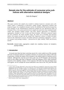
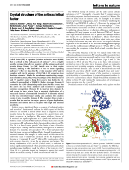
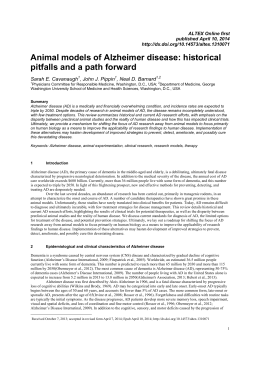
premio firenze university press
tesi di dottorato
–6–
Francesco Bemporad
Folding and aggregation studies
in the acylphosphatase-like family
Firenze University Press
2009
Folding and aggregation studies in the acylphosphataselike family / Francesco Bemporad. – Firenze : Firenze
University Press, 2009.
(Premio FUP. Tesi di dottorato ; 6)
http://digital.casalini.it/9788884539465
ISBN 978-88-8453-845-8 (print)
ISBN 978-88-8453-946-5 (online)
Progetto grafico di Alberto Pizarro Fernández
© 2009 Firenze University Press
Università degli Studi di Firenze
Firenze University Press
Borgo Albizi, 28
50122 Firenze, Italy
http://www.fupress.com/
Printed in Italy
To my mother
Contents
Chapter 1
Introduction
1
1.1 Different conformational states are populated during life-span of proteins
1
1.2 Protein folding
3
1.2.1 Definition of the protein folding problem
3
1.2.2 The early studies and the characterisation of intermediates
3
1.2.3 F value analysis and folding mechanisms: is there a unifying mechanism? 6
1.2.4 The role of topology in determining folding; the importance of
studying structurally related proteins
7
1.3 Protein misfolding
8
1.3.1 Definition and structure of amyloid fibrils
9
1.3.2 Mechanisms of amyloid aggregation
11
1.4 Aim of this thesis
12
1.4.1 The acylphosphatase-like family
12
1.4.2 Folding and aggregation in the acylphosphatase-like family
14
Chapter 2
Enzymatic activity in non-native Sso AcP
2.1 Introduction
2.2 Results
2.2.1 The partially folded state populated during folding of Sso AcP
shows acylphosphatase activity
2.2.2 The acylphosphatase activity observed in the Sso AcP partially
folded state is highly sensitive to mutations
2.2.3 Investigation of the partially folded and transition states of Sso
AcP using F value analysis
2.3 Discussion
2.3.1 Structure of the partially folded state and of the transition state
2.3.2 Enzymatic activity in the presence of a highly dynamic catalytic site
2.3.3 Biological function in the absence of a three-dimensional fold
2.4 Materials and methods
2.4.1 Mutagenesis
2.4.2 Protein expression and purification
2.4.3 Enzymatic activity essay
2.4.4 Development of enzymatic activity during folding
17
17
20
20
23
25
28
28
29
31
32
32
33
34
34
Francesco Bemporad, Folding and aggregation studies in the acylphosphatase-like family, ISBN
978-88-8453-845-8 (print) ISBN 978-88-8453-946-5 (online) © 2009 Firenze University Press
VIII
Francesco Bemporad
2.4.5 Equilibrium GdnHCl-induced unfolding curves
2.4.6 Folding kinetics
2.4.7 Unfolding kinetics
2.4.8 F value analysis
Chapter 3
Aggregation studies on Sso AcP
3.1 Introduction
3.1.1 Aggregation from native states
3.1.2 Aggregation of Sso AcP
3.2 Results
3.2.1 Sso AcP aggregates regardless of the position of the N-terminal segment
3.2.2 N-terminal segment does not induce Sso AcP aggregation via a
destabilising effect
3.2.3 A specific inter-molecular interaction between N-terminal
segment and globular Sso AcP leads to the formation of early aggregates
3.2.4 Formation of initial aggregates depends on protein concentration
3.2.5 Experiments with acrylodan
3.3 Conclusions: a possible aggregation mechanism for Sso AcP
3.4 Materials and methods
3.4.1 Materials
3.4.2 Mutagenesis, protein expression and purification
3.4.3 Far-UV Circular dichroism
3.4.4 Thioflavin T fluorescence
3.4.5 Aggregation kinetics followed by static light scattering
3.4.6 Congo red staining
3.4.7 Enzymatic activity measurements
3.4.8 Equilibrium unfolding
3.4.9 Dynamic light scattering
3.4.10 Cysteine labelling
Chapter 4
Role of π-stacking in amyloidoses
4.1 Aromatic residues and amyloidoses
4.1.1 Introduction
4.1.2 A possible role for p-stacking in amyloid-like aggregation
4.1.3 mt AcP as a model for investigating p-stacking
4.2 Results
4.2.1 Strategy employed
4.2.2 Phenylalanine 22
4.2.3 Tyrosine 25
4.2.4 Tyrosine 91
4.2.5 Tyrosine 98
4.2.6 Statistical analysis
4.3 Discussion
4.3.1 Aromatic residues promote amyloid aggregation of mt AcP due to
their hydrophobicity and b-sheet propensity
35
35
36
36
39
39
39
40
42
42
45
47
51
53
58
59
59
60
60
60
61
61
61
62
62
62
65
65
65
66
67
68
68
69
70
70
71
71
72
72
Contents
IX
4.3.2 Aromatic residues are frequent in the cross-b core of fibrils but are
not necessarily required
4.4 Conclusions
4.5 Materials and methods
4.5.1 Mutagenesis, protein expression and purification
4.5.2 Aggregation kinetics with Thioflavin T fluorescence
4.5.3 Data calculation
73
75
75
75
75
76
Chapter 5
Final remarks
5.1 Conformational states distinct from the fully folded structure can
present enzymatic activity
5.2 A comparison between the transition state ensembles of mt AcP and Sso AcP
5.3 Different regions of the sequence are involved in protein folding and
amyloid-like aggregation
5.4 Conclusions
Acknowledgements
77
77
78
80
82
83
Appendix A Equations and formulas
103
Appendix B Models for Sso AcP aggregation
111
Bibliography
119
Chapter 1
Introduction
1.1 Different conformational states are populated during life-span of proteins
Proteins are among the most important molecules in living organisms as they carry
out a variety of different and fundamental functions. The function of a given protein is
directly related to the unique features of its functional state, which determines partners,
ligands or substrates. Nevertheless, during their life-span proteins can adopt many different conformational states (Chiti and Dobson 2006).
With reference to figure 1.1, proteins are synthesized on ribosomes from the genetic information encoded in the cellular DNA. In some cases the protein is biologically active directly after translation, even though it populates an unfolded state
(Dunker et al. 2001). In many other cases proteins need to achieve a folded structure to
be functional. Folding in vivo is in some cases co-translational, implying that it is initiated before protein synthesis is completed, when the nascent chain is still attached to
the ribosome (Dobson 2003). Other proteins, however, undergo folding after release
from the ribosome, whereas others fold in specific compartments, such as mitochondria or the endoplasmic reticulum (ER), after trafficking and translocation through
membranes (Dobson 2003). In the majority of cases, folding in cytoplasm, ER and mytochondria is chaperon assisted (Bukau et al. 2006). The native state of the protein is attained through formation of partially folded conformations. Native proteins can then
interact with partners to form functional oligomers or polymers. Regulation of these
processes is crucial for cells as errors in folding can give rise to potentially dangerous
folds (Jahn and Radford 2005; Chiti and Dobson 2006). In physiological conditions
misfolded peptides are either refolded or degraded but, if this protein quality control is
impaired, they can expose sticky regions and form toxic oligomers. These species can
give rise to β-strand rich protofibrils and fibrils (see section 1.3). In the next sections
the processes of folding and misfolding, that represents topics of the present thesis, are
discussed in detail. The problems that will be discussed in the next chapters will also be
introduced.
Francesco Bemporad, Folding and aggregation studies in the acylphosphatase-like family, ISBN
978-88-8453-845-8 (print) ISBN 978-88-8453-946-5 (online) © 2009 Firenze University Press
2
Francesco Bemporad
Figure 1.1: Different conformational states can be populated by a polypeptide chain. A nascent protein folds to reach a native and biologically active state. During this process partially
folded conformations are transiently populated. Native states can form oligomers or polymers. At the end of its life-span a protein is degraded. The equilibrium between these species
is crucial as folded, partially folded and unfolded states can aggregate. These aggregates can be
either off-pathway (top of the figure) or on-pathway (bottom of the figure) and initially
maintain the structural features of the precursor conformational states. They can later reorganise to form β-sheet containing aggregates and then amyloid. Reprinted from (Chiti and
Dobson 2006).
Introduction
3
1.2 Protein folding
1.2.1 Definition of the protein folding problem
Protein folding is the physical process by which a polypeptide chain changes its conformation to reach a biologically active three-dimensional structure. After decades of
studies, protein folding has been recently named by Science among the 100 biggest unsolved problems in science (Editorial 2005). Traditionally, the protein folding problem is made by three distinct problems (Dill et al. 2007):
1. The thermodynamic question of how a native structure results from the interatomic forces acting on amino acid sequence (the folding code). This problem first
arose when Christian B. Anfinsen showed that the three-dimensional structure of
a protein is encoded by its amino acid sequence (Anfinsen et al. 1961; Anfinsen
1973). Although much work must be done to address this issue, it is now clear
that the folding code is distributed both locally and not locally in sequence, that its
dominant component is the hydrophobic interaction and that secondary structure
is more a consequence than a cause of folding (Dill 1999). Moreover, novel proteins are now being designed as variants of existing proteins (Dwyer et al. 2004;
Kaplan and DeGrado 2004).
2. The computational problem of how to predict three-dimensional structures
solely on the basis of the primary sequence of polypeptides. To address this issue, two different approaches have been proposed to date: (1) the development of
algorithms that use amino acid sequences as an input and produce, by homology
modelling, structures as an output. A major milestone in this field is CASP
(Critical Assessment of Techniques for Structure Prediction), a communitywide blind test to predict unknown structures (Moult 2006). Currently, structures of small globular proteins (i.e. about 90 residue long peptides) can be predicted within RMSD of 2-6 Å (Bradley et al. 2005; Zhang and Arakaki 2005). (2)
The development of physics-only methods aimed to understand the final structure without database-derived knowledge. Although these methods are limited
by huge computational requirements, using a distributed computing system
(Folding@Home) Pande et al. folded villin to a distance RMSD of 1.7 Å
(Zagrovic et al. 2002).
3. The kinetic question of how, and with which mechanism, can a protein fold so
quickly. In 1968 Cyrus Levinthal first pointed out that, if a given protein is to attain
its correctly folded configuration by sequentially sampling all the possible conformations, it would require a time longer than the age of the universe to arrive at its
correct native conformation (Levinthal 1969). Solving Levinthal’s paradox implies
that folding proceeds in a step-wise manner and that several intermediates with increasing native-like structure are populated along the folding coordinate. In the past
two decades major advances have been done in folding experiments and possible
mechanisms of protein folding have been proposed (sections 1.2.2 and 1.2.3).
1.2.2 The early studies and the characterisation of intermediates
As a consequence of the Levinthal’s paradox, the idea emerged that the characterization
of partially folded states transiently populated during folding would give important
4
Francesco Bemporad
Figure 1.2: (A) Partially folded states (I) can play different roles in folding. They can be (1) onpathway intermediates, (2) off-pathway partially folded states, (3) local minima transiently
populated in one of many parallel pathways that lead to the formation of the native state (F).
U refers to the unfolded state. (B) An example of energy landscape for folding of hen egg
lysozyme (reprinted from (Dinner et al. 2000)). In this graph free energy is reported versus the
number of native contacts in α (Qα) and β (Qβ) domains. Two parallel pathways are shown. A
fast pathway (yellow line) directly leads to the formation of the transition state and thus to
attaining the native state. A slow pathway (red line) leads to the formation of a conformation
that is in a local minimum, corresponding to a partially folded state in which only α domain is
structured.
insight into folding mechanism. At the end of 80s, the use of hydrogen exchange pulse
labelling coupled to NMR (Roder et al. 1988) and of protein engineering methods
(Matouschek and Fersht 1991) allowed structural characterization of partially folded
states. More recently, the development of new instrumentation, such as ultra-rapid
mixing devices (Shastry et al. 1998) and temperature jump relaxation techniques
(Mayor et al. 2003) allowed the measurements of events within dead-time of normal
stopped flow experiments. Finally, the destabilization of native state has allowed to increase the equilibrium population of partially folded states (Religa et al. 2005). This has
allowed solution NMR methods to be applied to solve the structure of folding intermediates of small proteins (Religa et al. 2005). Generally, it has been shown that in the intermediate states the overall topology resembles the native structure and that some regions are highly structured while other regions are more denatured-like (Matouschek
et al. 1989b; Salvatella et al. 2005).
Introduction
5
Figure 1.3: Models for protein folding (reprinted from (Nolting and Andert 2000)). Framework
model, hydrophobic collapse model and nucleation-condensation model are shown. These
model are described in the text.
The characterisation of partially folded states raised the question as to whether
these states are productive species en-route to the native state (true intermediates, see
model (1) in figure 1.2A) or kinetic traps that slow down the process (off-pathway partially folded conformations, see model (2) in figure 1.2A) (Bai 1999; Gianni et al.
2007b). If many proteins were shown to form on-pathway partially folded states (Bai
1999; Capaldi et al. 2001; Travaglini-Allocatelli et al. 2003; Jemth et al. 2004), recent
observation that non-native interactions may be observed for productive on-pathway
intermediates suggests that partial protein misfolding may be an obligatory step preceding native state consolidation (Capaldi et al. 2002; Religa et al. 2005). Moreover, in
many cases evidence emerged that parallel pathways can lead to the formation of native
states (Matagne and Dobson 1998) (see model (3) in figure 1.2A). This led to the description of energy landscapes for protein folding (figure 1.2B) (Dinner et al. 2000;
Dobson 2003; Vendruscolo and Dobson 2005). This “new view” uses the idea of an en-
6
Francesco Bemporad
ergy surface to describe the conformational ensemble accessible during folding. Unfolded molecules, structurally different, follow different pathways and populate different conformational states characterised by weak interactions (Matagne and Dobson
1998). Faster paths and local minima exist in the landscape, but Brownian motions allow each molecule to escape kinetic traps and continue the search for the native state. If
the trajectories can be numerous, the transition state is unique and all pathways lead to
the formation of native state as native interactions are the most stable (Dinner et al.
2000). Thus, energy landscapes are not in contrast with the concept of folding pathway;
they are instead able to join different pathways that can be detected with different experimental approaches.
1.2.3 Φ value analysis and folding mechanisms: is there a unifying mechanism?
Several methods have been developed in the two past decades to study folding (ZarrineAfsar and Davidson 2004; Dill et al. 2007). Single molecule measurements can now be
performed and FRET methods can watch directly the formation of particular contacts
(Schuler et al. 2002; Magg et al. 2006). A major milestone in the analysis of protein
folding mechanisms was the introduction, at the end of the 80s, of protein engineering
methods to perform Φ value analysis (Matouschek et al. 1989b). In this method a single
point mutation is introduced to remove a specific contact from the native state of a
given protein. The change in conformational stability upon mutation for the native
state and for the investigated state is calculated. The ratio between these two quantities
is a number (Φ value) that varies from0 to 1. A Φ value equal to 0 suggests that the contact removed by mutagenesis is not formed in the investigated state. A Φ value equal to
1 implies a native-like interaction in the investigated state for the removed group. In
this way the characterisation of conformational states populated along the folding coordinate is possible. Although the structural information contained in this energetic
parameter has been long discussed, protein engineering methods have been applied to
analyse protein folding transition states at atomic level, to find amino acids that control
folding speed (Matouschek et al. 1989b) and to investigate partially folded states
(Matouschek et al. 1989a; Matouschek et al. 1992). In this thesis Φ value analysis shall
be used to investigate a partially folded state transiently populated during folding of
the acylphosphatase from Sulfolobus solfataricus (see table 2.3).
Historically, the use of Φ value analysis, coupled to other techniques (Nolting and
Andert 2000; Daggett and Fersht 2003), allowed three mechanisms to be proposed for
protein folding (Nolting and Andert 2000):
• hydrophobic collapse: In this model (figure 1.3) the initial event of the reaction is
thought to be a relatively uniform collapse of the protein molecule, mainly driven
by the hydrophobic effect (Baldwin 1989; Dill 1990). Stable secondary structure
starts to grow only in the collapsed state, which narrows in a confined volume the
conformational search to the native state. Although this model was initially supported by the observation that the hydrophobic driving force provided by the expulsion of water from the burial of non-polar surfaces is substantial, the hydrophobic collapse presents a problem because an excess of non-native interactions
will hinder reorganisation of both the polypeptide chain and side chains (Daggett
and Fersht 2003).
Introduction
7
•
framework model: According to this model (figure 1.3) folding starts with the
formation of elements of secondary structure independently of tertiary structure,
or at least before tertiary structure is locked in place. These elements then assemble
into the tightly packed native tertiary structure either by diffusion and collision
(Karplus and Weaver 1994) or by propagation of structure in a stepwise manner
(Wetlaufer 1973). The validity of this model has been mainly shown for small helical proteins (Mayor et al. 2000; Myers and Oas 2001).
• nucleation-condensation: In the early 90s the previous model was challenged by
two observations: (1) some proteins fold in a two-state process without accumulation of secondary-structure intermediates (Jackson and Fersht 1991); (2) the use of
Φ value analysis (Matouschek et al. 1989b) showed that in the folding transition
state secondary and tertiary structure form in parallel (Otzen et al. 1994). This led
some authors to propose a model (figure 1.3) in which early formation of a folding
nucleus catalyses further folding (Fersht 1995; 1997). The nucleus primarily consists of a few adjacent residues that have some correct secondary and tertiary structure interactions but is stable only in the presence of further approximately correct
interactions. The presence of the folding nucleus allows the transition state to bear
a native-like topology (Lindorff-Larsen et al. 2005b) and the number of contacts
that must be sampled dramatically decreases. Many small α and α/β proteins were
shown to fold similarly (Clarke et al. 1997; Chiti et al. 1999b).
The three models mentioned above are in apparent contrast. Nevertheless, evidence
is now emerging that framework and nucleation-condensation represent extreme
manifestations of an underlying common mechanism. It was shown that when the helical propensity is increased, folding turns from nucleation condensation behaviour to
diffusion-collision behaviour, in which helical elements are fully preformed (Gianni et
al. 2003; White et al. 2005). In agreement with this observation, it was proposed that
significant secondary structure is present in the denatured state if such structure is sufficiently stable. Thus, the rate-limiting step involves docking of these elements
(Daggett and Fersht 2003). If instead the secondary structure is not stable in the unfolded state, a nucleation event is required to favour collapse of the structure (Daggett
and Fersht 2003). Finally, a PDZ domain was recently shown to recapitulate nucleation
condensation and diffusion-collision models in three steps: (1) the early formation of a
weak nucleus formed by few residues with fractional Φ values that determine the native-like topology of a large portion of the structure, (2) a global collapse of the entire
polypeptide chain, and (3) the consolidation of the remaining partially structured regions to achieve the native state conformation (Gianni et al. 2007a).
1.2.4 The role of topology in determining folding; the importance of studying structurally related proteins
If the fold is important in determining folding mechanism and speed, one can conclude
a major role for topology in protein folding. Perhaps the most dramatic evidence for
such a conclusion is the observation of a remarkable correlation between the experimental folding rates of a wide range of small proteins and the complexity of their folds,
measured by the contact order (Plaxco et al. 1998). The latter is the average separation
in the sequence between residues that are in contact with each other in the native structure. A correlation was also found between contact order and the position of the transi-
8
Francesco Bemporad
tion state along the folding coordinate (Plaxco et al. 1998). Many studies are now investigating the folding mechanism of different proteins sharing the same fold. The
folding of several cytochrome c proteins involves the formation of a partially structured intermediate and some essential structural features of the intermediate and transition states are highly conserved across this protein family (Travaglini-Allocatelli et
al. 2004). Five different PDZ domains have been shown to fold via an on-pathway intermediate and two transition states whose position is conserved along the folding coordinate (Chi et al. 2007). A comparison between Φ values of structurally related proteins with divergent sequence composition suggested that protein families with conserved transition states are confined to a single folding trajectory whereas protein
families with variable transition states have access to multiple pathways (ZarrineAfsar et al. 2005). As an addition to this simple rule, it has been proposed that a two
strand-helix (i.e. two β-strands docking against a single α-helix) motif is the minimal
folding nucleus, called foldon, and that pathway multiplicity is linked to the multiplicity of foldons within the protein structure (Lindberg and Oliveberg 2007).
Figure 1.4: Features of amyloid fibrils. (A) Fibrils have a long and unbranched shape. White
lines indicate a hypothetic enlargement of a fibril to show protofilaments. (B) Fibrils show a
cross-β structure. This scheme represents the typical appearance of fibrils when analysed with
X-ray fibre diffraction (reprinted from (Serpell 2000)). (C) Amyloid material shows green
Congo red birefringence under cross-polarised light. In this picture CR staining of amyloid
deposits of β2-microglobulin is shown (reprinted from (Ivanova et al. 2003)).
In this thesis a characterisation of a partially folded state and transition state populated
during folding of the acylphosphatase from Sulfolobus solfatricus shall be carried out (see
section 2.2.2). The results have implications in the study of biological function in the absence of a structured fold and will be discussed in section 2.2.3. Moreover, the obtained Φ
values shall be compared in chapter 5 with the results previously obtained on another
member of the same superfamily, human muscle acylphosphatase (Chiti et al. 1999b).
1.3 Protein misfolding
As mentioned above protein quality control is a crucial point of cell metabolism because incompletely folded proteins must inevitably expose to the solvent at least some
regions of structure that are buried in the native state and that are prone to inappropriate interaction with other molecules within the crowded intracellular or extracellular
Introduction
9
environments (Dobson 2003). Protein misfolding is the conversion of a protein into a
structure that differs from its native state (Chiti and Dobson 2006). This process is related to a set of human diseases (protein misfolding diseases), usually classified in three
distinct groups:
1. Diseases in which an impairment in the folding efficiency of a given protein results in a reduced amount of native folded protein. An example is cystic fibrosis,
where a mutated variant of a chloride channel (CFTR) populates a misfolded conformation that is degraded instead of being translocated to the cell membrane
(Thomas et al. 1995). The absence of the properly functioning protein accounts for
the symptoms of the disease (Thomas et al. 1995).
2. Diseases in which misfolding of a given protein results in improper trafficking. In
the case of early-onset enphysema, that we cite here as an example, mutations of
the gene encoding α1-antitripsyn determine the production of a protein that
forms polymers. These will be retained in the liver (where α1-antitripsyn is produced) and will not reach the lungs, where α1-antitripsyn is necessary for a constitutive inhibition of elastase. The absence of such an inhibitory effect gives rise
to the disease (Chiti and Dobson 2006).
3. The largest group of protein misfolding diseases is the group of pathological
states associated with the conversion of a given peptide or protein from its soluble
native state into highly organised fibrillar aggregates (Dobson 2003; Jahn and
Radford 2005; Chiti and Dobson 2006). These aggregates are usually referred to
as amyloid fibrils when they form in vivo outside the cell and intracellular inclusions when they form inside the cell. More than 40 human diseases are associated
to this process (for a complete list see (Chiti and Dobson 2006)). Importantly,
many of them have high social impact, such as Alzheimer’s disease, Parkinson’s
disease and type II diabetes mellitus. In the following sections structure of fibrils
and mechanisms of aggregation are discussed.
1.3.1 Definition and structure of amyloid fibrils
Proteins able to form amyloid aggregates do not share any sequence identity and structural homology. Despite that, some important structural features are common in amyloid fibrils formed by different peptides. Amyloid-like fibrils are defined on the basis
of peculiar physico-chemical properties when investigated with different techniques
(figure 1.4). In particular amyloid fibrils have a long and unbranched shape when observed with atomic force microscopy (AFM) or transmission electron microscopy
(TEM) (figure 1.4A). Fibrils usually consist of a number (typically from 2 to 6) of protofilaments, each about 2-5 nm in diameter (Serpell et al. 2000) (see figure 1.5A). The
protofilaments twist together and form rope-like fibrils that are typically 7-13 nm
wide (Sunde and Blake 1997; Serpell et al. 2000). When analysed by X-ray fiber diffraction (figure 1.4B), the various protein molecules are arranged so that the polypeptide
chains form β-strands that run perpendicular to the long axis of the fibril (cross-β
structure) (Sunde and Blake 1997). Finally, the fibrils have the ability to bind specific
dyes such as Thioflavin T (ThT) (Krebs et al. 2005) and Congo red (CR, figure 1.4C)
(Nilsson 2004), although the specificity of binding of CR to amyloid fibrils and the resulting green birefringence under cross-polarised light has recently been questioned
(Khurana et al. 2001; Bousset et al. 2004b).
10
Francesco Bemporad
Figure 1.5: Structure of amyloid fibrils and protofilaments. (A) Structure of Aβ fibrils; reprinted
from (Petkova et al. 2006). (B) Structure of human amylin fibrils; reprinted from (Kajava et al.
2005). (C) Structure of Sup35p protofilaments; reprinted from (Krishnan and Lindquist 2005).
High resolution structures of amyloid fibrils have been solved in recent years by
use of solid state NMR (SSNMR), site-directed spin labelling coupled to electron paramagnetic resonance (SDSL-EPR) and by X-ray diffraction analysis carried out on
nano- or microcrystals of small peptides having characteristics of amyloid fibrils
(Tycko 2004; Luhrs et al. 2005; Chiti and Dobson 2006; Petkova et al. 2006). In the case
of Aβ, the peptide whose aggregation is related to Alzheimer’s disease, SSNMR studies
led some authors to propose that each molecule forms two strands in the core of fibrils,
spanning residues 12-24 and 30-40. These strands are not part of the same β-sheet but
participate to the formation of two different parallel in register β-sheets that run parallel to the fibril axis (Antzutkin et al. 2000; Petkova et al. 2002). A single protofilament
has been proposed to be composed by four β-sheets (i.e. two Aβ molecules) separated
by distances of 10 Å (figure 1.5A). This structure has been confirmed by SDSL-EPR
studies that found residues 13-21 and 30-39 highly structured in the fibrils (Torok et
al. 2002). A peptide derived from the yeast prion Sup35p (GNNQQNY) and the peptide
KFFEAAAKFFE have been converted in three-dimensional crystals with features typical of amyloid fibrils. The structures of these crystals have been solved by X-ray crystallography (Makin et al. 2005; Nelson et al. 2005). In the case of the Sup35p fragment,
the crystal consists of pairs of parallel β-sheets in which each individual peptide molecule contributes a single β-strand. The stacked β-strands are parallel and in register in
both sheets. The two sheets interact with each other through the side chains of Asn2,
Gln4, and Asn6 (Nelson et al. 2005). Following these pioneering studies the X-ray
structure of other assembled peptides have been solved (Sawaya et al. 2007).
In some cases different approaches have been applied to solve amyloid fibril structures. A model for fibrils formed by human amylin, whose aggregation is related to
type II diabetes mellitus, has been recently proposed on the basis of different experimental evidences, such as protofilament diameter, the cross-β structure and the experimental evidence of a parallel and in register arrangement of the β-strands formed
by adjacent molecules (Kajava et al. 2005) (figure 1.5B). In the obtained model three
different β-strands, formed by residues 12-17, 22-27 and 31-37, participate to the formation of three β-sheets that run parallel to the fibril axis. In the case of the NM region
Introduction
11
of the yeast prion Sup35p, 37 single point mutations to cysteine were produced. These
variants were labelled with fluorescent probes (Krishnan and Lindquist 2005). The
wavelength maximum and the emission intensity of probes bound to different positions were then used to get information about burial of residues and possible interaction in fibrils. On the basis of these experiments a model was drawn in which NM
molecules interact via head-to-head (residues 25-38) and tail-to-tail (residues 107157) interactions (Krishnan and Lindquist 2005). The N-terminal (residues 1-20) and
the distal end (residues 158-250) are instead structurally heterogeneous and solvent
exposed (Krishnan and Lindquist 2005) (figure 1.5C). This approach has been used in
chapter 3 to get insight into the protofibril structure of the acylphosphatase from Sulfolobus solfataricus.
1.3.2 Mechanisms of amyloid aggregation
If formation of amyloid fibrils is related to the onset of several human diseases, the
ability to form amyloid-like aggregates in vitro was shown to be an inherent property
of the protein back-bone. Several proteins that are not involved in disease were shown
to form amyloid-like aggregates using particular cosolvents, temperature and salts
(Guijarro et al. 1998; Chiti et al. 1999c). Nevertheless, the fact remains that different
proteins show different tendencies and pathways leading to the formation of amyloidlike fibrils and that these differences can be explained on the basis of their sequence,
that is on the basis of the physico-chemical properties of the side chains of their amino
acids.
It is now clear that amyloid aggregation is a multi-step process in which different
states are transiently populated. The formation of amyloid fibrils has many characteristics of a “nucleation-growth” mechanism (Chiti and Dobson 2006). Conversion of
proteins into their fibrillar form follows two phases: (1) a lag phase, in which aggregation nuclei are formed and (2) a growth phase, in which further monomers or oligomers bind to the nuclei (Serio et al. 2000; Pedersen et al. 2004). This mechanism is
confirmed by the observation that the addition of aggregation nuclei (seeds) to the
sample shortens or abolishes the lag phase (Serio et al. 2000).
In the overall process of amyloid fibril formation various aggregates are thought
to form before mature amyloid fibrils accumulate. In the case of Aβ1-40 and Aβ1-42
oligomeric species made by 2-4 and 5-6 molecules have been observed, respectively
(Bitan et al. 2001; Bitan et al. 2003). In the case of Sup35p oligomers form rapidly and
these species only afterwards convert into species with extensive β-sheet structure able
to nucleate aggregation (Serio et al. 2000).
Other important species populated prior to the appearance of fibrils are protofibrils. These are isolated or clustered spherical beads 2-5 nm in diameter with β-sheet
structure (Chiti and Dobson 2006). These species usually can bind both ThT and CR
(Walsh et al. 1999). Species of this type have been observed for α-synuclein (Conway et
al. 2000), amylin (Kayed et al. 2004), transthyretin (Quintas et al. 2001) and the acylphosphatase from Sulfolobus solfataricus, one of the proteins object of this thesis
(Plakoutsi et al. 2004) (see chapter 3).
The study of oligomers has seen an increasing importance as these species have
been shown to be the most toxic for cells (Bucciantini et al. 2002). This is probably due
to the fact that these inherently misfolded species expose an array of groups that are
12
Francesco Bemporad
normally buried in globular proteins or dispersed in highly unfolded peptides. This is
likely to trigger aberrant events resulting from inappropriate interactions with cellular
components, such as membranes, small metabolites, proteins, or other macromolecules. These interactions cause impairment of oxidative stress, ion balance and other
factors that lead to cell death (Chiti and Dobson 2006).
It was also shown that, even if the final structure is conserved, different conformational states can be the starting point for amyloid-like aggregation (Bemporad et al.
2006) (see also figure 1.1). (1) A number of systems, including Aβ and α- synuclein, are
largely unfolded prior to aggregation (Bemporad et al. 2006; Chiti and Dobson 2006).
Short peptides have been shown to be able to form amyloid-like material (Lopez de la
Paz and Serrano 2004). In this case, since the whole protein sequence is fully exposed to
the solvent, aggregation is governed by simple physico-chemical factors, such as hydrophobicity, secondary structure propensities and net charge (Chiti et al. 2003) (for a
complete description of the determinants of aggregation from unfolded states see section 4.1.1). (2) In most cases globular proteins need to unfold, at least partially, to aggregate into amyloid-like fibrils. It is clear for example that conditions that promote
their partial unfolding, such as temperature, low pH, presence of organic cosolvents,
increase their propensity to aggregate (Guijarro et al. 1998) (Chiti et al. 2000; Gosal et
al. 2005). The aggregation of HypF-N can be initiated by a population of less than 1%
of a partially folded conformation in equilibrium with the native one (Marcon et al.
2005). (3) Although the “conformational change hypothesis” can account for the aggregation properties of many proteins, increasing evidence is now accumulating that native proteins retain a significant, albeit small, tendency to aggregate ((Bemporad et al.
2006) and chapter 3). Formation of amyloid-like fibrils of insulin at low pH is preceded by an oligomerization step in which a native-like α-helical content is retained,
while β-sheet rich aggregates form only later on (Bouchard et al. 2000). The acylphosphatase from Sulfolobus solfataricus aggregates from an ensemble of native-like conformations into early aggregates that retain enzymatic activity, while unfolding is two
orders of magnitude slower than aggregation (Plakoutsi et al. 2004; Plakoutsi et al.
2005).
In conclusion, the initial step of aggregation is the conversion of single molecules
populating an aggregation prone state -folded, partially folded or unfolded- into an
oligomer in which each monomer resembles the initial state. These oligomers convert
afterwards into β-sheet rich species that lead to formation of amyloid fibrils.
1.4 Aim of this thesis
1.4.1 The acylphosphatase-like family
In this thesis we focus our attention on proteins belonging to the acylphosphatase-like
structural family, the acylphosphatase from Sulfolobus solfataricus (Sso AcP) and the
acylphosphatase from human muscle (mt AcP).Acylphosphatase (AcP) is a small
(about 100 residue long) α + β protein belonging to the ferredoxin-like fold (that is a α
+ β sandwich with antiparallel β-sheet). The structure of the protein is highly conserved throughout the family. All the AcPs so far characterised show the same βαββαβ
topology that originates a β-sheet docking against the two helices (figure 1.6A to 1.6H)
Introduction
13
(Pastore et al. 1992; Thunnissen et al. 1997; Zuccotti et al. 2004; Miyazono et al. 2005;
Pagano et al. 2006). Sso AcP also bears an unstructured, N-terminal 11 residue long
segment that plays a major role in the aggregation mechanism of the protein
((Plakoutsi et al. 2006) and chapter 3). AcP is an enzyme (enzyme commission 3.6.1.7)
able to hydrolyse acylphosphates, with formation of a phosphate ion and a carboxylate
group (Stefani et al. 1997) (figure 1.6I). The mechanism of catalysis has been studied in
detail in mt AcP (Taddei et al. 1994; Taddei et al. 1996; Taddei et al. 1997). The catalytic
residues consist on an Arg and an Asn residues highly conserved within the family.
The catalytic cycle can be summarised as follows. (1) The Arg binds to the phosphate.
(2) The Asn residue stabilises the pentacovalent intermediate that forms following nucleophilic attack of a water molecule. (3) The products are released.
Figure 1.6: The structure of AcPs is highly conserved. AcPs are shown from Pyrococcus horikoshii (A, PDB entry 1W2I), Thermus thermophilus (B, PDB entry 1ULR), Drosophila melanogaster (C, PDB entry 1URR), Homo sapiens (muscolar type, D, PDB entry 1APS), Homo
sapiens (common type, E, PDB entry 2ACY), Escherichia coli (F, PDB entry 2GV1), Sulfolobus
solfataricus (H, PDB entry 1Y9O). HypF-N from Escherichia coli is also shown (G, PDB entry
1GXT), (I) Reaction catalysed by AcP on benzoyl-phosphate, a substrate largely used for AcP
enzymatic activity measurements (Stefani et al. 1997).
The function of the protein is yet not well understood. The initial observation that
the enzyme is active on 1,3-bisphosphoglycerate suggested for AcP a possible regulative role on glycolysis (Ramponi 1975). Nevertheless, different functions have been
proposed for this protein. The evidence that the enzyme is able to hydrolyse the αaspartyl-phosphate, which forms during action of membrane pumps, led some authors
to propose a role in regulation of membrane transport (Nediani et al. 1996). More recently it was shown the ability of AcP to induce apoptosis in HeLa cells (Giannoni et al.
2000). Since the protein structure is similar to the RNA binding domain of ribonucleoproteins (Swindells et al. 1993), it was proposed that the interaction of this protein with
nucleic acids has a physiological role. It was shown also a nuclear migration of mt AcP
and its interaction with other DNAses in response to various apoptotic stimuli either
in K562 or in Jurkat cells (Chiarugi et al. 1997). Finally, since the AcP levels increase
in several cell lines during differentiation and AcP is able to hydrolyse both γ and β
14
Francesco Bemporad
phosphate groups of ATP, it was proposed a role for AcP in regulation of differentiation through control of [ATP]/[ADP] levels (Paoli et al. 2000).
1.4.2 Folding and aggregation in the acylphosphatase-like family
In the last decade AcP has been largely used as a model for folding and amyloid-like
aggregation studies. The folding process shows high variability in the acylphosphatase-like family. Equilibrium and kinetic fluorescence studies suggest that the folding
of mt AcP is a two state process characterised by a rate constant (0.23 s-1) very low if
compared with both other members of the family and with other proteins (van Nuland
et al. 1998; Maxwell et al. 2005). The transition state for folding of mt AcP was also
characterised (Chiti et al. 1999b; Vendruscolo et al. 2001). The most structured region
corresponds to the central part of the β-sheet (β-strand 1 and 3). The second α-helix as
well plays an important role as it appears fully structured in the transition state (Taddei
et al. 2000). The other human AcP isoform, common type AcP (ct AcP) folds in a twostate process with a folding rate constant equal to 2.3 s-1. Although ct AcP bears a lower
conformational stability than mt AcP, this value is ten-fold higher than folding rate for
mt AcP, suggesting no correlation between folding rate and conformational stability
within the AcP family (Taddei et al. 1999). Interestingly, HypF-N from E. coli folds
with a rate constant equal to 70 s-1, two orders of magnitude higher than the folding rate
constant measured for mt AcP (Calloni et al. 2003). Folding proceeds via formation of
a partially structured state that forms on the sub-millisecond time-scale (Calloni et al.
2003). The folding of Sso AcP has been characterised in some detail. The protein folds
through formation of a partially folded state to reach the native structure with a folding
rate constant equal to 5.4 s-1 (see section 2.1 details). These studies, carried out on structurally related but evolutionary distant proteins, allowed some important parameters
for folding to be identified. Indeed, correlations within the acylphosphatase-like family
indicate hydrophobicity, relative contact order and α-helical propensity as important
determinants for folding rates (Chiti et al. 1999b; Taddei et al. 2000; Calloni et al. 2003;
Bemporad et al. 2004). Moreover, these studies clearly suggested that proline isomerism is not evolutionary conserved as proline residues that slow down folding are not
conserved in the family (Bemporad et al. 2004). In chapter 2 we shall present a characterisation of the partially folded state, transiently populated during folding, and of the
major transition state for folding of Sso AcP. The obtained results have important consequences in the study of biological function carried out in the absence of folded structures and will be discussed in section 2.3 and in section 5.2.
The amyloid aggregation processes of different AcPs have been studied as well. Interestingly, different conformational states can trigger the amyloid-like aggregation
process. In the case of mt AcP and HypF-N, it was shown that a partial unfolding is required to initiate the process (Chiti et al. 1999c; Marcon et al. 2005). By contrast, an
acylphosphatase from Drosophila melanogaster and Sso AcP have been shown to aggregate from a native-like state (Plakoutsi et al. 2004; Soldi et al. 2006a). The latter
mechanism is particularly important as it suggests that native states have significant
tendency to aggregate and that evolution has worked to keep under control this process
((Richardson and Richardson 2002) and paragraphs 1.3.2 and 3.1.1). The aggregation
mechanism of Sso AcP has been studied (Plakoutsi et al. 2004; Plakoutsi et al. 2005;
Plakoutsi et al. 2006). Although it was shown that an edge β-strand and the N-terminal
unstructured segment play an important role in the process, the mechanism of aggrega-
Introduction
15
tion of this system is still unclear (Plakoutsi et al. 2006). In chapter 3 we get some important clues into this process and we try to propose a possible model that summarises
all the experimental evidence so far observed including those described here and in our
previous work (see section 3.4). The model will be discussed with respect to the aggregation of other systems that aggregate starting from a native state.
The study of the aggregation of mt AcP has allowed several parameters that favour
aggregation to be proposed. An algorithm has been proposed to predict change in aggregation rate upon mutation that takes into consideration hydrophobicity, secondary
structure propensities and net charge as parameters (Chiti et al. 2003). Nevertheless,
several other parameters have been proposed as determinants for amyloid aggregation
(for details see section 4.1 and (DuBay et al. 2004; Pawar et al. 2005)). Among these parameters, the presence of aromatic residues has been proposed as an important determinant for aggregation rate constants (section 4.1). Nevertheless, several authors have
shown that aromatic residues play an important role only because of their hydrophobicity and secondary structure propensity (section 4.1). In chapter 4 the possible role
for aromaticity in amyloid aggregation is studied using mt AcP as a model. The results
are discussed in view of their possible implications in the development of new algorithms.
16
Francesco Bemporad
Chapter 2
Enzymatic activity in non-native Sso AcP
2.1 Introduction
Proteins are among the most abundant macromolecules in living systems and carry out
a vast number of functions including the catalysis of virtually every chemical transformation occurring in cell biology and the transduction of signals inside and between
cells. While it is well known that the flexibility of the native states of folded proteins is
crucial in the processes determining their function, increasing evidence is accumulating about the existence of proteins or protein domains that adopt unstructured but
functional states under physiological conditions (Dunker et al. 2001; Fink 2005). The
mechanisms, however, by which certain proteins are capable of being active and yet
natively unfolded is not completely understood (Fink 2005).
In this chapter we shall focus our attention on the acylphosphatase from the archaeon Sulfolobus solfataricus (Sso AcP, figure 2.1) and show that this protein retains
an ability to function as an enzyme when adopting a non-native state in which the catalytic site is largely unstructured and flexible. Sso AcP is a 101-residue protein belonging to the acylphosphatase-like structural family. The structure of the native state of
Sso AcP was recently determined by nuclear magnetic resonance (NMR) spectroscopy
and X-ray crystallography (Corazza et al. 2006). This protein shares the same βαββαβ
topology, typical of the ferredoxin-like fold, with the other acylphosphatases so far
characterised (Pastore et al. 1992; Thunnissen et al. 1997; Zuccotti et al. 2004; Miyazono et al. 2005; Pagano et al. 2006). By contrast to related proteins, however, Sso AcP
contains an unstructured, 12-residue N-terminal tail (Corazza et al. 2006). Sso AcP is
able to hydrolyse benzoyl-phosphate (BP; figure 1.6I), with kCAT and KM values of
198 ± 20 s−1 and 0.36 ± 0.04 mM, respectively, at pH 5.3 and 25 °C, and to be competitively inhibited by inorganic phosphate (Corazza et al. 2006). The kCAT value of the enzyme is low at 25 °C, but increases with temperature and reaches at 81 °C -the living
temperature for the Archaeon Sulfolobus solfataricus- a value close to those previously
reported for the mesophilic enzymes at 25 °C (Corazza et al. 2006). The native state of
Sso AcP is thermodynamically very stable with a free energy change of unfolding
(ΔGU-FH2O) of 47 ± 1 KJ mol−1 at 37 °C (Corazza et al. 2006). The midpoint of thermal
unfolding of the protein (Tm) is 100.8 ± 4.1 °C and at 81 °C the ΔGU-FH2O is as high as
20.6 ± 0.3 KJ mol-1, similar to that of human muscle acylphosphatase (mt AcP) at 28 °C
(Corazza et al. 2006).
The folding mechanism of Sso AcP was previously described at pH 5.5 and 37 °C
(Bemporad et al. 2004). After removal of the denaturant, the unfolded state of this protein collapses on the microsecond time scale into an ensemble of partially folded conFrancesco Bemporad, Folding and aggregation studies in the acylphosphatase-like family, ISBN
978-88-8453-845-8 (print) ISBN 978-88-8453-946-5 (online) © 2009 Firenze University Press
18
Francesco Bemporad
formations. This ensemble, which has a free energy of unfolding (ΔGU-IH2O) of 12.5 ± 2
KJ mol-1, is capable of binding the fluorescent dye 8-anilino-1-naphthalenesulfonic
acid suggesting the presence of hydrophobic clusters exposed to the solvent, and presents a far-UV mean residue ellipticity comparable to that of the fully native state, indicating that a native-like secondary structure is already formed in this state (Bemporad
et al. 2004). The partially folded ensemble converts into the fully folded state with a
rate constant of 5.4 ± 1.0 s-1; a small fraction of molecules (ca. 10%) folds slower with a
rate constant of ca. 0.2 s-1 as their folding process is rate-determined by the cis to trans
conversion of the Leu49-Pro50 peptide bond (Bemporad et al. 2004). The presence of a
relatively stable, partially folded state accumulating during folding of Sso AcP offers a
very favourable opportunity to study the function of a protein in a conformational state
different from the native and folded one.
Figure 2.1: Spectroscopic probes of Sso AcP. Trp4, Tyr17, Tyr21, Tyr45, Tyr61, Tyr86, Tyr91
and Tyr101 are depicted in orange. The figure has been drawn with VMD 1.8.3 for win 32
(Humphrey et al. 1996).
In this work the functional properties of the partially folded state of Sso AcP accumulating during folding are investigated using a procedure that allows the recovery of
enzymatic activity during folding to be determined in real time. The protein engineering method is then used to obtain information on the degree of structure formation, at
the level of the mutated residues, in both the partially folded and transition states of the
protein (Matouschek et al. 1989b). We shall show that the partially folded state of Sso
AcP accumulating during folding shows enzymatic activity comparable to that of the
native state. The experimentally obtained Φ values are used as restraints in molecular
dynamics simulations to obtain a model of the structures of the partially folded and
transition state ensembles (Vendruscolo et al. 2001; Gsponer et al. 2006). These procedures illustrate how this state is made up by an ensemble of conformations displaying a
native-like topology, but in which those regions of the sequence forming the active site
in the folded protein exhibit high structural heterogeneity. In spite of the high flexibility existing at the level of the active site, which is also indicated by ΦIH2O values close to
0 for mutations of residues in the catalytic loop, the native-like topology and the close
proximity between the main substrate binding residue (Arg30) and the main catalytic
residue (Asn48) ensures that this conformational state retains enzymatic activity.
Enzymatic activity in non-native Sso AcP
19
Figure 2.2: (A) Folding trace of Sso AcP recorded using intrinsic fluorescence as a probe in
0.275 M GdnHCl, 50 mM acetate buffer pH 5.5, 37 °C. The inset shows the first second of recording. (B) Observed folding/unfolding rate constant ln k versus denaturant concentration;
readapted from (Bemporad et al. 2004). The continuous line represents the expected plot for
a two-state model. (C) Observed folding rate constant ln k versus urea concentration. Above 5
M urea the plot is linear, suggesting two state folding in these conditions. (D) Time course of
BP absorbance recorded at 283 nm in the presence of native Sso AcP. (E) Time course of enzymatic activity of native Sso AcP, calculated as the opposite of the first order derivative of
the trace reported in panel D (see section 2.4). The continuous line represents the best fit to
equation 2.3. (F) Ratio between the main folding rate constant recorded in the presence of
phosphate (○) or phenyl phosphate (●) (kobs) and that recorded without substrate-analogue
in the sample (k0) plotted versus substrate-analogue concentration. (G) BP absorbance recorded at 283 nm after dilution of GdnHCl-unfolded Sso AcP into a refolding buffer; final
conditions are 0.275 M GdnHCl (continuous line) and 0.275 M GdnHCl, 7 M urea (dotted
line). The inset shows the first second of recording. (H) Development of relative enzymatic
activity, calculated as the opposite of the first order derivative of the traces reported in panel
G (see section 2.4), during Sso AcP refolding in the absence (●) and presence (□) of 7 M urea.
The continuous lines represent the best fits to equation 2.3. The activities of fully folded, partially folded and unfolded states are shown. (I) Comparison between the time courses of recovery of native conformation (reported as fraction folded, □) and enzymatic activity (reported as fraction of the native protein activity, ■). After 3.6milliseconds, when the partially
folded ensemble is populated more than 99%, Sso AcP exhibits already 79% of the native enzymatic activity.
20
Francesco Bemporad
2.2 Results
2.2.1 The partially folded state populated during folding of Sso AcP shows acylphosphatase activity
Sso AcP possesses one tryptophan in the N-terminal tail and seven tyrosines at various positions along the sequence, mainly positioned in the β-sheet (figure 2.1). Figure 2.2A shows the change of intrinsic fluorescence when one volume of Sso AcP unfolded in 5.5 M guanidinium hydrochloride (GdnHCl) is mixed with 19 volumes of
refolding buffer. This trace, which is in very good agreement with that previously
reported (Bemporad et al. 2004), indicates the presence of three phases in the folding
process. The first phase, which occurs within the dead-time (10ms) of the stoppedflow device utilised here, leads to an increase of the intrinsic fluorescence of the protein to a value that is about 40% higher than that of the native state. This initial phase
was shown to correspond to the conversion of the fully unfolded state into the partially folded state (Bemporad et al. 2004). The following rapid decrease of fluorescence with a rate constant (kI→F) of 5.3 ± 1.0 s-1, corresponds to the conversion of this
state into the fully native conformation, whereas the second slower decrease, with a
rate constant (k12) of 0.18 ± 0.04 s-1, corresponds to the cis-trans isomerisation of a
small fraction of protein molecules with the Leu49-Pro50 peptide bond initially in a
non-native cis configuration (Bemporad et al. 2004). In addition to the transient hyper fluorescence signal reported in figure 2.2A, the presence of a rapidly formed partially folded state is also indicated by the downward curvature at low GdnHCl concentrations in the Chevron plot showing ln kI→F H2O) versus denaturant concentration
(figure 2.2B).
We then studied the time course of recovery of enzymatic activity during Sso AcP
folding. Since the substrate BP, unlike its hydrolysis products benzoate and phosphate,
has a significant optical absorption at 283 nm, the rate of catalysed BP hydrolysis can
be accurately determined by measuring the decay rate of the absorbance at 283 nm (dA283 (t)/dt) in the presence of Sso AcP (Chiti et al. 1999a). In a first control experiment,
a solution containing native Sso AcP was mixed with the refolding buffer containing a
saturating concentration of BP and a non-denaturing concentration of GdnHCl. Final
conditions were 0.01 mg ml-1 protein, 10 mM BP, 0.275 M GdnHCl, 50 mM acetate
buffer, pH 5.5, 37 °C. The absorbance arising from BP at 283 nm was monitored in real
time and was found to decrease at a constant rate (figure 2.2D). This trace was analysed
to yield the time course of enzymatic activity, as described in section 2.4.4. As expected,
the enzymatic activity does not change with time (figure 2.2E).
To monitor the time course of enzymatic activity during Sso AcP refolding, a sample of GdnHCl-unfolded Sso AcP was mixed with the refolding buffer containing saturating BP. Final conditions were the same as those described above. The timedependent changes of absorbance at 283 nm and of the corresponding enzymatic activity were determined (figures 2.2G and 2.2H, respectively). Immediately after mixing,
when the partially folded state is maximally populated, the BP absorbance decay is occurring with considerable rate (see also the enlargement in the inset of figure 2.2G).
The enzymatic activity is therefore already present, corresponding to 79.3 ± 10% of
21
Enzymatic activity in non-native Sso AcP
Table 2.1: Catalytic parameters for a set of Sso AcP variants measured in 50 mM acetate buffer
at pH 5.5, 37 °C using BP as a substrate. Values reported in column 3 have been obtained by
best fits of data shown in figure 2.3 to equation 2.3. Values reported in column 4 have been
obtained combining data of column 1 and 2.
variant
native state kCAT (s )
Partially folded state
kCAT (s-1)
WT
V24A
V27A
R30A
N48A
P50A
G52A
K92A
222 ± 20
124 ± 10
13 ± 1
7±1
2±2
137 ± 14
178 ± 18
169 ± 20
175 ± 30
7±5
0 ± 10
n. d.
n. d.
0 ± 10
6±5
111 ± 30
-1
activity in the partially folded state
(% of that from native state)
79 ± 10
5.6 ± 10
0.0 ± 10
n. d.
n. d.
0.0 ± 10
3.3 ± 10
66 ± 10
that of the native protein under the same conditions (Figure 2.2H). The activity then
shows a small exponential increase with a rate constant of 3.53 ± 1.5 s-1 (figure 1F).
This value is in reasonable agreement with the rate constant of folding determined with
intrinsic fluorescence under these conditions (5.33 ± 1.0 s -1) and corresponding to the
conversion of the partially folded state into the native state. The kinetic traces of enzymatic activity and intrinsic fluorescence, both normalised to the values of the native
protein, are compared in figure 2.2I. After 3.6 milliseconds the activity is equal to 80%
of the value of the native protein, but the fraction of native protein is less than 4%.
In another experiment the GdnHCl-unfolded protein was diluted into a refolding
solution containing urea. Final conditions were the same described above, except for a
final urea concentration of 7 M. Under these conditions the native protein is still thermodynamically more stable than the unfolded state, making it possible to monitor the
kinetics of folding. In addition, the plot reporting the folding rate constant versus urea
concentration shows a downward curvature in the range of 0-5 M, indicating that in 7
M urea the partially folded state is destabilised and the protein folds according to a
two-state model (figure 2.2C). The results show that the activity is absent immediately
after mixing, when only the unfolded state is populated (figure 2.2G). Upon refolding,
the activity then increases with a rate constant of 0.13 ± 0.02 s-1, which is in good
agreement with the folding rate constant (0.16 ± 0.02 s -1) under these conditions (figure
2.2G). To rule out the possibility that a substantial fraction of the native state forms in
the dead time of the stopped flow experiment, a double-jump experiment was carried
out in which the GdnHCl-unfolded protein was diluted into the refolding buffer (first
jump, refolding) and then, after 10 ms, transferred again to solutions containing
GdnHCl at final concentrations ranging from 4.2 to 7 M (second jump, unfolding). Although such final conditions promote the unfolding of native Sso AcP and produce a
single exponential change of intrinsic fluorescence (Bemporad et al. 2004), no significant fluorescence changes were observed in any of these experiments, suggesting that
the native protein is not present 10 ms after the folding process was initiated when enzymatic activity is present.
22
Francesco Bemporad
Figure 2.3: The enzymatic activity of the partially folded state of Sso AcP is sensitive to mutations. (A, C, E, G) BP absorbance at 283 nm after dilution of the indicated GdnHCl-unfolded
mutants into a refolding buffer: the traces for wild type (A, continuous line), K92A (A, dotted
line), N48A (C, continuous line), R30A (C, dotted line), V24A (E, continuous line), V27A (E,
dotted line), P50A (G, continuous line) and G52A (G, dotted line) are shown. (B, D, F, H) Development of relative enzymatic activity, calculated as the opposite of the first order derivative
of the corresponding traces reported in the corresponding panels on the left (see section
2.4.4), during the refolding of the indicated Sso AcP mutants. The traces for wild type (B, ●),
K92A (B, □), N48A (D, ●), R30A (D, □), V24A (F, ●), V27A (F, □), P50A (H, ●) and G52A (H,
□) are shown. The continuous lines represent the best fits to equation 2.3. The various mutants are labelled in the panels.
Enzymatic activity in non-native Sso AcP
23
Figure 2.4: Folding thermodynamics and kinetics of Sso AcP variants. (A) Equilibrium unfolding curves for a set of Sso AcP variants in 50 mM acetate buffer, pH 5.5, 37 °C. The continuous
lines represent the best fits to the equation reported by Santoro and Bolen (Santoro and
Bolen 1988). The obtained parameters of conformational stability are reported in Table 2. (B)
Folding traces recorded in 0.275 M GdnHCl, 50 mM acetate buffer at pH 5.5, 37 °C. (C) Unfolding traces recorded in 6 M GdnHCl, 50 mM acetate buffer at pH 5.5, 37 °C. The inset
shows the first 5 seconds of recording. In all plots the traces refer to wild type (black), R71A,
(blue), A46G (orange), L65A (red) and V20A (green) Sso AcP.
In order to further rule out the possibility that folding of Sso AcP is accelerated by
BP and that the enzymatic activity observed at the beginning of the folding process is
due to an early substrate-induced folding of the protein, Sso AcP refolding was also
followed using intrinsic fluorescence in the presence of phosphate and phenyl phosphate, two competitive inhibitors of Sso AcP that are stable analogues of BP. Experimental conditions were the same as for the enzymatic activity experiments with no
added urea. The folding rate constant is not affected by either compound (figure 2.2F).
Taken together, these data indicate that the partially folded ensemble accumulating during folding of Sso AcP possesses significant enzymatic activity.
2.2.2 The acylphosphatase activity observed in the Sso AcP partially folded state is
highly sensitive to mutations
The recovery of enzymatic activity during folding was also recorded for 7mutants of
Sso AcP. The K92A variant showed behaviour similar to that of the wild type protein
with enzymatic activity detected for both the partially folded and native states (figures
2.3A and 2.3B; table 2.1). By contrast, the traces recorded for the N48A and R30A variants showed full inactivation of their partially folded and native states (figures 2.3C
and 2.3D; table 2.1). These results confirm the key role of these two highly conserved
residues in the catalysis of the native state of acylphosphatases (Stefani et al. 1997) and
suggest their major role in the catalytic mechanism of the partially folded state as well.
The partially structured ensembles of the V24A and V27A mutants do not show significant residual enzymatic activity (figures 2.3E and 2.3F; table 2.1). In the case of
V24A, the enzymatic activity increases upon folding with a rate constant of 3.2 ± 1.0 s-1,
a value that is, within experimental error, similar to that measured by following folding of the mutant using intrinsic fluorescence (table 2.3). Similarly, two variants with
mutations within the 49-52 loop (P50A and G52A) display fully inactive partially
folded states, but native structures with significant activity that is recovered with rate
24
Francesco Bemporad
constants highly consistent with those measured for folding using intrinsic fluorescence (figures 2.3G and 2.3H; tables 2.1 and 2.3). Hence, unlike the wild type protein,
the recovery of enzymatic activity for the V24A, P50A and G52A mutants occurs concomitantly with the conversion of the partially folded ensemble into the native state.
These data show that the ability to hydrolyse the substrate of the partially folded ensemble is more sensitive to mutations than the native state and provide in sight into regions of the structure that are more important for enabling enzymatic activity.
Table 2.2: Equilibrium unfolding data for a set of Sso AcP variants. ΔΔG U-F H2O values have
been calculated according to equation 2.4, using the average m value over the m values reported in column 3.
variant
WT
R15A
M16A
A18G
R19A
V20A
V24A
V27A
F29L
R30A
A37G
I42V
A46G
N48A
L49A
P50A
G52A
V54A
A58G
E59A
Y61A
Y61L
L65A
L68A
R71A
I72V
P76A
P77A
V81A
V84A
F88A
S89A
K92A
G93A
F98L
position
β-sheet 1
β-sheet 1
β-sheet 1
β-sheet 1
β-sheet 1
loop
loop
α-helix 1
α-helix 1
α-helix 1
loop
β-sheet 2
β-sheet 2
β-sheet 2
loop
loop
β-sheet 3
β-sheet 3
β-sheet 3
β-sheet 3
β-sheet 3
α-helix 2
α-helix 2
α-helix 2
α-helix 2
loop
loop
β-sheet 4
β-sheet 4
β-sheet 4
β-sheet 4
loop
loop
β-sheet 5
C m (M)
4.23±0.07
3.88±0.07
3.10±0.07
3.21±0.07
3.56±0.07
3.78±0.07
3.28±0.07
3.78±0.07
3.58±0.07
4.41±0.07
3.36±0.07
4.11±0.07
3.07±0.07
4.34±0.07
3.73±0.07
4.20±0.07
3.34±0.07
3.11±0.07
3.89±0.07
3.52±0.07
4.18±0.07
4.18±0.07
2.59±0.07
2.54±0.07
4.52±0.07
3.78±0.07
3.73±0.07
4.19±0.07
3.13±0.07
3.26±0.07
2.92±0.07
4.17±0.07
4.07±0.07
4.05±0.07
3.01±0.07
m (KJ mol-1 M-1)
11.3±1.1
11.9±1.2
11.0±1.1
10.3±1.0
10.4±1.0
11.7±1.2
9.7±1.0
11.1±1.1
10.9±1.1
12.2±1.2
10.1±1.0
11.5±1.1
9.9±1.0
11.2±1.1
11.2±1.1
11.8±1.2
11.3±1.1
10.6±1.1
10.6±1.1
12.0±1.2
14.5±1.4
14.0±1.4
10.3±1.0
10.0±1.0
12.6±1.3
11.8±1.2
12.1±1.2
10.1±1.0
10.0±1.0
10.6±1.1
12.7±1.3
11.2±1.1
10.1±1.0
10.8±1.1
8.2±0.8
ΔΔG U-F H2O (KJ mol-1)
3.90±1.10
12.59±1.13
11.36±1.12
7.46±1.11
5.01±1.11
10.58±1.12
5.01±1.11
7.24±1.11
-2.00±1.10
9.69±1.12
1.34±1.10
12.92±1.13
-1.23±1.10
5.57±1.11
0.33±1.10
9.91±1.12
12.47±1.13
3.79±1.10
7.91±1.11
0.56±1.10
0.56±1.10
18.27±1.15
18.82±1.15
-3.23±1.10
5.01±1.11
5.57±1.11
0.45±1.10
12.25±1.12
10.80±1.12
14.59±1.13
0.67±1.10
1.78±1.10
2.00±1.10
13.59±1.13
25
Enzymatic activity in non-native Sso AcP
2.2.3 Investigation of the partially folded and transition states of Sso AcP using Φ value
analysis
To characterise the folding pathway of Sso AcP and obtain structural information on
the partially folded and transition states we carried out a Φ value analysis using 34 single mutants (see table 2.2 for a complete list). Mutations were chosen to probe (1) the
hydrophobic core (18 mutations involve residues contributing to the hydrophobic core
of the native protein), (2) the active site (4 of the investigated mutations, namely R30A,
N48A, V24A and V27A involve the catalytic site of the acylphosphatases and were
found here to abolish or decrease dramatically the enzymatic activity of Sso AcP), (3)
the salt bridges that are present on the protein surface and contribute to the enhanced
structural stability of the protein (we studied R15A and E59A to probe the cluster
formed by Lys14, Arg15, Glu59, Glu62, and Glu94; R19A to probe the cluster formed
by Tyr17,Arg19, Tyr45, Lys47,Asp51, andGlu55; R71Ato probe the charged interactionwithGlu70; R30A to follow the salt bridge with the C-terminal carboxylate of the
polypeptide chain (Corazza et al. 2006)).
Table 2.3: Folding and unfolding kinetics data for a set of Sso AcP protein variants. Φ values
have been calculated according to equation 2.7 and 2.8.
variant
WT
R15A
M16A
A18G
R19A
V20A
V24A
V27A
F29L
A37G
A46G
L49A
G52A
V54A
A58G
E59A
L65A
L68A
R71A
I72V
P76A
V81A
V84A
F88A
F98L
kI→F H2O (s-1)
5.436±0.272
3.012±0.151
7.179±0.359
4.249±0.212
0.652±0.033
1.477±0.074
4.290±0.214
6.473±0.324
6.421±0.321
4.996±0.250
0.855±0.043
1.422±0.071
0.181±0.009
0.754±0.038
1.567±0.078
2.343±0.117
2.354±0.118
1.064±0.053
10.090±0.504
4.364±0.218
5.003±0.250
4.761±0.238
4.729±0.236
5.176±0.259
4.211±0.211
kF→U H2O (s-1)
(6.10±0.30)10 6
(2.07±0.10)10 5
(2.04±0.10)10 4
(3.21±0.16)10 5
(2.25±0.11)10 5
(3.30±0.17)10 6
(1.94±0.10)10 4
(4.43±0.22)10 5
(1.47±0.07)10 4
(1.43±0.07)10 4
(1.66±0.08)10 4
(1.49±0.07)10 5
(1.21±0.06)10 5
(1.81±0.09)10 4
(6.14±0.31)10 6
(5.49±0.27)10 5
(2.78±0.14)10 4
(3.35±0.17)10 4
(7.01±0.35)10 6
(2.22±0.11)10 5
(3.83±0.19)10 4
(7.72±0.38)10 4
(2.98±1.49)10 4
(2.01±0.10)10 3
(7.61±3.80)10 3
Φ I H2O
-0.20±0.35
0.34±0.06
0.57±0.05
-0.18±0.18
0.64±0.09
0.10±0.10
0.07±0.21
-0.07±0.17
0.14±0.10
-0.03±0.09
-0.03±0.21
-0.06±0.12
-0.11±0.10
0.15±0.26
0.01±0.14
0.34±0.04
0.23±0.05
0.62±0.15
0.22±0.18
-0.96±0.39
-0.05±0.10
0.04±0.10
-0.03±0.08
0.04±0.08
Φ‡ H2O
0.19±0.23
0.28±0.07
0.62±0.04
0.55±0.07
1.31±0.08
0.16±0.09
-0.02±0.23
-0.13±0.18
0.16±0.10
0.34±0.06
0.59±0.09
0.82±0.03
0.30±0.06
1.00±0.05
0.28±0.10
0.46±0.04
0.45±0.04
1.11±0.07
0.34±0.15
-0.92±0.38
-0.02±0.09
0.07±0.10
-0.02±0.08
0.08±0.08
An equilibrium GdnHCl-induced denaturation experiment was carried out for
each mutant to yield the change in conformational stability upon mutation ΔGU-FH2O)
(see section 2.4.5). Final conditions were 50 mM acetate buffer, pH 5.5, 37 °C. Figure
26
Francesco Bemporad
2.4A shows representative equilibrium unfolding curves for the wild type protein, the
only mutant found to be stabilised relative to the wild type (R71A), and three destabilised mutants, V20A, A46G, L65A. All plots have been analysed with equation 2.4 (see
section 2.4.5). The results show that several mutants are destabilised (table 2.2).
The destabilised (or stabilised) mutants with ΔGU-FH2O values higher than 3.2 KJ
mol or lower than -3.2 KJ mol were analysed to obtain the Φ values of the corresponding mutations for the partially folded (ΦIH2O) and transition states (Φ‡H2O). For
each of these mutants kinetic traces for folding and unfolding were acquired at various
denaturant concentrations, using intrinsic fluorescence and far-UV ellipticity as probes
for folding and unfolding, respectively. Figures 2.4B and 2.4C show representative
traces for folding and unfolding, respectively. All mutants showed, at low denaturant
Figure 2.5: Native Sso AcP colour-coded to show the obtained Φ I H2O (left) and Φ‡H2O (right)
values. Residues are shown in yellow if their Φ value is lower than 0.3, orange if the value is between 0.3 and 0.8 and red if the value is greater than 0.8. In the latter case the residue is labelled and shown also in a ribbon representation. The figures have been drawn with VMD
1.8.3 for win32 (Humphrey et al. 1996).
concentrations, a downward curvature in the folding limb of the plot reporting the
folding/unfolding rate constant versus denaturant concentration. This deviation from
the two-state model is similar to that observed for the wild type protein (figure 2.2B)
and suggests that the partially folded ensemble forms in all the mutants that we studied.
For each mutant, the various kinetic traces were analysed as described in the (see sections 2.4.6 and 2.4.7) to yield the folding and unfolding rate constants in the absence of
denaturant (kI→FH2O refers to the rate of formation of the native state regardless of the
on- or off-pathway nature of the partially folded state, while kF→UH2O refers to the unfolding rate constant). A complete list of the kI→FH2O and kF→UH2O values obtained for all
the analysed mutants is reported in table 2.3. The thermodynamic and kinetic data were
combined to obtain the Φ values for the partially folded and transition states. The Φ
values for the partially folded ensemble (ΦIH2O) are generally lower than the corresponding ones for the transition state (Φ‡H2O), showing a gain of structure along the
folding coordinate (table 2.3). In the partially folded state the catalytic site does not appear to be fully structured. The R30A and N48A variants were not analysed, due to
their low ΔGU-FH2O value. Nevertheless, the ΦIH2O values obtained for the V24A and
V27A variants are close to 0, suggesting that the catalytic 22-28 loop does not display a
Enzymatic activity in non-native Sso AcP
27
Figure 2.6: Structural properties of the partially folded (PFE) and transition state (TSE) ensembles of Sso AcP. (A) Profiles of the experimentally determined Φ values (Φexp) and those calculated from the simulations (Φcalc) for PFE and TSE of Sso AcP. The ensemble average Φcalc values
of the PFE and TSE are shown in red and blue, respectively. Φexp values of the PFE and TSE are
indicated as green and yellow diamonds, respectively. (B) Comparison of the representative
structures of the four biggest clusters in the PFE (left) and TSE (middle) with the X-ray structure
of the native state (right); the “scaffold region” (residues 13-23 and 60-90) is shown in red, the
region around the catalytic site (residues 24-59) in blue; residues Arg30 and Asn48 are highlighted
in yellow. (C) Energy maps of PFE (left) and TSE (right). The pair wise interaction energies of the
native state are shown above the diagonal, those of the PFE and TSE below the diagonal. The
interaction between Arg30 and Asn48 is highlighted by blue squares.
28
Francesco Bemporad
native like structure in this ensemble. These data suggest that the catalytic properties of
the partially folded ensemble do not arise from an overall native-like structural formation of the active site in this state. However, even if the double-jump experiment described above rules out the occurrence of global folding associated with binding, it is
possible that the substrate directly participates to this positioning by binding the catalytic residues in the partially folded state and hence determining a local folding of the
active site.
Moreover, the analysis of all mutants used in this study enables the folding pathway of
Sso AcP to be characterised. The region that appears most structured in the ensemble of
partially folded conformations is the interface between the first β-strand and the second
α-helix (figure 2.5). The rest of the molecule shows Φ values close to 0. The transition
state displays a more compact structure, with generally increased Φ values (figure 2.5;
table 2.3). In this state, the establishment of native contacts is propagated to the βhairpin formed by β-strands 2 and 3. Four residues (Val20, Gly52, Ala58 and Arg71)
appear to drive structure formation in the transition state ensemble (figure 2.5). Interestingly, Arg71 does not appear important for the overall structure stabilisation, suggesting a specific role for this residue in transition state formation.
Figure 2.7: The high heterogeneity of the partially folded state is due to the catalytic region.
(A, B) Distribution of the Cα-RMSD of the entire sequence (black), the scaffold region (red),
and the catalytic region (blue) from the X-ray structure in PFE (A) and TSE (B). (C): The C αRMSD per residue of the PFE (red) and TSE (black) from the native state. The region around
the catalytic residue Asn48 is structurally very different from the native state in PFE and becomes more native-like in the TSE.
2.3 Discussion
2.3.1 Structure of the partially folded state and of the transition state
In a collaboration with the University of Cambridge (UK) we have used molecular dynamics simulations with Φ value restraints (Vendruscolo et al. 2001; Gsponer et al.
2006) to generate two ensembles of structures representing the partially folded and the
transition states, respectively (figure 2.6A and 2.6B and section 2.4.8). The
CHARMM22 force-field (Mac Kerell et al. 1998), an all-atom protein representation,
the TIP3P water model and periodic boundary conditions have been used. The partially
folded state is characterised by a significant structural heterogeneity and by the presence of several non-native interactions. The native topology is, however, rather well
Enzymatic activity in non-native Sso AcP
29
preserved, as shown by the comparison of the structures of the intermediate and the native state (figure 2.6B) and by the energy maps, which provide an illustration of the
most strongly interacting regions (figure 2.6C). The catalytic site (formed by residues
in α-helix 1, β-strand 2 and the loop between β-strand 1 and α-helix 1) is very flexible,
although, importantly, theArg30 andAsn48 residues, which are the most important for
the catalytic activity, remain in more than 20% of the structures closer than 6 Å to each
other. Overall, the native architecture is particularly well conserved in the region of αhelix 2 and β-strand 1 and 4. By contrast, the regions corresponding to α-helix 1, βstrand 2, 5 and 3 are much less well structured. To quantify these observations, we have
considered the protein structure as divided into two parts, the first one corresponding
to α-helix 1 and β-strands 5, 2 and 3 (catalytic region) and the second one corresponding to α-helix 2 and β-strands 1 and 4 and we calculated the probability distributions
of the Cα -carbon root mean square distance from the native state (Cα-RMSD) (figure
2.7A and 2.7B). The results show that the high heterogeneity of the partially folded
state is due to the catalytic region (blue), which shows distances comparable to that of
the entire protein (black), while the remainder of the molecule (red) virtually shows a
native-like fold, with an average distance from the native state equal to about 2 Å. We
refer to this region as the “scaffold region”. The presence of this scaffold region implies
that, although the catalytic residues and the catalytic loop are highly dynamic in the
partially folded ensemble, the overall topology of the protein is already formed in this
state and this decreases the number of its accessible conformations, that is its entropy.
Thus, the structure determination that we present here provides a result that is not apparent from an immediate inspection of the experimental Φ values that are all small in
this region. This analysis provides a structural basis for rationalising the maintenance
of the catalytic activity in the partially folded state. For comparison, the transition state
is much more native-like and comprises a particularly well structured region that includes parts of β-strands 1, 2, 3 and 4, α-helices 1 and 2, bearing a more folded catalytic
site (figure 2.6C and 2.7C).
2.3.2 Enzymatic activity in the presence of a highly dynamic catalytic site
We have shown that the partially structured ensemble of Sso AcP accumulating during
folding prior to the formation of the native state is enzymatically active. This conclusion was reached through the series of experiments that we summarise here. We first
used a technique based on the monitoring of the absorbance of the substrate to assess
the catalytic activity in real time. Prior to performing experiments on the activity of
the intermediate we monitored the time course over 20 seconds of the substrate absorbance under native conditions, which provided the reference rate of disappearance of
the substrate (figures 2.2D and 2.2E). We then followed for a similar time the same absorbance signal under refolding conditions, which resulted in a similar trend (figures
2.2G and 2.2H). In particular, after the first four milliseconds of the refolding process
less than 1%of the protein molecules are fully folded and about 80% of the native enzymatic activity was recorded (figures 2.2G, 2.2H and 2.2I). As a control experiment
we carried out the refolding reaction in 7 M urea. Under these conditions the folding of
Sso AcP takes place without intermediates, and, correspondingly, we detected a development of the enzymatic activity with a rate very close to the folding rate (figures 2.2G
and 2.2H). As a second control experiment we performed a double-jump experiment
that ruled out the presence of a significant fraction of fully folded molecules during the
30
Francesco Bemporad
first 10ms of the refolding process. The possibility of a substrate induced folding was
also ruled out through a third control experiment in which two competitive inhibitors
of enzymatic activity of Sso AcP were shown to leave the folding rate unaffected (figure
2.2F).
A few systems have been shown to bear enzymatic activity in the absence of folded
structure. An intermediate in the folding of ribonucleaseT1 is characterised by extensive secondary and tertiary structure, a hydrophobic core with low solvent accessibility and partial enzymatic activity (Kiefhaber et al. 1992). A monomeric chorismate mutase obtained by topological redesign of a dimeric helical bundle enzyme from Methanococcus jannaschii shows properties typical of a molten globule state. This protein is
enzymatically active and its activity is coupled with a substrate-induced folding
(Vamvaca et al. 2004). Moreover, the complex formed upon binding of substrate retains high flexibility (Pervushin et al. 2007). Two variants of dihydrofolate reductase
are enzymatically active and show molten globule features. They gain native-like structure in the presence of methotrexate and NADPH (Uversky et al. 1996). The catalytic
site of Sso AcP that we characterised here is highly heterogeneous in the partially
folded ensemble. This conclusion follows from the observation that Φ values of residues in the catalytic 22-28 loop are close to 0. Moreover, the overall structural analysis
carried out with all of the experimentally determined Φ values confirms that the catalytic site of the protein is not yet fully structured, although it may become so upon
binding of the ligand. Importantly, the folding is not accelerated in the presence of substrate analogues, suggesting that no global substrate-induced folding occurs, even
though a local reorganisation of the catalytic region cannot be ruled out.
These findings indicate that an enzyme can be an efficient catalyst even in the absence of its stable native conformation and provides clues to the characterisation of the
structural and dynamic features of a protein that allow the catalysis to take place in the
absence of a fully structured catalytic site. Indeed, we have shown that the presence of a
scaffold region, whose structure is already formed in the partially folded state, determines the topology of the entire molecule; thus, the catalytic residues, albeit highly dynamic, remain in close proximity and can hydrolyse the substrate. We have found that
the activity of the partially folded state is highly sensitive to mutations, suggesting that
in the partially structured ensemble a small number of native contacts stabilises the
overall structure and enables the protein to hydrolyse the substrate; substitutions may
therefore cause a higher degree of flexibility and allow a full inactivation of the enzyme
due to an increase in entropy.
The importance of conformational changes and flexibility in enzyme catalysis has
been widely recognised (Osborne et al. 2001; Eisenmesser et al. 2002; Benkovic and
Hammes-Schiffer 2003; Poulsen et al. 2003; Garcia-Viloca et al. 2004). The detection
of enzymatic activity in the absence of a structured catalytic site and the ability to determine the distribution of structures in these non-native states represent a key step
forward in the elucidation of the protein dynamics that are required for enzyme catalysis. In addition, the identification of a highly dynamic functional site in the presence of
a scaffold region that restricts the conformational space of the flexible region suggests
a potentially important concept in molecular biology. It has been shown, for example,
that it is possible to change the catalytic activity of an existing protein scaffold by substituting several loops and then introducing point mutations to tune the enzyme activity (Park et al. 2006). Moreover, it has been proposed that the order of formation of na-
Enzymatic activity in non-native Sso AcP
31
tive contacts during folding recapitulates the emergence of topology in molecular evolution (Nagao et al. 2005). The fact that the catalytic site of Sso AcP is highly heterogeneous in the partially folded ensemble and folds afterwards -i.e. concomitantly to the
formation of the native state- indicates the presence of an efficient evolutionary mechanism in which the scaffold of a protein is maintained and the regions or residues directly involved in function (for example in catalysis or binding) are allowed to mutate
without compromising the overall stability of the structure. This mechanism may help
to increase the rate of development of new activities and could explain how proteins
with the same topology can possess very different functions.
2.3.3 Biological function in the absence of a three-dimensional fold
It has been proposed that a considerable fraction of eukaryotic proteins are either fully
unstructured or contain significant portions of their sequence -i.e. regions longer than
50 residues- in an unstructured state (Dunker et al. 2001; Fink 2005). Intriguingly,
these natively unfolded polypeptide chains are mostly involved in fundamental biological processes, such as transcription, translation and regulation of the cell cycle
(Nakayama et al. 2001). The existence of such natively unfolded regions, however, is at
first sight surprising as proteins have a high propensity to aggregate into deleterious
misfolded structures under these conformational states (Dobson 2003). It has been
suggested that, because of their high flexibility, these polypeptide chains are able to
bind many substrates and interact with many targets (Uversky 2002); this is consistent
with the observation that many “hubs” (i.e. proteins with a large number of interaction
partners) in protein interaction networks are constituted by proteins either completely
or partially disordered in solution (Dunker et al. 2005). It has also been proposed that,
since natively unfolded proteins need to fold before binding, they can couple high
specificity with a low affinity (Dunker et al. 2001). These proteins overcome steric restrictions, giving rise to interaction surfaces larger than those obtained from a native
state (Dunker et al. 2005) and their flexibility increases the rate of specific macromolecular association (Uversky 2002). The disorder of these peptides speeds up their
turnover and this favours rapid response to cell signalling in fundamental points of the
protein network (Fink 2005). The widespread presence of natively unfolded proteins in
living organisms suggests that proteins do not have to adopt necessarily compact
globular structures to be functional, at least for the molecular recognition of their targets.
In this work we have reported evidence that a conformational state structurally distant from the native structure -particularly in those loops or residues that form the
substrate binding and catalytic site- is able to bind substrates, carry out catalysis and
release products. These findings extend the spectrum of possible biological functions
carried out in the absence of a folded state to include enzyme catalysis. If confirmed, the
results that we have presented will suggest an extension of the paradigm that specific
biological functions can only be associated with unique three-dimensional folds, and
thus provide an important conceptual tool for a better understanding of the complex
network of biological functions, protein-protein interactions and regulatory mechanisms that are at the basis of the function of the cell.
32
Francesco Bemporad
2.4 Materials and methods
2.4.1 Mutagenesis
The gene encoding wild type Sso AcP was initially inserted in a pGEX-2T plasmid.
Mutants of Sso AcP were produced by using the Quick Change site-directed mutagenesis kit® from Stratagene. In particular, the following protocol was applied to achieve desired mutation (figure 2.8):
1. Wild type plasmid was extracted from DH5-α E. coli cells using the QIA
quick extraction kit (see figure 2.8B).
Figure 2.8: Agarose gel at 1% (w/v) showing the steps of mutagenesis. (A) 1Kb standard; length
values corresponding to bands are shown on the left. (B) The extracted, supercoiled, template
plasmid. (C) Result of the amplification reaction: the product is not supercoiled; a band is
visible corresponding to supercoiled plasmid due to the presence of the template. (D) Result
of treatment with Dpn I; the band of the template is now not visible. (E) Plasmid extracted
from transformed cells. The plasmid is supercoiled.
2. A mutagenic PCR was carried out with 10 ng of pGEX-2T plasmid carrying
the gene encoding wild type Sso AcP as template. Primers were designed to
contain the desired mutation in the middle with about 15 bases of correct sequence on both sides. The melting temperature Tm was calculated to be ≥ 78 °C
according to the following formula:
where %GC is the relative content of G and C bases and N is the primer length
in bases. 125 ng of both primers and 2.5 UI of Pfu Turbo DNA polymerase
were used. 19 polymerisation cycles were carried out with the following pro-
Enzymatic activity in non-native Sso AcP
33
tocol: (1) 30 seconds at 95 °C; (2) one minute at 55 °C; (3) 10 minutes at 68 °C
(figure 2.8C).
3. After amplification reaction, the sample was treated with Dpn I (10 UI/μ l) at
37 °C for one hour to digest the parental supercoiled double strand DNA (figure 2.8D).
4. XL1-Blue supercompetent cells were transformed with 1 ml of the sample.
Cells were grown on LB plates containing 0.1 μg ml-1 ampicillin.
5. The plasmid carrying the desired mutation was extracted from colonies (figure
2.8E). Presence of the desired mutation was assessed by DNA sequencing.
Cells were stored in glycerol at -80 °C.
Figure 2.9: SDS-polyacrilammide gel electrophoresis showing the different steps of purification. (A) Sigma wide range standard; mass values in kDa corresponding to bands are shown on
the left. (B and C) Pellet (B) and supernatant (C) separated by centrifugation after cell lysis;
the band at 37 kDa corresponds to the GST-Sso AcP fusion protein (see text). (D) Flowthrough harvested at the bottom of the column after application of the supernatant containing GST-Sso AcP fusion protein. The 37 kDa band has disappeared as fusion protein is bound
to the resin. (E and F) Two washing steps with PBS (E) and TRIS (F) buffer. After the second
step non-specific proteins are not present in the column. (G) Sso AcP eluted after thrombin
cleavage. (H) GST eluted after washing the column with glutathione.
2.4.2 Protein expression and purification
Expression and purification of wild type protein and mutants were carried out by affinity chromatography as previously described (Modesti et al. 1995). In particular the following protocol was applied (figure 2.9):
1. Plasmid carrying the gene encoding the Sso AcP protein variant was extracted
and transformed into BL21 E. coli competent cells.
2. Cells were grown over night in a LB medium with 0.1 μg ml-1 ampicillin.
3. Expression of Glutathione-S-transferase/Sso AcP fusion protein (GST-Sso
AcP) was induced with isopropyl β-D-1-thiogalactopyranoside (IPTG, Inalco,
34
Francesco Bemporad
4.
5.
6.
7.
Milan, Italy) at a final concentration equal to 50 mg ml -1. After three hours of
expression cells were separated from medium by centrifugation (15 minutes at
7,000g) and resuspended with a 20 mM sodium phosphate and 250 mM
NaCl buffer at pH 7.3 (PBS) with ethylenediaminetetraacetic acid (EDTA)
1mM, β-mercaptoethanol 1 mM and phenylmethylsulfonyl fluoride (PMSF)
0.1 mM. Cells were stored at -20 °C.
Cell lysis was carried in three steps: (1) thawing cells; (2) adding lysozyme to a
final concentration of 1 mg ml-1; (3) 6 sonication cycles of 30 seconds. The obtained sample was centrifuged for 40minutes at 39,000g (figure 2.9B and
2.9C).
The supernatant was applied to a column containing glutathione-agarose resin
(Sigma) (figure 2.9D).
After two washing steps with buffers (figure 2.9E and 2.9F), 10 ml of a 50 mM
2-amino-2-hydroxymethyl-1,3-propanediol (TRIS) and 150 mM NaCl buffer
at pH 8.0 containing 50 UI of human thrombin (Sigma) were added to the column. Thrombin cleavage was carried out over night.
The eluted proteinwas concentratedwith centriplus (Millipore) and checked by
sodium dodecyl sulfate polyacrylamide gel electrophoresis (SDS-PAGE) and
electrospray mass spectrometry (figure 2.9G and 2.9H). Protein concentration
c was measured according to the Lambert-Beer law:
Where A280 is the absorbance at 280 nm measured in a JascoV-630 spectrophotometer (Tokio, Japan) spectrophotometer, ε280 is the extinction coefficient calculated as reported (Gill and von Hippel 1989) and l is the cell length. Protein
was stored at -20 °C.
2.4.3 Enzymatic activity essay
Enzymatic activity of native Sso AcP was measured in a continuous optical test at 283
nm using benzoyl-phosphate (BP) as a substrate ((Ramponi et al. 1966) and figure 1.6I)
with a Lambda 4V Perkin Elmer spectrophotometer (Wellesley, Massachusetts). Experimental conditions were 2.0 μg ml-1 Sso AcP, 5.0 mM BP, 50 mM acetate buffer at
pH 5.5, 37 °C. BP was synthesised as previously described (Camici et al. 1976) and
freshly dissolved before enzymatic activity measurements.
2.4.4 Development of enzymatic activity during folding
A Bio-logic SFM-3 stopped-flow device (Claix, France) coupled with an absorbance
detection system and thermostated with a RTE-200 water circulating bath from Neslab
(Newington, New Hampshire) was used to measure the recovery of enzymatic activity
during folding. Sso AcP was initially unfolded at a concentration equal to 0.4 mg ml-1
in 5.5 M GdnHCl (Sigma-Aldrich), 50 mM acetate buffer, pH 5.5, at 37 °C. 20 μl aliquots of this sample were mixed with 380 μl of a solution containing 5.27 mM BP, 50
mM acetate buffer, pH 5.5. Final conditions in the assay test were 0.02 mg ml -1 Sso
AcP, 0.275 M GdnHCl, 10 mM BP, 50mMacetate buffer, pH5.5, 37 °C. The experimental dead time ranged from 4 to 20 ms. The cuvette length was 1 cm. The signal at
283 nm (A283) was acquired during protein refolding. Since the decrease of this signal is
Enzymatic activity in non-native Sso AcP
35
proportional to the enzyme activity, for each obtained trace the decay rate of absorbance at 283 nm was calculated, after a smoothing on a 0.02 s sliding window, as the
opposite of the first order derivative (-dA283(t)/dt). The obtained values were normalised to the activity of the native wild type protein, plotted versus time and fitted to the
following equation:
where t is the time, -dA283(t)/dt is the enzymatic activity measured during folding, Ctot
is the total protein concentration, kCATF and kCATI are kCAT of native and partially
folded states and kI→F is the main folding rate. Derivation of equation 2.3 is reported
in appendix A (see section A.4).
2.4.5 Equilibrium GdnHCl-induced unfolding curves
For each mutational variant of Sso AcP, 28 samples containing 0.2 mg ml-1 of the
tested protein, 50mMacetate buffer, pH 5.5 and a GdnHCl concentration ranging
from 0 to 7.2 M, were incubated for 2 h at 37 °C to reach equilibrium. After this time,
the circular dichroism (CD) signal at 222 nm was acquired for all samples using a 0.1
cm path-length cuvette in a Jasco J-810 CD spectropolarimeter (Great Dunmow, Essex, United Kingdom) thermostated with a C25P Thermo Haake water circulating
bath (Karlsruhe, Germany). The mean residue ellipticity was plotted versus GdnHCl
concentration. The obtained plots were fitted to a two state transition according to
the equation described by Santoro and Bolen (Santoro and Bolen 1988) to obtain the
free energy difference between the unfolded and the native states in the absence of
denaturant !GU-FH2O), the dependence of the !GU-FH2O on GdnHCl concentration (m
value) and the midpoint of denaturation (Cm).
The fraction folded at each GdnHCl concentration was calculated as described
(Chiti et al. 1998). For each mutant, the change in conformational stability upon mutation, !!GU-FH2O was calculated according to
where C’m and Cm are the mid-denaturation concentrations for the considered mutant and the wild type, respectively, and m is the average m value over the wild type
and 34 mutants that we studied here. The average m value was used following
(Matouschek and Fersht 1991), since the m value obtained by the best fit of a single
mutant to the Santoro & Bolen model arises from the few points in the transition
zone of the plot (see figure 2.4A) and is therefore highly sensitive to the experimental
error. Cm, !GU-FH2O and individually calculated m values are reported in table 2.2.
2.4.6 Folding kinetics
Folding experiments were carried out using the Bio-logic SFM-3 stopped flow device
(Claix, France) equipped with a fluorescence detection system and thermostated with a
RTE-200 water circulating bath from Neslab (Newington, New Hampshire). An excitation wavelength of 280 nm and a band-pass filter to monitor emitted fluorescence above
320 nm were used. The cuvette path-length was 0.15 cm. 20 μl aliquots of 0.4 mg ml-1 protein unfolded in 5.5 M GdnHCl were mixed with 380 μl of refolding buffer. Final condi-
36
Francesco Bemporad
tions were 0.02 mg ml-1 protein, 50 mM acetate buffer, pH 5.5, a GdnHCl concentration
ranging from 0.2 to 2.5 M, 37 °C. The experimental dead time was 10.4 ms. The obtained
traces were fitted to a double exponential equation of the following form:
where f(t) is the fluorescence as a function of time t, A1 and A2 are amplitudes, q is the
equilibrium signal and k12 and kI→F are folding constants. Derivation of equation 2.5 is
reported in appendix A (section A.2.1). The main folding rate constant was then plotted versus denaturant concentration to extrapolate the folding rate constant in the absence of denaturant (kI→FH2O) as reported in appendix A (section A.2.3). However,
kI→FH2O refers to the rate of formation of the native state regardless of the on- or offpathway nature of the partially folded state. In another set of experiments 20 μl aliquots
of 0.4 mg ml-1 wild type Sso AcP unfolded in 5.5 MGdnHCl were mixed with 380 μl of
refolding buffer containing different concentrations of inorganic phosphate and phenyl
phosphate. Final conditions were 0.02 mg ml-1 Sso AcP, 0.275 M GdnHCl, 50 mM acetate buffer, pH 5.5, a phosphate and phenyl phosphate concentration ranging from 0.1
to 10 mM, 37 °C. The resulting traces were fitted to equation 2.5.
2.4.7 Unfolding kinetics
Unfolding experiments were carried out using a Bio-logic SFM-20 stopped flow device (Claix, France) equipped with a Jasco J-810 circular dichroism detection system
(Great Dunmow, Essex, United Kingdom) and thermostated with a C25P Thermo
Haake water circulating bath (Karlsruhe, Germany). The signal was recorded at 230
nm with a slit window of 4.0 nm. The cuvette path-length was 0.2 cm. 85 μl aliquots
of 1.4 mg ml-1 protein in 1.0 M GdnHCl were mixed with 215 μl of a solution containing 8.0 M GdnHCl, 50 mM acetate, pH 5.5. Final conditions were 0.4 mg ml-1
protein, 50 mM acetate buffer, pH 5.5, 6.0 M GdnHCl, 37 °C. The experimental dead
time was 74 ms. To obtain the unfolding rate constant (kF→U), 6 to 10 traces were averaged and fitted to a single exponential equation of the following form:
where ["](t) is the CD signal as a function of time t, A is the amplitude and q is the equilibrium signal. Derivation of equation 2.6 is reported in appendix A (section A.2.2). The
unfolding rate constant in the absence of denaturant (kF→UH2O) was obtained with a linear
extrapolation method using a previously obtained slope (Bemporad et al. 2004).
2.4.8 # value analysis
A # value analysis was carried out on the partially folded state and the transition state ensemble populated during the Sso AcP folding process. The # values for the transition
state ensemble (Matouschek and Fersht 1991), #‡H2O, were calculated according to
where C’m and Cm are the mid-denaturation concentrations for the considered mutant
Enzymatic activity in non-native Sso AcP
37
and the wild type, respectively, R is the ideal gas constant (8.314 J mol -1 K-1), T is the
temperature in K (310.15 K), k’F→UH2O and kF→UH2O are the unfolding rate constants in the
absence of denaturant for the mutant and the wild type protein, respectively. Derivation
of equation 2.7 is shown in appendix A (see section A.3). Φ values for the partly folded
state, ΦIH2O (Matouschek et al. 1992) were calculated according to
where k’I→FH2O and kI→FH2O are the folding rate constants in the absence of denaturant for
the mutant and the wild type protein, respectively. Derivation of equation 2.8 is shown
in appendix A (see section A.3).
38
Francesco Bemporad
39
Chapter 3
Aggregation studies on Sso AcP
3.1 Introduction
3.1.1 Aggregation from native states
As introduced in section 1.3.2 the “conformational change hypothesis” can account for
the aggregation of most peptides. Nevertheless, evidence is now emerging that native
folded states retain a significant, albeit small, propensity to aggregate (section 1.3.2 and
(Bemporad et al. 2006; Chiti and Dobson 2006)). Indeed, edge β-strands are potentially
dangerous as they are already in the right conformation to interact with any other βstrand they encounter. This can be the initial step that triggers the formation of amyloid-like aggregates. This natural tendency to aggregate of proteins and edge β-strands
is kept under control by several evolutionary strategies (Richardson and Richardson
2002; Monsellier and Chiti 2007; Monsellier et al. 2007). For example, α-helix proteins cover their β-sheet ends with loops of different length and structure. In many
cases, edge β-strands are protected by presence of particular structure, such as β-bulges
or particular residues, such as proline, that force the strand in a conformation with low
propensity to aggregate (Richardson and Richardson 2002). Finally, in some cases edge
β-strands are very short and this prevents their aggregation (Richardson and
Richardson 2002).
Despite the existence of evolutionary strategies to keep under control this process,
some systems are able to aggregate starting from an ensemble of native-like conformations (Bemporad et al. 2006; Chiti and Dobson 2006). In the case of insulin, for example, aggregation at low pH is preceded by an oligomerization step in which a nativelike content of α-helical structure is almost completely retained, and aggregates with a
morphology reminiscent of amyloid protofibrils and with a high content of βstructure appear only later in the process (Bouchard et al. 2000). In the case of ataxin-3,
the protein associated with spinocerebellar ataxia type-3, a polyglutamine insertion
strongly enhances aggregation propensity but does not affect native state stability. This
led the authors to propose a model for amyloid aggregation in which the pathways of
unfolding and misfolding are distinct and separate (Chow et al. 2004). In addition, the
S6 protein from Thermus thermophilus adopts a quasi-native state at pH 2.0, 0.4 M
NaCl, and 42 °C. Under stirring, the protein grows into fibrils after several days. Interestingly, kinetic analysis revealed that longer lag phases in aggregation correlate with
faster unfolding rates in a number of variants, suggesting that the native-like state
rather than an ensemble of highly fluctuating conformations, participates in the nucleation of the fibrillation process (Pedersen et al. 2004). Finally, in the case of the yeast
Francesco Bemporad, Folding and aggregation studies in the acylphosphatase-like family, ISBN
978-88-8453-845-8 (print) ISBN 978-88-8453-946-5 (online) © 2009 Firenze University Press
40
Francesco Bemporad
prionUre2p, it was shown that a native-like conformation is retained also in the fibrils,
suggesting a native-like state as the initial conformer that initiates amyloid-like aggregation (Bousset et al. 2002; Bousset et al. 2004a; Bousset et al. 2004b).
Figure 3.1: Aggregation of Sso AcP; reprinted from (Plakoutsi et al. 2005). (A) The aggregation
process of Sso AcP. An ensemble of native-like conformations gives rise to the formation of
early aggregates in which the native structure is retained. These convert afterwards in amyloidlike protofibrils. (B) Electron micrograph at two different magnifications of negatively stained
Sso AcP aggregates formed after one hour in 20% (v/v) TFE, 50 mM sodium acetate at pH 5.5
and 25 °C. Small aggregates consisting of globules and short thin fibrils are visible throughout
the samples and form the background of the grid. These species have diameters of 3-5 nm.
Elongated protofibrils are indicated by arrows.
The ability to aggregate of native conformations is not surprising if we consider
that native states are actually ensembles of a multitude of conformers (Lindorff-Larsen
et al. 2005a). Some of these conformers will be only transiently populated but could be
significant for aggregation just as they are for the hydrogen exchange of their mainchain amide groups (Chiti and Dobson 2006).
3.1.2 Aggregation of Sso AcP
Two members of the acylphosphatase family (section 1.4.1) aggregate starting from an
ensemble of native-like conformations (Bemporad et al. 2006). The acylphosphatase
from Drosophila melanogaster (AcP Dro2) forms amyloid-like fibrils under conditions in which the protein has initially a secondary structure, hydrodynamic diameter,
catalytic activity, and packing around hydrophobic residues indistinguishable from
Aggregation studies on Sso AcP
41
those of the native state (Soldi et al. 2006a). Importantly, conformational stabilities
measured in native and aggregation promoting conditions do not significantly differ
and the protein does not need to unfold to initiate aggregation (Soldi et al. 2006a).
Figure 3.2: Structure of Sso AcP. Ribbon representation of the Sso AcP structure (Corazza et
al. 2006). The regions that were shown to be important in amyloid-like aggregation are depicted in red. These correspond to N-terminal segment and fourth β-strand (Plakoutsi et al.
2006). The figure has been drawn with VMD 1.8.3 for win32 (Humphrey et al. 1996).
The acylphosphatase from Sulfolobus solfataricus (Sso AcP) aggregates starting
from an ensemble of native-like conformations as well. The protein is able to give rise
to amyloid-like protofibrils in about one hour, in 15-25 % 2,2,2-Trifluoroethanol
(TFE) at pH 5.5 and 25 °C (Plakoutsi et al. 2004; Plakoutsi et al. 2005). Importantly, in
these conditions, before aggregation occurs, Sso AcP has a considerable enzymatic activity (Plakoutsi et al. 2004) and native-like far- and near-UV circular dichroism (CD)
spectra (Plakoutsi et al. 2006). Moreover, folding is faster than unfolding (Plakoutsi et
al. 2004). Aggregation occurs from this native-like state in two phases (see figure 3.1).
1. In a first phase the Sso AcP molecules interact giving rise to an aggregated
species, referred to as early aggregates, that retain native-like CD spectra and
enzymatic activity ((Plakoutsi et al. 2005) and figure 3.1A). Importantly, this
species does not bind to Congo red (CR) and Thioflavin T (ThT) dies, suggesting no amyloid-like conformation (Plakoutsi et al. 2005). Finally, early aggregates do not show any increase in β-structure, as observed by means of Fourier
transform infrared (FTIR) spectroscopy (Plakoutsi et al. 2005).
2. Only afterwards, in a second phase, the early aggregates convert into amyloidlike protofibrils (figure 3.1B). These species do not show enzymatic activity
(Plakoutsi et al. 2005). Importantly, they bind both ThT and CR dies and possess extensive β-structure (Plakoutsi et al. 2005). The rate of this phase does
not seem to depend on protein concentration, suggesting that this phase is an
intra-molecular reorganisation rather than an elongation phase (Plakoutsi et
al. 2005). Finally, protofibrils appear as thin filaments with a diameter of 3-5
nm(figure 3.1B),when analysed with transmission electron microscopy (TEM)
(Plakoutsi et al. 2005).
42
Francesco Bemporad
In a recent study, we tried to investigate in detail the mechanism of aggregation of Sso
AcP and the regions that promote aggregation of the protein. As introduced in section
1.3.2 Sso AcP possesses an 11 residue, unstructured N-terminal segment ((Corazza et
al. 2006) and figure 3.2). Importantly, neither the unstructured segment nor the globular part of Sso AcP without segment (ΔN11 Sso AcP) are able to aggregate in conditions that promote aggregation of the wild type protein (Plakoutsi et al. 2006). This
clearly suggests that the N-terminal segment plays a major role in promoting aggregation of Sso AcP. Moreover, limited proteolysis data, coupled to hydrogen/deuterium
exchange experiments and equilibrium unfolding experiments carried out on a set of
Sso AcP protein variants, allowed the fourth β-strand of the molecule to be identified
as an important region in promoting the aggregation of the molecule (Plakoutsi et al.
2006). Importantly, this strand is an edge β-strand of Sso AcP, confirming the importance of protecting these regions in prevention of native state aggregation.
Despite the identification of these two regions as major determinants of the amyloid-like aggregation of Sso AcP (see figure 3.2), the aggregation mechanism of the
protein is still unclear. Moreover, it was shown that ΔN11 Sso AcP has a conformational stability higher than wild type protein (Plakoutsi et al. 2006). This could suggest
that this segment induces aggregation of the molecule through a destabilisation effect.
Finally, no information on the structure of early aggregates and protofibrils has been
so far collected. In the following sections we shall get further insight into the role of the
N-terminal segment in the process studying the effect of changing its position in the
sequence on the aggregation rate and mechanism. Moreover, we shall investigate the
ability of this segment to affect wild type and ΔN11 Sso AcP behaviour in aggregation
promoting conditions. The role of the destabilisation induced by the N-terminal segment and the dependence of the rates of the two aggregation phases on Sso AcP concentration shall be studied. Finally, we shall use a fluorescent probe, acrylodan, to get further insight into the regions buried in early aggregates and protofibrils. The obtained
results allow to rule out several models reported in appendix B and shall be discussed
on section 3.3 to obtain a model for the aggregation of Sso AcP that recapitulates all the
experimental evidences collected so far on this system.
3.2 Results
3.2.1 Sso AcP aggregates regardless of the position of the N-terminal segment
As mentioned above, the aggregation properties of Sso AcP suggest a major role in the
process for the 11 residue unstructured N-terminal segment and for the fourth βstrand, positioned at the edge of the protein (figure 3.2 and (Plakoutsi et al. 2006)).
However, the role played by these two regions in the aggregation process is still unclear (Plakoutsi et al. 2006). To get insights into the mechanism of amyloid-like aggregation of Sso AcP, we have produced a mutant in which the unstructured segment is
moved from N-terminus to C-terminus. Importantly, in this mutant the primary sequence of both segment and the globular part of Sso AcP do not change. However, Nterminus and C-terminus are far from each other (figure 3.2) and this different positioning offers an unique opportunity to check possible intra-molecular interactions be-
Aggregation studies on Sso AcP
43
tween unstructured segment and globular part of Sso AcP. We will refer to this mutant
as C-tail Sso AcP.
Figure 3.3: Properties of native C-tail Sso AcP. (A) Far UV circular dichroism spectra of wild
type (continuous line) and C-tail (dashed line) Sso AcP in 10 mM TRIS buffer at pH 8.0, 25 °C.
Mean residue ellipticity is shown versus wavelength. The overlapping of spectra suggests similar secondary structure contents in native states of these protein variants. (B) Dynamic light
scattering spectra of wild type (continuous line) and C-tail (dashed line) Sso AcP in 10 mM
TRIS buffer at pH 8.0, 25 °C. An apparent diameter equal to 3.7 ± 0.1 nm has been measured
for both monomers. (C) Equilibrium unfolding curves of wild type (filled circles) and C-tail
(empty circles) Sso AcP carried out in 50 mM acetate buffer at pH 5.5, 37 °C. The relative
amount of native protein (folded fraction) is reported versus denaturant concentration. Continuous and dashed lines represent best fits of experimental data to the Santoro & Bolen
model for wild type and C-tail Sso AcP, respectively ((Santoro and Bolen 1988) and section
A.1.1).
Since the aggregation of Sso AcP starts from an ensemble of native-like conformations
we have checked the effect of the mutation on the native state of the protein. Figure 3.3A
shows a comparison between far-UV CD spectra of wild type Sso AcP and C-tail Sso
AcP recorded in 10 mM TRIS buffer at pH 8.0 and 25 °C. Both spectra are typical CD
spectra of α + β globular proteins. Moreover, the overlapping of these spectra suggests
that moving the 11 residue segment from N-terminus to C-terminus does not affect the
secondary structure content of the globular part of the molecule. In a second experiment we have measured the hydrodynamic diameter of wild type Sso AcP and C-tail
Sso AcP in 10 mM TRIS buffer at pH 8.0 and 25 °C (figure 3.3B). Both proteins show a
peak at 3.7 ± 0.1 nm. This value is consistent, within the experimental error, with the
average diameter determined by 1H-NMR (Corazza et al. 2006). This result shows that
the mutation inserted in C-tail Sso AcP does not affect the compactness of the native
state. Then, we have measured the conformational stability of C-tail Sso AcP in an
equilibrium unfolding experiment carried out in 50 mM acetate buffer at pH 5.5 and 37
°C. The obtained plot is reminiscent of a two-state cooperative transition. Thus, it has
been analysed with the method provided by Santoro & Bolen ((Santoro and Bolen 1988)
and section A.1.1; figure 3.3C). Results of this analysis show an m value equal to 11.4 ±
0.5 KJ mol-1 M-1 for both wild type and C-tail Sso AcP. The concentration of middle
denaturation (Cm) is 4.2 ± 0.1 M and 3.8 ± 0.1 M for wild type and C-tail Sso AcP, respectively. The free energy change upon denaturation (ΔGU-FH2O) 43.0 ± 2 KJ mol-1 for
C-tail Sso AcP. The ΔGU-FH2O value for wild type Sso AcP determined in the same conditions is equal to 48.0 ± 2.0 KJ mol-1 (Bemporad et al. 2004; Corazza et al. 2006). This
shows that moving the unstructured segment from N-terminus to C-terminus induces
44
Francesco Bemporad
a slight destabilisation of the protein. Finally, we have measured the enzymatic activity
of C-tail Sso AcP in 50 mM acetate buffer at pH 5.5 and 25 °C using benzoylphosphate (BP) as a substrate. The obtained values are 190 ± 20 s-1 for wild type Sso
AcP (Corazza et al. 2006) and 152 ± 20 s-1 for C-tail Sso AcP. This decrease in enzymatic activity is probably due to a steric effect induced by moving the unstructured
segment to the C-terminus, which is much closer to the catalytic site of Sso AcP than
N-terminus (Corazza et al. 2006). Taken together, these results show that moving the
unstructured segment from the N-terminus to the C-terminus does not affect the structural parameters of the globular part of Sso AcP.
Figure 3.4: Aggregation of C-tail Sso AcP. (A) ThT fluorescence during aggregation of wild
type (●) and C-tail (○) Sso AcP 34 μM in 50 mM acetate buffer at pH 5.5, 20% (v/v) TFE, 25
°C. The ThT signal has been normalised to the plateau value. Lines represent best fits of experimental data to equation 3.3. (B) Circular dichroism spectra, reported as mean residue ellipticity versus wavelength, of wild type (continuous lines) and C-tail (dashed lines) Sso AcP.
Spectra are shown for native proteins (black lines) in 10mMTRIS buffer at pH 8.0 and 25 °C,
early aggregates (dark grey lines) and protofibrils (light grey lines) in 50 mM acetate buffer at
pH 5.5, 20% (v/v) TFE 25 °C. Early aggregates spectra have been extrapolated using equation
3.2 as reported in section 3.4. The inset shows the change in mean residue ellipticity at 208 nm
over aggregation time for wild type (●) and C-tail (○) Sso AcP 34 μM in 50 mM acetate buffer
at pH 5.5, 20% (v/v) TFE, 25 °C. Continuous lines represent best fits of experimental data to
equation 3.2. (C) CR staining of C-tail Sso AcP. Spectra for CR alone, aggregates alone and CR
red in the presence of protofibrils are labelled. The inset shows the spectra obtained subtracting the contributions of CR alone and aggregates alone from the spectrum of CR in the presence of aggregates, for wild type (continuous line) and C-tail (dashed line) Sso AcP. The presence of the peak at 540 nm suggests ordered aggregates.
We have studied the behaviour of C-tail Sso AcP in conditions that induce amyloid-like aggregation of wild type protein, that is 34 μM protein in 50 mM acetate
buffer at pH 5.5, 20% (v/v) TFE, 25 °C (figure 3.4). Both first and second phase have
been investigated. The results show that, in aggregation conditions, C-tail Sso AcP induces the same increase in ThT fluorescence as the wild type protein ((Plakoutsi et al.
2004; Plakoutsi et al. 2005) and figure 3.4A). Aggregation rate constants, determined
by best fits of experimental data to equation 3.3 are (3.7 ± 0.4) · 10-3 s-1 for wild type Sso
AcP and (2.5 ± 0.3) · 10-3 s-1for C-tail Sso AcP (figure 3.4A). Moreover, the species
populated by C-tail Sso AcP at the plateau of the ThT kinetic experiment binds to CR
dye inducing the same shift in peak wavelength as the wild type protein (Plakoutsi et al.
2004) (figure 3.4C). To monitor formation of early aggregates, aggregation of C-tail
Sso AcP has been followed also with circular dichroism. Similarly to wild type protein, recorded traces show two distinct phases (inset in figure 3.4B). The first phase
Aggregation studies on Sso AcP
45
corresponds to the formation of early aggregates. Aggregation rate constants are (2.5 ±
0.3) · 10-2 s-1 for wild type Sso AcP and (2.6 ± 0.3) · 10-2 s-1 for C-tail Sso AcP. The second phase corresponds to the process monitored by ThT kinetics. Aggregation rate
constants determined by best fits of experimental data to equation 3.2 are (3.0 ± 0.3) ·
10-3 s-1 for wild type Sso AcP and (2.9 ± 0.3) · 10-3 s-1 for C-tail Sso AcP. These values
are consistent, within the experimental error, with values determined by ThT kinetics.
CD spectrum of early aggregates formed by C-tail Sso AcP shows properties similar
to the spectrum recorded for early aggregates formed by wild type Sso AcP, with a single negative peak at about 224 nm (figure 3.4B). Taken together, these data show that
the positioning of the unstructured segment of Sso AcP does not affect the aggregation
process. These observations rule out possible models for the aggregation mechanism
of the protein in which a specific intra-molecular interaction is supposed to be the fundamental step that leads to the formation of an aggregation prone monomer (see models B.2.5 and B.2.11 in appendix B). In fact, N-terminus and C-terminus are far from
each other and moving the segment to a different position should affect any specific interaction.
Figure 3.5: Investigation on the effect on Sso AcP aggregation of destabilisation induced by Nterminal segment. (A) Equilibrium unfolding curves of wild type (●) ΔN11 (■) and I72V-ΔN11
(◆) Sso AcP carried out in 50 mM acetate buffer at pH 5.5, 37 °C. The relative amount of native protein (folded fraction) is reported versus denaturant concentration. Continuous lines
represent best fits of experimental data to the Santoro & Bolen model (Santoro and Bolen
1988). Elimination of a methyl group from the hydrophobic core of the Sso AcP globular part
results in a protein variant more destabilised than the protein lacking the unstructured Nterminus. (B) Determination of Ki of Sso AcP and phosphate. Michaelis-Menten plot of 5 μM
ΔN11 Sso AcP in 50 mM acetate buffer pH 5.5 with 20% (v/v) TFE in the absence (●) and in
the presence (◆) of 1.5 mM phosphate. Apparent KM values are shown. (C) ThT fluorescence
during aggregation of Sso AcP in different conditions. Traces are shown for wild type 34 μM
in 50 mM acetate buffer at pH 5.5, 20% (v/v) TFE and 25 °C (●), wild type 34 μM in 44.1 mM
acetate and 4.8 mM phosphate buffer at pH 5.5, 20% (v/v) TFE and 25 °C (▲), I72V-ΔN11 34
μM in 50 mM acetate buffer at pH 5.5, 20% (v/v) TFE and 25 °C (◆). The inset shows the first
hour of recording. Continuous lines represent best fits of experimental data to equation 3.3.
3.2.2 N-terminal segment does not induce Sso AcP aggregation via a destabilising effect
As mentioned above, we previously showed that the Sso AcP protein variant lacking
the 11 residue N-terminal tail (ΔN11 Sso AcP) is characterised by a conformational
stability higher than wild type Sso AcP (figure 3.5Aand (Plakoutsi et al. 2006)). ΔGUH2O
values are equal to 48.0 ± 2.0 KJ mol-1 and 52.0 ± 1.7 KJ mol-1 for wild type
F
(Bemporad et al. 2004) and ΔN11 (Plakoutsi et al. 2006) Sso AcP, respectively. Since
the latter protein variant is not able to aggregate in conditions that promote aggrega-
46
Francesco Bemporad
tion of wild type Sso AcP, it is possible that the role played by the unstructured segment in the aggregation process is related to its destabilising effect on the protein. To
verify this hypothesis, we have studied Sso AcP aggregation both in conditions that decrease the conformational stability of ΔN11 protein variant and in conditions that increase the conformational stability of wild type protein.
In order to destabilise ΔN11 Sso AcP we have applied a protein engineering strategy (figure 3.5). In particular, we have introduced an isoleucine to valine single point
mutation in the hydrophobic core of the molecule to eliminate a methyl group from
Ile72. We will refer to the obtained mutant as I72V-ΔN11 Sso AcP. The equilibrium
unfolding curve carried out in 50 mM acetate buffer at pH 5.5 and 37 °C on this mutant
shows that the elimination of the methyl group results in a significant destabilisation
of the protein (figure 3.5A). The ΔGU-FH2O value is equal to 43.1 ± 1.6 KJ mol-1. Thus,
I72V-ΔN11 Sso AcP is a protein variant without unstructured segment with a conformational stability lower than wild type protein. The change in ThT fluorescence over
time after dilution of this mutant in the buffer that induces aggregation of wild type
protein (see above) is shown in figure 3.5B. This mutant induces the same change in
fluorescence as wild type protein does. However, it is important to observe that the
process is three orders of magnitude slower than the one observed for wild type protein. The aggregation rate constant, determined by best fit of experimental data to equation 3.3, is equal to (8.1 ± 0.8) · 10-6 s-1. The species populated at the plateau of the kinetic experiment binds to CR dye (data not shown).
Stabilisation of wild type protein has been achieved taking advantage of the catalytic properties of Sso AcP. This protein is an enzyme able to hydrolyse phosphoanhydridic bonds of acylphosphates (Corazza et al. 2006). All proteins belonging to acylphosphatase superfamily follow standard Michaelis-Menten kinetic theory and phosphate ion is a well known competitive inhibitor of their activity (Stefani et al. 1997). In
the presence of phosphate the amount of native protein will increase and the resulting
stabilisation ΔΔGU-FPi can be calculated as follows:
where CPi is the phosphate concentration, Ki is the affinity constant of phosphate, R is
the ideal gas constant and T is the temperature. Derivation of this equation is shown in
appendix A (see section A.1.2). Equation 3.1 allows the phosphate concentration to be
calculated that induces the desired stabilisation on Sso AcP. We have determined the
affinity constant of Sso AcP for phosphate ion in the aggregation promoting conditions. In these conditions Ki is equal to 1.12 ± 0.1 mM (data not shown). Then we have
followed ThT fluorescence in the presence of 34 μM wild type protein in 44.1 mM acetate and 4.8 mM phosphate buffer at pH 5.5, 20% (v/v) TFE and 25 °C (figure 3.5B). In
these conditions wild type protein has the same conformational stability as ΔN11 Sso
AcP, as determined by equation 3.1, while the overall ionic strength does not vary. The
fluorescence of the dye increases to reach a plateau in a single exponential phase. Aggregation rate constant, determined by best fit of experimental data to equation 3.3, is
equal to (2.6 ± 0.3) · 10-6 s-1. The species populated at the plateau of the kinetic experiment binds to CR dye (data not shown).
These experiments show that phosphate slows down amyloid-like aggregation of
wild type Sso AcP. This is probably due to the ability of this ion to bind to the catalytic
site and to decrease the conformational fluctuations (Soldi et al. 2006b). Moreover, de-
Aggregation studies on Sso AcP
47
stabilising ΔN11 Sso AcP induces its aggregation. Nevertheless, aggregation rate constant determined for the stabilized wild type protein is significantly higher than the one
obtained for destabilised ΔN11 Sso AcP. These observations rule out models for the
aggregation mechanism of the protein in which the fundamental force that leads to
formation of early aggregates is the destabilisation induced by the N-terminal unstructured segment on the globular part of Sso AcP (see models B.2.10 and B.2.13 in appendix B).
Figure 3.6: Aggregation of wild type Sso AcP in the presence of ΔN11 Sso AcP. (A) Aggregation of 34 μM Sso AcP in 50 mM acetate buffer at pH 5.5, 20% (v/v) TFE and 25 °C monitored
by means of mean residue ellipticity at 208 nm. Although the total amount of protein is constant, the traces show different relative amounts of protein with N-terminal segment (wild
type Sso AcP) and without N-terminal segment (ΔN11 Sso AcP). Continuous lines represent
best fits of experimental data to equation 3.2. (B) Aggregation of 34 μM Sso AcP in 50 mM
acetate buffer at pH 5.5, 20% (v/v) TFE and 25 °C monitored by means of ThT fluorescence.
The signal is shown relative to ThT fluorescence in the presence of the blank solution, i. e. 50
mM acetate buffer at pH 5.5, 20% (v/v) TFE and 25 °C. Although the total amount of protein
is constant, the traces show different relative amounts of protein with N-terminal segment
(wild type Sso AcP) and without N-terminal segment (ΔN11 Sso AcP). Continuous lines represent best fits of experimental data to equation 3.3. (C) Plateau circular dichroism (●) and ThT
fluorescence (■) versus wild type relative content in the experiments reported in panels (A)
and (B). Continuous lines represent best linear fits of experimental data.
3.2.3 A specific inter-molecular interaction between N-terminal segment and globular
Sso AcP leads to the formation of early aggregates
We showed in our previous experiments that neither the globular part nor the Nterminal segment of Sso AcP are able to aggregate when separated from the remaining
of the molecule (Plakoutsi et al. 2006). However, whether or not the unstructured segment gives rise to specific interactions in the early aggregates and the intermolecular
or intra-molecular nature of these interactions is still unclear. To get insight into the
aggregation mechanism of Sso AcP we have studied the behaviour, in conditions that
promote aggregation, of solutions containing different relative amounts of wild type
Sso AcP, ΔN11 Sso AcP and four short peptides.
In a first set of experiments we have checked if the aggregation nucleus whose formation is led by the N-terminal segment is able to sequester the protein variant lacking
this portion (figure 3.6). That is, we have investigated the effect of mixing wild type
Sso AcP with ΔN11 Sso AcP on the two phases of the process. In this experiment the
Sso AcP concentration remains equal to 34 μM in all tested samples. However, the
relative amount of the two protein variants varies ranging from 0% to 100% of wild
48
Francesco Bemporad
type. These two latter conditions represent control experiments. Figure 3.6A shows
aggregation kinetics followed by mean residue ellipticity at 208 nm. Mixing different
relative amounts of the protein variant carrying the N-terminal segment with the one
lacking this segment does not affect the rate of formation of protofibrils, as determined
by best fits of experimental data to equation 3.2 (data not shown). There is instead a
significant correlation between aggregation rate constant k1 and wild type content.
Most importantly, the secondary structure content present at the end of the kinetic experiment, as inferred from the value reached at the plateau of the trace, increases as the
relative amount of ΔN11 Sso AcP in the sample increases. Similar results have been
obtained following the process by ThT fluorescence (figure 3.6B). The aggregation rate
constant k2, determined by best fits of experimental data to equation 3.3, does not show
significant changes as the relative amounts of the protein variants change. However, an
increase of the wild type content in the sample results in a higher fluorescence at the
plateau of the experiment (figure 3.6B). Plateau values measured in ThT and mean residue ellipticity kinetics have been plotted versus wild type relative content in figure
3.6C. The figure shows a significant linear correlation between these values and the
content of protein carrying the N-terminal segment. This confirms that only wild type
Sso AcP is able to aggregate and that this variant is not able to hijack the protein lacking the N-terminal segment into initial aggregates. Thus, presence of this segment is
required not only in nucleation of the process, but also in the elongation of early aggregates. Moreover, these observations rule out models based on the idea that the Nterminal segment leads to the formation of large early aggregates in which globular
portion of the molecules interact as well (see model B.2.3 and B.2.4 in appendix B).
To get further information into the role of the N-terminal segment we have studied
the effect on Sso AcP aggregation of the four following molecules:
1. An 11 residue peptide bearing the sequence of the Sso AcP N-terminal segment, MKKWSDTEVFE. We will refer to this peptide as tail-11.
2. A 14 residue peptide bearing the sequence of the Sso AcP N-terminal segment
and the initial three residues of the first β-strand, MKKWSDTEVFEMLK. We
will refer to this molecule as tail-14.
3. An 11 residue peptide bearing the same amino-acid content as tail-11 but a different sequence, TMFKDWESEKV. We will refer to this molecule as scrambled peptide.
4. An 11 residue peptide designed to be soluble and charged at pH 5.5. The sequence of the peptide is KSRAHNGKSAQ. We will refer to this molecule as
control peptide.
The peptide tail-14 has been used to check the possibility that in the aggregation promoting conditions a partial unfolding occurs that exposes to the solvent a portion of
the molecule longer than the segment. The scrambled peptide has been used to check if
the possible effects induced by tail-11 are due to its primary sequence or to its overall
physico-chemical properties. Finally, the control peptide has been used to control possible effects on the aggregation due to molecular crowding.
We have investigated in detail possible effects induced by tail-11 and tail-14 peptides on the behaviour shown by ΔN11 Sso AcP. In fact, this protein variant does not
undergo any conformational modification in conditions that promote aggregation of
wild type protein. The hydrodynamic diameter of the molecule shows a small decrease
Aggregation studies on Sso AcP
49
Figure 3.7: Effect of peptides on the aggregation of Sso AcP. (A) Dynamic light scattering spectra of
34 μM ΔN11 Sso AcP in 10mMTRIS buffer at pH8 and 25 °C (black continuous line), in 50 mM
acetate buffer at pH 5.5, 20% (v/v) TFE and 25 °C (black dashed line), in 50 mM acetate buffer at
pH 5.5, 20% (v/v) TFE and 25 °C with a 4 fold molar excess of tail-11 (red line) and scrambled (blue
line) peptides. The inset shows circular dichroism spectra of 34 μM ΔN11 Sso AcP in 10 mM TRIS
buffer at pH 8 and 25 °C (continuous line) and in 50 mM acetate buffer at pH 5.5, 20% (v/v) TFE
and 25 °C (dashed line). (B) ThT fluorescence, reported as fold-increase relative to the blank (50
mM acetate buffer at pH 5.5, 20% (v/v) TFE) during incubation of ΔN11 Sso AcP in 50 mM acetate
buffer at pH 5.5, 20% (v/v) TFE and 25 °C in the presence of different molar excesses of tail-11
(filled symbols) and tail-14 (empty symbols) peptides. The trace for wild type Sso AcP in the same
conditions (●) is shown for comparison. Continuous line represents best fit of experimental data to
equation 3.3. (C) First phase of aggregation of 34 μM wild type Sso AcP in 50 mM acetate buffer at
pH 5.5, 20% (v/v) TFE and 25 °C in the presence of 4 fold molar excesses of peptides. Static light
scattering signal recorded at 208 nm normalised to the maximum value is shown versus time in this
panel. Traces are shown for Sso AcP alone (●), Sso AcP and tail-11 peptide (○), Sso AcP and tail-14
peptide (●), Sso AcP and scrambled peptide (●), Sso AcP and soluble peptide (○). Continuous lines
represent best fits of experimental data to equation 3.4. (D) Second phase of aggregation monitored by ThT fluorescence normalised to the plateau value, of 34 μM wild type Sso AcP in 50 mM
acetate buffer at pH 5.5, 20% (v/v) TFE and 25 °C in the presence of 10 fold molar excesses of peptides. Traces are shown for Sso AcP alone (●), Sso AcP and tail-11 peptide (○), Sso AcP and tail-14
peptide (●), Sso AcP and scrambled peptide (●), Sso AcP and soluble peptide (○). Continuous lines
represent best fits to equation 3.3 of experimental data.
50
Francesco Bemporad
when a sample containing native protein is diluted into a 50 mM acetate buffer at pH
5.5, TFE 20% (v/v) and 25 °C. This is probably due to a change in solvent conditions
and suggests that no partial unfolding or decrease in compactness (figure 3.7A) occurs.
The secondary structure content of the protein, as assessed by far-UV CD, does not
significantly change (inset to figure 3.7A). Thus, we have checked the effect of tail-11
and scrambled peptide on the hydrodynamic diameter of the protein.
The results show that incubating ΔN11 Sso AcP in the presence of an equimolar
amount of both peptides does not induce increase in protein dimensions compatible
with dimeric or oligomeric species (figure 3.7A). The observed slight increase relative
to the control can be due to imprecision in data fitting induced by the presence of the
peptides or to the interaction between peptide and ΔN11 molecule. Since ΔN11 Sso AcP
is stable in the aggregation conditions, we have checked whether increasing the relative
amount of peptides is able to induce aggregation of the protein. In particular, we have
followed the ThT fluorescence induced by samples containing 34 μM ΔN11 Sso AcP in
50 mM acetate buffer at pH 5.5, 20% (v/v) TFE, 25 °C and in the presence of tail-11 and
tail-14 molar excesses ranging from 1 fold to 25 fold. The results show that, while the
aggregation of wild type protein reaches a plateau in about one hour, there are no significant changes in ThT fluorescence when incubating ΔN11 Sso AcP in the presence of
these peptide excesses for a week (figure 3.7B). These observations allow to rule out
aggregation mechanisms based on a bridging effect of the N-terminal segment between
two Sso AcP molecules (models B.2.2, B.2.3, and B.2.9 in appendix B) and models based
on intra-molecular interactions between unstructured segment and globular part of Sso
AcP that give rise to an aggregation prone state (models B.2.5, B.2.6, B.2.11 and B.2.12
in appendix B). Finally, the absence of oligomers when ΔN11 Sso AcP is incubated in
aggregation conditions rules out models in which two molecules interact through their
globular parts (models B.2.7 and B.2.8 in appendix B).
The effect of peptides has been also studied on the aggregation of wild type Sso
AcP. We have followed both first phase of aggregation via static light scattering signal
at 208 nm and second phase via ThT fluorescence (see section 3.4). In the presence of a
fourfold molar excess of tail-11 and tail-14 formation of early aggregates is significantly slower (figure 3.7C). Aggregation rate constants k1, determined by best fits of
experimental data to equation 3.4, are equal to (3.4 ± 0.3) · 10-2 s-1, (1.8 ± 0.2) · 10-2 s-1
and (1.7 ± 0.2) · 10-2 s-1for wild type alone, wild type in the presence of a fourfold molar
excess of tail-11and wild type in the presence of a fourfold molar excess of tail-14, respectively. Most importantly, the scrambled peptide is not able to induce the same effect on the process (figure 3.7C). Aggregation rate constant k1 for wild type protein in
the presence of a fourfold molar excess of scrambled peptide is (3.0 ± 0.3) · 10-2 s-1. This
value is, within the experimental error, compatible with aggregation rate constant of
wild type protein alone. Finally, we have also checked the effect of molecular crowding
on formation of early aggregates. Aggregation rate constant k1 for wild type protein in
the presence of a fourfold molar excess of control peptide is (3.4 ± 0.3) · 10-2 s-1, showing no effects on the process. Similar conclusions can be made for the second phase of
aggregation, which has been studied by means of ThT kinetics (figure 3.7D). Adding a
10 molar excess of tail-11 or tail-14 peptides significantly slows down formation of
protofibrils. Aggregation rate constants k2, determined by best fits of experimental to
data equation 3.3, are equal to (3.7 ± 0.3) · 10-3 s-1 and (8.5 ± 0.8) · 10-4 s-1for wild type
alone and wild type in the presence of a 10 fold molar excess of either peptide, respec-
Aggregation studies on Sso AcP
51
tively. In fact, no significant differences have been found between the effects of tail-11
and tail-14. By contrast, the scrambled peptide does not show a similar effect on the aggregation of the protein. Aggregation rate constant k2 for wild type Sso AcP in the
presence of a 10 fold molar excess of scrambled peptide is (4.7 ± 0.4) · 10-3 s-1, showing
a light, albeit significant, acceleration of the process.
Figure 3.8: Dependence of k1 and k2 on Sso AcP concentration. In this figure the ratio is
shown between the observed first phase (■) and second phase (●) aggregation rate constants
at a given protein concentration and the value measured at 0.1 mg ml-1. Continuous lines represent best linear fits of experimental data.
However, this effect is probably due to the crowding in the solution as the control
peptide shows a similar effect. Aggregation rate constant k2 for wild type Sso AcP in
the presence of a 10 fold molar excess of control peptide is (5.8 ± 0.5) · 10-3 s-1.
Taken together, these results show that tail-11 and tail-14 peptides compete with
the N-terminal Sso AcP segment for binding to a specific sequence as aggregation is
affected by the presence of either molecule and that this interaction leads to amyloid
aggregation. The interaction seems to be an inter-molecular one as peptides induce a
deceleration instead of an acceleration of the process.
3.2.4 Formation of initial aggregates depends on protein concentration
The analysis of the experiments reported in figure 3.6A suggests that formation of
early aggregates depends on protein concentration while formation of protofibrils does
not. To get further insight into the aggregation process, we have studied the dependence
of the two aggregation rate constants k1 and k2 on protein concentration. In this experiment only wild type protein is present in the samples, albeit in different concentrations.
The process has been followed by means of mean residue ellipticity at 208 nm (data not
shown) and the recorded traces have been analysed with equation 3.2. Figure 3.8 shows
the ratio between measured aggregation rate constants at a given protein concentration,
k1 (squares) and k2 (circles), and the corresponding value measured at the lowest investigated concentration, 0.1 mg ml-1. The results confirm that, as we previously showed
52
Francesco Bemporad
(Plakoutsi et al. 2005), for protein concentrations ranging from 0.1 to 1 mg ml-1 k2 values do not significantly change. However, a different trend has been observed for formation of early aggregates. Aggregation rate constant k1 linearly increases as Sso AcP
concentration increases. This experiment suggests that formation of early aggregates is
an inter-molecular process whose rate depends on the concentration of free molecules
in the solvent. By contrast, the second phase of the process, the formation of protofibrils, is likely to be an intra-molecular process in which the native-like fold of the protein converts into a ThT binding state characterised by increase in β-structure.
Figure 3.9: Properties of 6-acryloyl-2-dimethylaminonaphthalene (acrylodan). (A) Structure of
acrylodan and its reaction with sulphydrylic group of cysteine. (B) Determination of the molar
extinction coefficient of acrylodan. The figure shows dependence of absorbance at 360 nm
(●) and 390 (■) nm on acrylodan concentration. Measurements have been performed in 50
mM phosphate buffer at pH 7, 25 °C. (C) Fluorescence spectra of 34 μM acrylodan bound
and unbound to glutathione in 50 mM acetate buffer at pH 5.5 and 25 °C. (D) Fluorescence
spectra of 34 μM acrylodan in 50 mM acetate buffer at pH 5.5 and different TFE concentrations, ranging from 0% (v/v) to 30% (v/v).
Aggregation studies on Sso AcP
53
3.2.5 Experiments with acrylodan
The experiments presented in the previous sections give kinetic results that allow the
role of the tail in the aggregation of Sso AcP to be clarified. Moreover, they can be used
to propose a possible model for the aggregation mechanism of this protein (see section
3.3). Nevertheless, these experiments do not give any information about the most
structured regions in the early aggregates and amyloid-like protofibrils.
Figure 3.10: Preliminary aggregation studies on acrylodan labelled Sso AcP protein variants. (A)
ribbon representation of Sso AcP to show the labelled residues. (B) Absorbance spectrum of a
cysteine variant of Sso AcP labelled with acrylodan. E70C variant is shown. The degree of labelling, calculated as reported in section 3.4, is equal to 1.10 ± 0.05. (C) and (D) Preliminary
aggregation studies on labelled proteins. Data for E70C (●) and wild type (●) are shown. First
phase (C) has been monitored with normalised light scattering; second phase (D) has been
monitored with ThT fluorescence.
To perform a structural analysis on the species transiently populated along the aggregation pathway of Sso AcP, we performed fluorescence studies using 6-acryloyl-2dimethylaminonaphthalene (acrylodan; figure 3.9A). This molecule emits fluorescence
between 400 and 600 nm when excited at 390 nm and has peculiar properties. First, it is
able to react with sulphydrylic groups of cysteine residues while it does not react with
side chains of other amino acids. This is particularly important in the view that wild
type Sso AcP does not possess any cysteine and therefore that different amino acids can
54
Francesco Bemporad
be substituted with a cysteine through a single point mutation. This feature allows labelling with acrylodan in specific positions of the sequence to be performed without
risk of non-specific binding. Thus, the fluorescence of the bound probe can be studied
during aggregation of the protein. The presence of the probe can be verified by absorbance measurements as the probe has an extinction coefficient at 360 nm equal to 14500
± 200 M-1 cm-1 (see figure 3.9B). A second important property of this molecule is that
fluorescence of acrylodan bound to cysteine residues is more than 100-fold higher than
fluorescence of unbound acrylodan (figure 3.9C). This is fundamental in order to avoid
artefacts in the measurements due to the presence of unbound probe that has not been
removed from the sample.
Table 3.1: Aggregation kinetic data for a set of Sso AcP cysteine variants labelled with acrylodan. Aggregation rate constants k1 have been determined by best fits of experimental data to
equation 3.4. Aggregation rate constants k2 have been determined by best fits of experimental
data to equation 3.3.
protein variant
WT
D6C
G26C
K31C
K47C
P50C
E70C
P77C
D85C
E96C
E99C
position
tail
loop α1-β1
α1
β2
loop β2-β3
α2
loop α2-β4
β4
loop β4-β5
β5
k1 (s-1)
(3.4 ± 0.4) · 10-2
(3.6 ± 0.4) · 10-2
(2.9 ± 0.6) · 10-1
(7.4 ± 0.7) · 10-2
(1.1 ± 0.2) · 10-1
(5.6 ± 0.6) · 10-2
(2.2 ± 0.3) · 10-2
(2.2 ± 0.3) · 10-1
(4.0 ± 1.0) · 10-1
(8.4 ± 0.8) · 10-2
(1.1 ± 0.2) · 10-1
k2 (s-1)
(3.7 ± 0.4) · 10-3
(2.2 ± 0.2) · 10-3
(3.2 ± 0.4) · 10-3
(2.4 ± 0.2) · 10-2
(2.0 ± 0.3) · 10-2
(1.1 ± 0.2) · 10-3
(6.7 ± 0.7) · 10-3
(2.0 ± 0.3) · 10-2
(1.2 ± 0.2) · 10-2
(1.4 ± 0.2) · 10-3
(2.2 ± 0.2) · 10-2
The fluorescence of acrylodan is sensitive to the local environment. In particular,
when the molecule is in a polar environment it emits fluorescence with a peak of about
520 nm. When the probe is instead in an apolar environment, its fluorescence shows
significant blue shift (with a peak that reaches 470 nm) and increase in intensity
(Krishnan and Lindquist 2005; Sun et al. 2007). This allows the local environment of
the regions labelled with the probe to be investigated. In particular, in the case of amyloid aggregation studies, this feature allows the region buried in the different phases of
aggregation to be found. However, it is important to observe that the fluorescence of
acrylodan is not only sensitive on the solvent exposure of the labelled moiety. The signal depends also on the physico-chemical properties of the solvent. Figure 3.9D shows
the dependence of acrylodan fluorescence spectra on TFE concentration. The graph
shows that increasing the concentration of this cosolvent, which possesses two aliphatic carbons, induces an increase and a blue shift of the probe fluorescence. This behaviour must be taken into account during analysis of the data as TFE is the solvent
used to induce aggregation of Sso AcP.
We have produced 10 variants of Sso AcP carrying a cysteine in different positions
(figure 3.10A, table 3.1). Residues to be mutated have been chosen to have the following
properties: (1) Mutated residues are exposed to the solvent. Solvent accessibility is important to reach the equilibrium of the labelling reaction in which the entire protein
population is labelled. Moreover, the aggregation of Sso AcP starts from an ensemble
55
Aggregation studies on Sso AcP
of native-like conformations and the regions of the sequence that initiate the process
are likely to be solvent exposed in the native state. (2) Mutated residues span different
secondary structure elements of Sso AcP. Mutations have been chosen in order to investigate the two edge β-strands, the two α-helices and the N-terminal unstructured
segment. (3) Mutated residues are positioned in loops. Four of the 10 investigated positions are loop residues. Labelling reaction has been carried out as reported in section
3.4.10. Presence and purity of the labelled protein has been verified by MALDI-TOF
mass spectrometry; labelling degree has been calculated with equation 3.5. In all cases
the degree of labelling has been higher than 98% (figure 3.10B). Purity of samples is
important to avoid the presence of two subpopulations of the protein in the sample that
can show two different aggregation mechanisms.
Table 3.2: Peaks of acrylodan fluorescence spectra of cysteine mutants of Sso AcP. Data for
native proteins have been acquired in 10mMTRIS buffer at pH 8.0 and 25 °C. Data for early
aggregates and protofibrils have been acquired at the beginning and at the plateau of the ThT
kinetics, respectively. Conditions have been 20% (v/v) TFE and 50 mM acetate buffer at pH
5.5, 25 °C.
protein variant
D6C
G26C
K31C
K47C
P50C
E70C
P77C
D85C
E96C
E99C
Peak in native
protein (nm)
504.2 ± 0.5
508.4 ± 0.5
513.9 ± 0.5
514.8 ± 0.5
520.3 ± 0.5
517.1 ± 0.5
503.7 ± 0.5
519.5 ± 0.5
519.3 ± 0.5
518.2 ± 0.5
Peak in early
aggregates (nm)
503.5 ± 0.5
495.6 ± 0.5
496.5 ± 0.5
498.1 ± 0.5
505.0 ± 0.5
503.9 ± 0.5
499.1 ± 0.5
501.3 ± 0.5
503.1 ± 0.5
499.9 ± 0.5
Peak in protofibrils
(nm)
496.5 ± 0.5
495.3 ± 0.5
494.0 ± 0.5
492.2 ± 0.5
496.2 ± 0.5
501.2 ± 0.5
497.2 ± 0.5
500.3 ± 0.5
496.9 ± 0.5
497.7 ± 0.5
In order to verify that the presence of the probe on the protein surface does not affect the amyloid-like aggregation process, a set of preliminary experiments has been
carried out on the labelled protein variants (table 3.1). In particular:
• Static light scattering kinetics: To monitor the first phase of the process, aggregation of 34 μM labelled protein variants in 20% (v/v) and 50 mM acetate buffer at pH
5.5 and 25 °C has been followed by static light scattering signal at 208 nm (figure
3.10C). The results (table 3.1) show that all but one variants show a significant, in
some cases remarkable, increase of the aggregation rate constant k1 determined with
equation 3.4. This effect is not surprising as the presence of a large, hydrophobic
moiety on the surface of the globular part of Sso AcP increases the
ability to interact of two Sso AcP molecules. Moreover, it is possible that the mutation introduced has a destabilising effect on native Sso AcP and it has been observed
that the extent of destabilisation is correlated with the aggregation rate of the protein (Plakoutsi et al. 2006).
• ThT fluorescence kinetics: To monitor the second phase of the process, aggregation of 34 μM labelled protein variants in 20% (v/v) and 50 mM acetate buffer at pH
5.5 and 25 °C has been followed by ThT fluorescence at 485 nm (figure 3.10D). As
verified for the first phase, in most cases kinetic rate constants k2 determined by
56
Francesco Bemporad
best fits to equation 3.3 show that the presence of the probe on the protein surface
speeds up the process (table 3.1). However, this increase in aggregation rate k2 is
likely to be an effect of the increase of k1 constant, as we have shown that the rate of
the second phase does not depend on peptide concentration (see section 3.2.4). Importantly, all protein variants show the same increase in ThT fluorescence that has
been observed for wild type protein, suggesting that the species populated at the
plateau of the ThT kinetic experiment is not affected by the presence of acrylodan.
• CR staining: The species populated at the plateau of the ThT kinetics have been
stained with CR. In all cases presence of a peak at 550 nm can be observed (data not
shown). Similar behaviour was shown for wild type protein (Plakoutsi et al. 2004),
suggesting that protein variants labelled with acrylodan give rise to protofibrils
with regular structure.
Taken together, these results show that the presence of the probe on the protein
surface speeds up the aggregation process but does not affect the aggregation mechanism that leads to the formation of protofibrils.
Once investigated the aggregation properties of the cysteine variants produced and
labelled, we have studied fluorescence of the probe during aggregation (table 3.2). In
particular, the following spectra have been acquired for each mutant:
• native protein: A spectrum between 400 nm and 700 nm with an excitation wavelength of 390 nm has been acquired for each mutant. Conditions are 34 μM protein
in 10 mM TRIS at pH 8.0 and 25 °C (table 3.2). In these conditions the protein variants are enzymatically active (data not shown). Thus, this spectrum is important to
verify the fluorescence of the probe in conditions in which the protein is native and
monomeric.
• early aggregates: For each mutant a set of spectra has been acquired over time in
conditions that induce aggregation of Sso AcP, that is 34 μM protein in 20% (v/v)
TFE and 50 mM acetate buffer at pH 5.5, 25 °C. Fluorescence has been recorded between 450 nm and 550 nm, with an excitation wavelength of 390 nm (table 3.2).
Since the fluorescence at every wavelength has shown exponential behaviour, the
value at the beginning of the experiment (corresponding to the early aggregates) has
been extrapolated using equation 3.3.
• protofibrils: Finally, the spectrum of protofibrils has been obtained using the same
set of spectra used for early aggregates. For each wavelength, the plateau value of the
probe fluorescence has been measured in order to obtain the plateau spectrum (table
3.2).
Native proteins show a fluorescence peak similar to the peak of acrylodan bound to
glutathione in aggregation promoting conditions (518.0 ± 0.5 nm). In these conditions
acrylodan is completely exposed to the solvent. This confirms that in the native states,
after labelling, the probe is still on the protein surface. In the early aggregates we observe, as expected, a blue shift in the fluorescence peak. This shift ranges from 13 nm
for G26C to 20 nm for P50C (table 3.2). In protofibrils, further blue shift of the fluorescence peak can be observed (table 3.2). Nevertheless, it must be noticed that the observed changes are not position dependent. All investigated regions show similar behaviours. This clearly suggests that the structural reorganisation that the globular part
of Sso AcP undergoes during aggregation is a general one. The first phase of aggregation consists of a general collapse of the structure in which the entire globular Sso AcP
is sequestered from the solvent. Moreover, the observed values are not remarkable, albeit significant. In the case of Sup35p, residues labelled with acrylodan and buried in
Aggregation studies on Sso AcP
57
protofilaments show fluorescence peak equal to about 470 nm (Krishnan and Lindquist
2005). In the case of prion PrP all labelled residues show fluorescence peaks in fibrils
that range from 460 nm to 470 nm (Sun et al. 2007). This is compatible with the fact
that in the two mentioned cases experiments were performed on mature fibrils, while
in the case of Sso AcP we have worked on prefibrillar aggregates. Finally, the increase
in fluorescence during aggregation is not remarkable. This is probably due to the presence of water molecules in the aggregates, which act as quencher of fluorescence and
thus decrease its quantum yield. During the second phase, part of these molecules is expelled from aggregates and the fluorescence increases. However, it is also possible that
the light is scattered by the aggregates that form clusters and that the probe in the aggregates is excited to a lower extent than in the native monomeric state.
Figure 3.11: A possible model for aggregation of Sso AcP. (A) in the first phase the unstructured N-terminal segment interacts with a region of the globular part of Sso AcP (for instance
the fourth β-strand is shown) and this interaction leads to the formation of the early aggregates. Fourth β-strand and unstructured segment are depicted in red. (B) The early aggregates
are characterised by a global collapse of the structure and all the regions of the sequence retain a native-like conformation. In the second phase an intra-molecular reorganisation leads
to the formation of protofibrillar aggregates that bind ThT, CR and possess extensive β-sheet
structure. The unstructured N-terminal segment and the fourth β-strand are depicted in blue
and red, respectively.
58
Francesco Bemporad
3.3 Conclusions: a possible aggregation mechanism for Sso AcP
In this chapter we have investigated the aggregation mechanism of Sso AcP. The results
can be summarised as follows. (1) The N-terminal unstructured segment of Sso AcP is
able to induce aggregation of the molecule regardless of its position (section 3.2.1). (2)
The N-terminal unstructured segment of the protein is able to induce aggregation only
when it is bound to the globular part of the molecule. In fact, this segment is not able to
restore aggregation of ΔN11 Sso AcP, even if it is added in large excess (section 3.2.3).
(3) The aggregation properties of the N-terminal segment of Sso AcP are not due to the
destabilising effect that this peptide has on the molecule as a stabilised variant of the
protein with N-terminal segment aggregates faster than a destabilised protein variant
lacking the segment (section 3.2.2). (4) A peptide carrying the sequence of N-terminal
segment slows down the aggregation of wild type Sso AcP and this feature is due to its
primary sequence, as a scrambled peptide does not show the same properties (section
3.2.3). (5) The first phase of the process depends on protein concentration while the
second one does not (section 3.2.4). This suggests inter-molecular recognition as a major event in the first phase and intra-molecular reorganisation as the mechanism that
leads to the formation of protofibrils in the second phase. (6) During aggregation of
Sso AcP the whole sequence collapses and participates to the formation of the early aggregates. Water molecules are present in the aggregates and are expelled only later on
(section 3.2.5).
These results allow a possible model for the aggregation of Sso AcP to be proposed. The model (this model is not discussed in appendix B) can be summarized in
three points:
1. Once diluted in aggregation promoting conditions, Sso AcP undergoes conformational modifications that lead to the formation of a native-like ensemble.
It is possible that in this ensemble some regions buried in the native state are
exposed to the solvent. Otherwise, it is possible that some regions undergo
conformational modifications and become more flexible and prone to aggregate. According to previous experiments carried out in our lab the fourth βstrand plays a major role in the aggregation (Plakoutsi et al. 2006). It is possible that this region is important in the initial conformational modification
event.
2. The native-like ensemble gives rise to the early aggregates. Our experiments
suggest that the major event in this phase is a specific inter-molecular interaction between the unstructured N-terminal segment of one molecule of Sso AcP
and the globular part of another molecule (figure 3.11A). The interaction is an
inter-molecular one because a peptide corresponding to the N-terminal segment slows down the process. Moreover, if the interaction were intramolecular, the fact that shifting the N-terminal segment does not affect the
process would be in contrast with the evidence that the interaction is specifically due to the sequence of the segment. The interaction cannot be between
two unstructured segments as we previously showed that the segment alone is
not able to aggregate (Plakoutsi et al. 2006). Finally, the interaction is specifically due to the particular sequence of the segment. This is clearly shown by
the fact that a scrambled peptide is not able to slow down the process as the
peptide corresponding to the N-terminal segment does. Figure 3.11A shows
an interaction between the unstructured segment and the fourth β-strand. Al-
Aggregation studies on Sso AcP
59
though this interaction cannot be ruled out on the basis of our experiments, it
must be noticed that it is possible that residues on this segment are fundamental only for the initial conformational modification that leads to native-like
ensemble, and that in this state another region gets exposed to the solvent and
flexible. Thus, figure 3.11A proposes only one of the possible interactions between unstructured segment and globular part. The early aggregates that form
do not possess extensive β-structure and do not bind CR and ThT (Plakoutsi
et al. 2004; Plakoutsi et al. 2005). Sso AcP molecules are instead still nativelike and enzymatically active (Plakoutsi et al. 2004; Plakoutsi et al. 2005). Our
experiments with acrylodan suggest that the entire globular part of Sso AcP is
collapsed in their structure (figure 3.11B) and that water molecules are still
present between Sso AcP molecules.
3. In the second phase of aggregation early aggregates undergo intra-molecular
modifications (figure 3.11B). This is shown by the fact the kinetic rate constant
of this phase does not depend on protein concentration. During this phase Sso
ACP molecules loose their native-like topology (Plakoutsi et al. 2005). The
aggregates loose enzymatic activity and acquire extensive β-sheet structure
and ability to bind CR and ThT dies (Plakoutsi et al. 2004; Plakoutsi et al.
2006). Finally, our data show that part of the water molecules are expelled during this phase as acrylodan fluorescence increases during the process.
The model presented in this section for the aggregation mechanism of Sso AcP is consistent with all the experimental evidences presented in this chapter and in our previous work (Plakoutsi et al. 2004; Plakoutsi et al. 2005; Plakoutsi et al. 2006). Although
the model is in contrast with the evidence that in most fibrils parallel β-sheets form
between the same segments of different molecules (Tycko 2004; Kajava et al. 2005;
Krishnan and Lindquist 2005; Chiti and Dobson 2006), an interaction between two unstructured segments is impossible in our case on the basis of our experiments and on
the basis of the experiments that we previously carried out (Plakoutsi et al. 2006). The
target region of the segment remains still unclear. Nevertheless, only further experiments will allow to find out, at atomic level, the regions that interact in the early aggregates and in the protofibrils formed by Sso AcP. However, these experiments are particularly important as they give detailed information for studies of aggregation starting from native states. Moreover, the data presented here are relevant for the study of
protein dynamics in solution.
3.4 Materials and methods
3.4.1 Materials
Guanidine hydrochloride, 2,2,2-trifluoroethanol, ThT and CR were purchased from
Sigma. The synthetic peptides were purchased from Genscript Corporation (Piscataway, New Jersey, USA) and had amidated C-termini. The purity of samples was in
all cases ≥ 95%. Benzoyl-phosphate was synthesised as previously described (Camici
et al. 1976). Oligonucleotides for site directed mutagenesis were purchased from
MWG (Ebersberg, Germany). 6-acryloyl-2-dimethylaminonaphthalene (acrylodan)
was purchased from Molecular Probes.
60
Francesco Bemporad
3.4.2 Mutagenesis, protein expression and purification
Wild type Sso AcP and !N11 Sso AcP were expressed and purified as previously described ((Bemporad et al. 2004; Plakoutsi et al. 2006) and section 2.4.2). The genes encoding for C-tail Sso AcP and I72V-!N11 Sso AcP were obtained starting from the pGEX-2T
plasmid carrying the gene of !N11 Sso AcP and using Quick Change Site-Directed
Mutagenesis Kit from Stratagene (see section 2.4.1). In the case of C-tail Sso AcP three
consecutive insertions were made to reach the final insertion of 11 residues. The desired
mutations were verified by DNA sequencing. The encoded proteins were expressed and
purified as wild type Sso AcP (Bemporad et al. 2004). Purity of samples was checked by
SDS-PAGE and mass spectrometry. Protein concentration was calculated using extinction coefficients at 280 nm (Gill and von Hippel 1989) and Lambert-Beer law.
3.4.3 Far-UV Circular dichroism
Far-UV CD spectra were acquired at 25 °C with a Jasco J-810 spectropolarimeter (Tokyo, Japan) equipped with a thermostated cell holder and a quartz cell of 1 mm path
length. Spectra of wild type Sso AcP, !N11 Sso AcP and Sso AcP were acquired at a
protein concentration equal to 34 μM in a 10 mM TRIS buffer at pH 8.0 and 25 °C.
Spectra of !N11 Sso AcP were also acquired at a protein concentration equal to 34 μM
in 50 mM acetate buffer at pH 5.5 with 20% (v/v) TFE and 25 C. In another set of experiments the change of signal at 208 nm over time was recorded in the 1mmcell using
the J-810 instrument during aggregation of three types samples. (1) Samples containing
different relative amounts of wild type and !N11 Sso AcP. In this case the total protein
concentration was 34 μM in all cases. (2) Samples containing wild type Sso AcP in the
presence of four fold molar excesses of the four peptides. Sso AcP concentration was 34
μM. (3) Samples containing different amounts of wild type Sso AcP ranging from 0.1 to
1.0 mg ml-1. The recorded traces were analysed using the following equation:
where ["]208(t) represents the CD signal at 208 nm as a function of time, q represents
the plateau signal, A1, A2, k1 and k2 are amplitudes and rate constants of the first and
second aggregation phase, respectively. Best fits of experimental data to equation 3.2
allowed quantitative estimations of aggregation rate constants to be obtained.
3.4.4 Thioflavin T fluorescence
crAggregation kinetics followed by ThT fluorescence were performed as previously reported (Plakoutsi et al. 2004; Plakoutsi et al. 2005; Plakoutsi et al. 2006). The experiments were carried out in 50 mM acetate buffer at pH 5.5 with 20% (v/v) TFE and 25
°C on the following samples: (1) 34 μM wild type Sso AcP alone; (2) 34 μM wild type
Sso AcP in the presence of 10 fold molar excesses of the four peptides; (3) 34 μM !N11
Sso AcP in the presence of the four peptides in molar excesses ranging from 1 fold to 25
fold. In another experiment ThT fluorescence was measured in the presence of wild 34
μM type Sso AcP incubated in a 44.1 mM acetate and 4.8 mM phosphate buffer at pH
5.5, 20% (v/v) TFE and 25 °C. Two different types of plots were produced. (1) In figures
3.4 and 3.5 ThT fluorescence intensity in the absence of protein was subtracted from all
the fluorescence measurements in the presence of the tested sample and
Aggregation studies on Sso AcP
61
the resulting values were normalised so that the final fluorescence intensity at the endpoint of the kinetic trace was 100%. (2) In figures 3.6 and 3.7 ThT fluorescence intensity in the presence of the tested sample was directly reported as fold increase relative
to signal in the presence of blank solution. The obtained plots were analysed using the
following equation:
where f(t) represents the fluorescence as a function of time, A is the amplitude of the observed phase and k2 is the kinetic rate constant for the conversion of early aggregates
into amyloid-like protofibrils. Best fits of experimental data to equation 3.3 gave quantitative estimations of aggregation rate constant k2.
3.4.5 Aggregation kinetics followed by static light scattering
The presence of peptides resulted in a significant increase of the absorbance of samples
and thus in a reduction of the signal to noise ratio of the traces recorded with the Jasco
spectropolarimeter. As a consequence, formation of early aggregates in the presence of
these molecules could not be followed by means of mean residue ellipticity. Formation
of early aggregates in the presence of peptides was followed with static light scattering
at 208 nm in a Jasco V-630 spectrophotometer (Tokio, Japan) with a 0.1 cm cuvette. The
signal was followed after dilution of wild type Sso AcP into an aggregation buffer. Final conditions were 34 μM Sso AcP in 50 mM acetate buffer at pH 5.5 with 20% (v/v)
TFE at 25 °C. In another set of experiments the signal was followed for wild type protein in the presence of a fourfold molar excess of the tested peptides. The obtained trace
was normalised to the observed linear decrease in signal due to precipitation of sample.
The plot was analysed with the following equation:
where A208(t) is the absorbance as a function of time, A is the amplitude of the observed
phase, k1 is the rate of formation of the early aggregates and q is the equilibrium value.
Best fits of experimental data to equation 3.4 allowed the aggregation rate constant k1 to
be measured.
3.4.6 Congo red staining
Species populated at the plateau of the ThT kinetics were tested for CR binding. Aliquots of 60 μl of each protein solution were mixed with 440 μl of solutions containing
20 μM CR, 5 mM phosphate buffer, 150 mM NaCl, pH 7.4. After sample equilibration,
optical absorption spectra were acquired from 400 to 700 nm with the Jasco spectrophotometer. A 5 mm path length cuvette was used. Solutions without tested sample and
solutions without CR were used as controls.
3.4.7 Enzymatic activity measurements
Enzymatic activity of wild type and C-tail Sso AcP protein variants was measured in a
continuous optical test at 283 nm using BP as a substrate (Ramponi et al. 1966) with a
62
Francesco Bemporad
Lambda 4V Perkin Elmer spectrophotometer (Wellesley, Massachusetts). Experimental conditions were 2.0 μg ml-1 Sso AcP, 5.0 mM BP, 50 mM acetate buffer at pH 5.5, 37
°C. BP was freshly dissolved before enzymatic activity measurements. In another set of
experiments enzymatic activity of 2.0 μg ml-1 ΔN11 Sso AcP was measured at 25 °C in
50 mM acetate buffer at pH 5.5, TFE with 20% (v/v) and a BP concentration ranging
from 0.2 to 8 mM. The experiment was carried out both in the absence and in the presence of 1.5 mM phosphate ion. Affinity constant for phosphate was calculated using
standard Michaelis-Menten theory.
3.4.8 Equilibrium unfolding
28 samples of the tested protein variant (8.5 μM) were incubated for 1 hr in 50mM acetate at pH 5.5, 37 °C, and different concentrations of GdnHCl ranging from 0 to 7.2 M.
The values of mean residue ellipticity at 222 nm ([Θ]222) of the samples were measured
with the Jasco J-810 CD. The cell length was 1 mm. The plot of [Θ]222 versus GdnHCl
concentration was fitted to a two-state transition equation, as described ((Santoro and
Bolen 1988) and section A.1.1) to determine the free energy change upon unfolding in
the absence of denaturant (ΔGU-FH2O), the dependence of ΔGU-FH2O on GdnHCl concentration (m value), and the midpoint of unfolding (Cm).
3.4.9 Dynamic light scattering
Hydrodynamic diameter was measured for wild type Sso AcP, C-tail Sso AcP and
ΔN11 Sso AcP in 10 mM TRIS buffer at pH 8.0 and 25 °C. In another set of experiments DLS measurements were performed on 34 μM ΔN11 Sso AcP in the absence and
in the presence of equimolar amounts of tail-11 and tail-14 peptides. Experimental
conditions were 50mMacetate buffer at pH5.5, TFE with 20% (v/v). Size distributions
by light scattering intensity were acquired with a Zetasizer Nano S DLS device from
Malvern Instruments (Malvern, Worcestershire, UK). Low volume 12.5 45 mm
disposable cuvettes were used. A Peltier thermostating system maintained the temperature at 25 °C. The viscosity and refractive index parameters were set for each solution. The buffer and stock protein solutions were centrifuged (16,000×g for 5 minutes)
and filtered with 0.02 mm Anotop 10 filters (Whatman, Maidstone, UK) before the
measurements.
3.4.10 Cysteine labelling
Ten Sso AcP protein variants were produced as reported above (see section 3.4.2).
These were labelled with 6-acryloyl-2-dimethylaminonaphthalene (acrylodan). The
following protocol was applied for each mutant:
1. Reaction between acrylodan and free cysteine was carried out with 300 μM
protein in a solution containing 3mM acrylodan, 600 μM trycarboxyethilphosphine (TCEP), 10% (v/v) dimethylformammide (DMF) 50 mM phosphate at pH 7. Samples were incubated for three hours at 25 °C. The use of
TCEP is recommended to avoid contemporaneously dimerisation of the protein, which carries a free cysteine on its surface, and reaction between the reducing agent and cysteine. Unlike β-mercaptoethanol and DTT, TCEP is able
Aggregation studies on Sso AcP
63
to reduce dimerised cysteine without binding the protein. Thus, TCEP does
not compete with acrylodan for labelling reaction.
2. Samples were centrifuged for 8 minutes at 16,000×g in order to separate supernatant from the pellet, that contained both precipitated probe and Sso AcP
variant.
3. Unlabelled probe was removed from samples through size exclusion chromatography. SephadexTM G-25 medium in a PD-10 desalting column (GEHealthcare) was used. Labelled protein was diluted into 10mMTRIS buffer at pH 8.0,
in which Sso AcP is stable.
4. Labelling of the protein was assessed in two ways. (1) AMALDI-TOF mass
spectrum was acquired for each mutant to show the presence of the labelled
protein. In all cases no unlabelled protein was detected, suggesting that at the
equilibrium only labelled protein was present. (2) An absorbance spectrum
was acquired for each labelled protein between 250 nm and 500 nm using the
Jasco V-630 spectrophotometer (see figure 3.10B). The concentration of bound
acrylodan was calculated from the signal at 360 nm using Lambert-Beer law
and a molar extinction coefficient ε360 equal to 14500 M-1 cm-1. The contribution of the probe at 280 nm was then subtracted by comparing the acquired
spectrum with a reference spectrum of acrylodan alone. Finally, the protein
concentration was calculated as reported above (section 3.4.2). The degree of
labelling r was calculated according to the following equation:
where [acrylodan] and [SsoAcP] are molar concentrations of probe (supposing
that all the acrylodan molecules are bound to the protein) and Sso AcP. In all
cases r resulted ≥98%.
5. Labelled samples were stored at -80 °C.
64
Francesco Bemporad
65
Chapter 4
Role of π-stacking in amyloidoses
4.1 Aromatic residues and amyloidoses
4.1.1 Introduction
As introduced in section 1.3, a wide range of human diseases is associated with the conversion of specific peptides or proteins from their soluble state into highly organised
aggregates known as amyloid fibrils (Stefani and Dobson 2003; Dobson 2004). These
include Alzheimer’s disease, type 2 diabetes mellitus, and several systemic amyloidoses. The fibrillar aggregates in these diseases show some typical features, such as a
long and unbranched morphology, a cross-β X-ray pattern diffraction pattern (Sunde
and Blake 1997; Jimenez et al. 1999), and peculiar tinctorial properties upon binding
with Congo Red (CR) and Thioflavin T (ThT) ((Klunk et al. 1989; LeVine III 1995) and
section 1.3.1). While it has been widely demonstrated that under appropriate conditions many, if not all, polypeptide chains can convert into amyloid-like fibrils
(Guijarro et al. 1998; Chiti et al. 1999c), it is also clear that they do so with very different propensities. Therefore, an understanding of the parameters that modulate the aggregation propensity of a polypeptide chain and of the mechanism by which it forms
fibrils is fundamental to gain insight into the pathogenesis of protein deposition diseases and to better understand the process of amyloid formation of polypeptide chains
more generally. A great effort has been expended in the past few years to predicting the
key determinants of the aggregation propensity and the aggregation-prone regions of a
given sequence. The hydrophobic content of a sequence has been suggested as a determinant of the aggregation rate of an unstructured polypeptide chain (Calamai et al.
2003; Chiti et al. 2003; DuBay et al. 2004). Other authors have shown that sequences
designed to share an identical pattern of alternating polar and non-polar residues are
able to promote aggregation into amyloid-like fibrils (West et al. 1999). Indeed, this
particular pattern is highly prone to form β-sheets, the underlying type of secondary
structure observed in amyloid fibrils. The role of the propensity to form secondary
structure has been extensively investigated by many other investigators. It has been
shown that several proteins forming amyloid structures under physiological conditions present a α-helix in a segment that has rather a high propensity to form a βstrand according to secondary structure predictions (Kallberg et al. 2001). Various
mutants of the activation domain of human procarboxypeptidase A2 (ADA2h) designed to increase the local stability of the two helical regions have been found to be
less prone to form fibrils and this is due to a decreased aggregation propensity of the
unfolded state (Villegas et al. 2000). It has also been shown that destabilising the αFrancesco Bemporad, Folding and aggregation studies in the acylphosphatase-like family, ISBN
978-88-8453-845-8 (print) ISBN 978-88-8453-946-5 (online) © 2009 Firenze University Press
66
Francesco Bemporad
conformer of a given sequence is not enough to start the aggregation and that only an
increase of the β-sheet propensity can favour aggregation (Ciani et al. 2002).
Moreover, it has been demonstrated that a highly significant inverse correlation
exists between the rates of aggregation in a set of protein mutants under denaturing
conditions and their overall net charge, clearly suggesting the protein charge as a major
determinant for amyloid formation (Chiti et al. 2002a). Accordingly, it has been demonstrated that the tetrapeptide KFFE is able to aggregate whereas KFFK and EFFE are
not (Tjernberg et al. 2002). Taking into account the parameters mentioned above, some
algorithms have been proposed to predict the effects of mutations on the aggregation
rate of an unstructured polypeptide chain (provided that the mutation falls within the
aggregation prone regions) (Chiti et al. 2003; Tartaglia et al. 2004), the absolute aggregation rate (DuBay et al. 2004), and the aggregation-prone regions of an unfolded protein (Fernandez-Escamilla et al. 2004; Pawar et al. 2005) solely on the basis of its primary structure.
4.1.2 A possible role for π-stacking in amyloid-like aggregation
It was also suggested that the presence of aromatic residues, particularly phenylalanine
and tyrosine, promotes amyloid formation and stabilises the resulting amyloid fibrils
(Azriel and Gazit 2001; Chelli et al. 2002; Gazit 2002; Porat et al. 2003; Porat et al.
2004). Usually, the interactions formed by several aromatic ring planes that are parallel
to each other are referred to as π-stacking (Gazit 2002). A possible role of π-stacking in
protein aggregation has been initially prompted by the observation that substitution of
Phe23 with alanine in the most amyloidogenic segment of amylin (the segment
NFGAILSS, corresponding to residues 22-29) results in a dramatically decreased aggregation propensity (Azriel and Gazit 2001). A molecular dynamics simulation has
suggested that Phe23 may potentially allow a coherent association between sheets by
cementing the macromolecular assemblies due to its low conformational flexibility
when interacting with other aliphatic residues (Zanuy et al. 2004). Given the high frequency of aromatic residues in aggregation-prone fragments deriving from diseaserelated proteins, it was concluded that aromatic residues play a major role in the molecular recognition, which is likely to be a fundamental step in amyloid formation
(Gazit 2002). Based on these data, an algorithm which predicts the change of aggregation rate upon mutation has been developed by taking into account aromaticity as an
additional parameter (Tartaglia et al. 2004).
As evidence was accumulating on the possible role of π-stacking in amyloid formation, other authors have reported results that induce the importance of aromaticaromatic interactions in amyloid assembly to be reconsidered (Tracz et al. 2004). A Phe
to Leu substitution in the NNFGAILSS amylin segment (residues 21-29) does not prevent aggregation and formation of fibrils as observed with Fourier transform infrared
spectroscopy (FTIR), transmission electron microscopy (TEM), and CR staining (Tracz
et al. 2004). The F23L variant of the NFGAIL segment also results in amyloid formation at low and high pH values (Tracz et al. 2004). Substitution of the phenylalanine
with an alanine results in a segment that is less prone to form fibrils than the corresponding wild type and F23L variants (Tracz et al. 2004). Aromatic residues are characterised by a high hydrophobicity and propensity to form β-sheet structure. Both
Role of π-stacking in amyloidoses
67
Figure 4.1: (A) The sequence of mt AcP. Regions in rectangles have been demonstrated to be
important for the aggregation of the protein from a partially unfolded ensemble (Chiti et al.
2002b). Apart fromPhe94, which does not seem to play a role in aggregation, the aromatic
residues belonging to these two segments and mutated in the present analysis are shown in
bold and italic. (B) The proposed aggregation pathway of mt AcP. Aggregation starts from an
ensemble of partially unfolded conformations and involves formation of β-structured and
ThT-binding protofibrils that convert later into long protofilaments and fibrils; reprinted,
with permission, from (Chiti et al. 1999c), copyright 1993-2005 by the National Academy of
Sciences of the USA. No detailed structural model has been proposed for the protofibrils; for
descriptive purposes they are depicted in this figure as parallel β-sheets rich oligomers.
leucine and alanine residues are non-aromatic; the only difference is that leucine possesses a hydrophobicity and a β-sheet propensity greater than those of alanine; based on
these results it was proposed that aromatic residues only affect aggregation because of
these features rather than for their ability to form π-stacking. These conflicting reports
raise the question as to whether aromaticity performs any role in the mechanism of
amyloid formation and on the nature of the stabilising interactions in the resulting
amyloid fibrils.
4.1.3 mt AcP as a model for investigating π-stacking
To answer to these questions we have focused our attention on human muscle acylphosphatase (mt AcP), a 98 residue enzyme introduced above (section 1.4.1 and
(Pastore et al. 1992; Stefani et al. 1997)) whose sequence is shown in figure 4.1A. As introduced in section 1.4.2, although mt AcP is not associated with any known human
disease, it can form aggregates which show an extensive β-sheet structure, as revealed
by circular dichroism (CD) and FTIR spectroscopy, tinctorial properties typical of
amyloid, such as a yellow-green birefringence under cross-polarised light in the presence of CR and a high fluorescence in the presence of ThT and a fibrillar appearance as
detected with TEM (Chiti et al. 1999c). Initially, when incubated in a solution containing moderate concentrations of 2,2,2-Trifluoroethanol (TFE), mt AcP converts, on a
time scale of a few seconds, into an ensemble of partially unfolded conformations with
a far-UV CD spectrum indicative of extensive α-helical content. Within 1-2 h the protein shows a transition from this state to an aggregated state that appears to be rich in
68
Francesco Bemporad
β-sheet structure, with increased fluorescence with ThT, but lacking evidence for extended fibrils. No structural models have been proposed for these aggregates and it is
not still clear whether their strands are parallel or anti-parallel. After 1-2 months electron micrographs show isolated as well as bundles of 3-5 nm wide protofilaments,
which display CR birefringence (Chiti et al. 1999c). This sequential appearance of species during mt AcP aggregation is shown in figure 4.1B. A protein engineering approach has allowed the regions of the sequence that promote the conversion of the partially unfolded ensemble into β-structured aggregates to be determined (Chiti et al.
2002b). All the mutations that significantly alter the aggregation rate have been found
in two regions of the primary structure corresponding to residues 16-31 and 87-98
(figure 4.1A). These two segments correspond to two insoluble peptides when dissected from the remainder of the sequence (Chiti et al. 2002b). Moreover, they have
values of β-sheet propensity and hydrophobicity above the average values calculated
from the entire mt AcP sequence (Chiti et al. 2002b) and appear to be solvent-exposed
and/or flexible in the initial partially unfolded state (Monti et al. 2004).
The ability of mt AcP to convert into β-structured oligomers and fibrils, under
conditions in which the protein is initially in a partially unfolded state, and the presence of aromatic residues within the two segments that promote such conversion make
mt AcP a good model-system to study the importance of aromaticity in amyloid aggregation. Here we have determined the aggregation rate for a series of single point
mutants of mt AcP in which aromatic residues have been substituted with other residues. The data have been analysed to assess the importance of aromatic residues in aggregation and to distinguish between aromaticity and other more generic effects in the
possible aggregation-promoting action of these residues.
4.2 Results
4.2.1 Strategy employed
Two regions of the sequence of mt AcP have previously been found to promote the
conversion of the partially unfolded ensemble populated in the presence of moderate
concentrations of TFE into structured amyloid aggregates (Chiti et al. 2002b). These
encompass residues 16-31 and 87-98 (figure 4.1A). These two segments contain five
aromatic residues: Phe22, Tyr25, Tyr91, Phe94, and Tyr98. Phe94 was suggested to
play a critical role in the folding mechanism of the molecule and in the stabilisation of
the folding transition state and native structures (Chiti et al. 1999b; Vendruscolo et al.
2001). However, it does not seem to play a significant role in promoting aggregation
directly, probably because the side chain of this residue is buried in the partially unfolded ensemble (Chiti et al. 2002b). In contrast, the four remaining aromatic residues
appear to significantly influence aggregation, with the rate of assembly changing when
they are mutated to other residues (Chiti et al. 2002b). For each of these four residues a
set of single mutants has been produced with the wild type residue substituted with a
large hydrophobic (leucine), a small hydrophobic (alanine), a hydrophilic (serine or
glutamine), and a charged (arginine) residue. Table 1 reports a list of the produced variants.
The rate of conversion of the TFE-denatured state into aggregates rich in β-sheet
and able to bind ThT was measured for each variant. This was achieved by incubating
Role of π-stacking in amyloidoses
69
all variants separately in 50 mM acetate buffer, 25% TFE, pH 5.5, 25 °C and monitoring the formation of aggregates using the ThT assay. Under this condition wild type
and destabilised variants of mt AcP are known to denature rapidly on the time scale of a
few seconds (Chiti et al. 1999c). The process of aggregation that is therefore monitored
in the following minutes and hours consists in the conversion of the denatured ensemble into aggregates. The apparent change of aggregation rate following mutation is
hence fully attributable to the effect of the amino acid substitution on this process
rather than on the conformational destabilisation of the native state.
Figure 4.2: Aggregation kinetics for mutants of Phe22 (A), Tyr25 (B), Tyr91 (C), and Tyr98 (D).
All panels show data for the wild type protein (●) and for mutations to leucine (○), alanine
(■), a hydrophilic residue (□), and arginine (▲). The insets show the first day of recording. The
lines represent best fits of collected data to single exponential equations (see section 4.5.2 and
equation 4.1). The obtained rate constants v (s-1) are shown in table 4.1.
4.2.2 Phenylalanine 22
Four mutants have been purified for this residue, with substitutions to leucine, alanine,
serine, and arginine (table 4.1). The results show that the substitution of the phenyla-
70
Francesco Bemporad
lanine at this position results in a dramatically decreased aggregation rate (figure
4.2A).
The kinetic constants measured for all of the four mutants are significantly lower
than that of the wild type (table 4.1). The most intense effect is observed for the F22R
mutant, while the least effective deceleration is obtained when substituting phenylalanine with leucine. Mutations to alanine and serine result in similar effects, although
substitution of the wild type residue with a hydrophilic one displays a slightly greater
change in the aggregation rate.
Table 4.1: Kinetic data for a set of aromatic mutants of mt AcP. Observed ln (vmu t /vwt) values
have been obtained from best fits of experimental data to equation 4.1. Theoretical ln
(vmu t /vwt) values have been obtained from equation 4.2.
protein variant
experimental v (s-1)
WT
F22L
F22A
F22S
F22R
Y25L
Y25A
Y25S
Y91A
Y91Q
Y91R
Y98A
Y98Q
Y98R
(8.9 ± 1.2) · 104
(1.7 ± 0.2) · 104
(3.2 ± 0.4) · 105
(2.5 ± 0.3) · 105
(1.5 ± 0.2) · 105
(1.2 ± 0.2) · 104
(4.8 ± 0.6) · 105
(2.7 ± 0.3) · 105
(3.4 ± 0.4) · 105
(3.2 ± 0.4) · 105
(2.0 ± 0.3) · 105
(3.0 ± 0.4) · 105
(8.5 ± 1.1) · 105
(1.5 ± 0.2) · 105
observed
ln (vmu t /vwt)
1.7 ± 0.3
3.3 ± 0.3
3.6 ± 0.3
4.1 ± 0.3
2.0 ± 0.3
2.9 ± 0.3
3.5 ± 0.3
3.3 ± 0.3
3.3 ± 0.3
3.8 ± 0.3
3.4 ± 0.3
2.3 ± 0.3
4.1 ± 0.3
theoretical
ln (vmu t /vwt)
0.87
2.31
2.14
5.02
0.00
1.80
1.69
2.31
2.16
5.00
1.66
2.41
4.57
4.2.3 Tyrosine 25
This residue has been substituted with leucine, alanine, and serine. Results are similar
to those obtained for phenylalanine 22, with all substitutions resulting in slower aggregation (figure 4.2B; table 4.1). By comparing the Y25L and Y25S mutants we can see
that the latter is much less prone to aggregate. Indeed, the kinetic constants v mut are reduced by factors equal to 7.4 and 33, respectively. Similar decelerations were obtained
for the F22L and F22S variants.
4.2.4 Tyrosine 91
Tyr91 is located in the second aggregation-promoting segment (figure 4.1A). For this
residue we have collected the kinetic profiles for the Y91A, Y91Q, and Y91R mutants
(figure 4.2C). We have also designed an Y91L mutant, but it was not analysed due to
purification problems. The results of the analysis of these mutants are shown in Figure
2C. The replacement of Tyr91 with a small hydrophobic residue or with a hydrophilic
one results, within experimental error, in an identical effect (table 4.1). Interestingly,
the order of aggregation speed is similar for the variants involving Phe22 and Tyr91:
substitution of either residue to alanine or a hydrophilic group leads to decelerations
Role of π-stacking in amyloidoses
71
that are fairly similar to each other and comparable in the two sets of variants. In addition, in both cases, replacement to arginine leads to the most remarkable deceleration
effect, with the aggregation reaction reaching equilibrium after many days.
Figure 4.3: Calculated Vs observed change of aggregation rate upon mutation ln (vmu t /vwt) for
a set of mutants of mt AcP involving substitution of aromatic residues (●) along with a set of
other mutants of mt AcP involving non-aromatic residues (○) (Chiti et al. 2003). The ln
(vmu t /vwt) data have been calculated using equation 4.2. The continuous and dotted lines represent the best linear fits obtained using all and only non-aromatic mutants, respectively.
4.2.5 Tyrosine 98
The aggregation kinetic profiles for mutants Y98Q, Y98A, and Y98R were also recorded (figure 4.2D). As for tyrosine 91, the Y98L variant could not be purified due to
aggregation into inclusion bodies after expression in Escherichia coli. Although all
mutations resulted in slower aggregation, along the lines observed for the other sets of
variants, mutation to glutamine results in a less marked deceleration compared to the
mutation of Tyr98 to alanine and also of Tyr91 to glutamine (table 4.1). Since tyrosine
98 is the C-terminal residue, it is possible that the C-terminal carboxyl group masks
some of the effects observed for mutations at other positions. Apart from this effect, it
is still evident that the most anti-amyloidogenic mutation is obtained if a charge residue is added, i.e., in the Y98R variant.
4.2.6 Statistical analysis
The results presented here clearly showthat aromatic residues play a fundamental role
in promoting aggregation of mt AcP. Indeed, in all cases elimination of an aromatic
residue results in a significant, sometimes remarkable, decrease of the aggregation rate.
72
Francesco Bemporad
Nevertheless, it is important to understand if aromatic residues favour amyloid formation because of their high hydrophobicity and propensity to form β-structure, or by
forming aromatic-aromatic interactions. To address this issue, we have compared the
experimentally obtained aggregation rates with those calculated theoretically by using
a previously described algorithm (Chiti et al. 2003). The equation is reported in section
4.5.3 as equation 4.2 and allows the natural logarithm of the ratio of the aggregation
rates for the mutant and wild type, ln (vmut/vwt), to be calculated. The equation contains
terms for hydrophobicity, net charge and free energy variation when a residue changes
its conformation from an α-helix to a β-strand. Moreover, the equation was derived by
considering mainly mutations that did not involve aromatic residues (Chiti et al.
2003).
A summary of the experimental and theoretical values of ln (vmut/vwt) are reported
in Table 1 and figure 4.3. In figure 4.3, we compare the data for the mutants which we
have examined here (●) with those for other mt AcP mutants involving non-aromatic
residues (○). These mutants have been previously used to develop the algorithm mentioned above (Chiti et al. 2003). Thus, if the experimental ln (vmut/vwt) values for aromatic mutants deviated from the expected behaviour, our conclusion would be that
aromatic residues play a role in amyloid formation different from that expected on the
basis of their hydrophobicity and ability to form secondary structure. By using equation 4.2 we notice that some experimental values of ln (vmut/vwt) are slightly lower or
slightly higher than those calculated by taking into consideration only hydrophobicity,
ability to form secondary structure, and net charge as parameters (figure 4.3). More
importantly, the calculated aggregation rates of the mutants are not systematically
higher than the observed values (figure 4.3). The data points for aromatic mutants are
scattered around the line of best fit to a degree comparable with those of the nonaromatic variants. The p-parameter calculated for the best linear fit carried out with
aromatic residues (continuous line) is lower than 0.05% (figure 4.3); the same result is
obtained if the data from aromatic mutants are not included in the analysis (dotted line).
A χ2 test has also been carried out for the data set. The test yields the probability to obtain the distribution if the studied phenomenon is well represented by the tested law. A
probability value lower than 5% has been obtained when equation 2 has been used. This
suggests that it is not necessary to add an aromaticity term in the equation to improve
the correlation between experimental and theoretical values.
4.3 Discussion
4.3.1 Aromatic residues promote amyloid aggregation of mt AcP due to their hydrophobicity and β-sheet propensity
The aim of this work is to characterise the role of aromatic residues in amyloid formation and to understand whether the capability of aromatic residues to promote aggregation arises from their aromaticity or rather from their high hydrophobicity and βsheet propensity. On the one hand the side chains of phenylalanine and tyrosine contain
seven carbons. These residues, particularly phenylalanine, are ranked as highly hydrophobic according to all scales of hydrophobicity so far edited (Creighton 1993). On the
other hand, it has been shown that the avoidance of steric clashes between the side chain
Role of π-stacking in amyloidoses
73
and its local backbone causes the β-sheet propensity of aromatic residues to be particularly high (Street and Mayo 1999). The high occurrence of phenylalanine and tyrosine
residues in natural β-sheets of proteins also confirms the high propensity of these residues to form β-structure (Chou and Fasman 1974). In addition to having a high hydrophobicity and a high propensity to form β-sheet structure, phenylalanine and tyrosine
residues also contain planar rings with six covalently bonded carbon atoms and a delocalised π system. This chemical characteristic, termed aromaticity, has been proposed
to be responsible for the ability of these residues to promote amyloid fibril formation.
Our results show that substitution of any of the four aromatic residues present in
the aggregation promoting stretches of mt AcP invariably results in a slower aggregation process of the entire molecule (figure 4.2; Table1). These observations confirm
previous reports that phenylalanine and tyrosine side chains promote amyloid aggregation very effectively. However, our analysis rules out that aromaticity is responsible
per se for the aggregating potential of these residues. An equation derived from mutations involving predominantly non-aromatic residues and considering physicochemical factors other than aromaticity can account for the observed reductions of aggregation rates when the aromatic residues of mt AcP are substituted with others (figure
4.3). The observed decelerations can be entirely attributed to the decrease of hydrophobicity and β-sheet propensity (and increase of net charge if applicable) following substitution. The effectiveness of aromatic residues in promoting amyloid formation has
probably to be sought in these, rather than other, chemical characteristics of their side
chains. Importantly, the scales of hydrophobicity and β-sheet propensity values for the
20 amino acid residues used in the algorithm were derived from partition coefficients
between water and octanol (Creighton 1993) and effective stabilities when adopting a
β-sheet structure in monomeric proteins (Street and Mayo 1999), respectively. These
factors and the resulting algorithm are not therefore indirectly influenced by πstacking or other types of aromatic-aromatic interactions.
4.3.2 Aromatic residues are frequent in the cross-β core of fibrils but are not necessarily required
Although aromaticity does not seem to be the origin of the efficacy of aromatic residues to promote aggregation, the fact remains that they can establish a network of hydrophobic interactions and can pay the minimal entropic cost in formation of the
cross-β structure. For these reasons they may have a significant stabilising effect in the
resulting fibrils and it is not surprising that they are highly recurrent in amyloidogenic sequences. X-ray studies have led to a detailed characterisation of the crystals
formed by assemblies of an amyloidogenic 12-mer peptide (Makin et al. 2005) and a 7residue peptide derived from the yeast prion Sup35p (Nelson et al. 2005). In the first
case stacking between phenylalanine residues is found to stabilise the inter-sheet packing of the structure (Makin et al. 2005), while in the second report tyrosine side chains
stack on the solvent-exposed faces of the two sheets, presumably forming stabilising
interactions (Nelson et al. 2005).
Several peptides containing residues 16-20 of the amyloid β peptide (Aβ) readily
form fibrils: The sequence of this segment, KLVFF, presents two aromatic residues
(Tjernberg et al. 1999). A search for the residues that promote the aggregation of the
entire Aβ peptide has shown the importance of Phe19 (Wurth et al. 2002). More inter-
74
Francesco Bemporad
estingly, solid state nuclear magnetic resonance (SS-NMR) experiments have led some
authors to a model of the Aβ 1-40 protofilaments in which two β-strands formed by
residues 12-24 and 30-40 give rise to two in register parallel β-sheets which interact
through side chain-side chain contacts (Petkova et al. 2002). This structure clearly
shows that Phe19 and Phe20 form inter-strand π-stacking (Petkova et al. 2002).
The contacts between phenylalanine residues in fibrils formed from the amylinderived NFGAIL peptide are thought to be important stabilising interactions (Azriel
and Gazit 2001; Gazit 2002; Porat et al. 2003; Porat et al. 2004). In the recently proposed structure of fibrils from full-length amylin any individual peptide molecule
contributes to three β-strands, each of which is parallel and in register with the corresponding ones from other molecules (Kajava et al. 2005). In this structure not just
Phe23, but also Phe15, His18, and Tyr37 are stacked along the axis of the fibril to form
long rows of intermolecular interactions (Kajava et al. 2005).
Phe23 and Tyr37 form additional intra-molecular interactions in each peptide. It
was suggested that in the NFGAIL segment the phenylalanine residue directs ordered
β-sheet stacking through both specific interactions between aromatic rings and nonspecific clustering of phenylalanine with other hydrophobic residues (Wu et al. 2005).
This suggests that the hydrophobic properties of this residue are able to favour the aggregation of the entire segment without invoking its ability to form specific interactions.
If many peptides and proteins aggregate via interactions of aromatic residues,
many others have been shown to promptly aggregate without any involvement of aromatic residues. A saturation mutagenesis analysis on the de novo designed amyloid
peptide STVIIE has allowed an aggregation prone sequence pattern to be determined
(Lopez de la Paz and Serrano 2004). Although each position of this sequence can be mutated to aromatic residues without losing the ability of the hexapeptide to aggregate, the
presence of aromatic residues does not seem to be an essential requirement for the aggregation of the hexapeptide (Lopez de la Paz and Serrano 2004). In α-synuclein, a protein whose aggregation is related to Parkinson’s disease, several short segments of 10
residues have been found to aggregate even if separated from the remainder of the sequence, for example the 71-82 (Giasson et al. 2001), 66-74 (Du et al. 2003), and 69-79
sequences (El-Agnaf and Irvine 2002). These three segments, whose sequences largely
overlap to each other allowing an aggregation-prone region of the entire protein to be
identified, do not contain aromatic residues. It has been proposed that the 31-37 region
of the Aβ sequence is important in aggregation of the full-length Aβ peptide as mutations to proline in this region cause a significant decrease in the stability of the resulting fibrils (Williams et al. 2004). Accordingly, other authors have shown that the segment spanning approximately residues 30-38 gives rise to a β-strand in fibrils
(Petkova et al. 2002; Torok et al. 2002). This segment does not contain aromatic residues.
Recently, the structure of the amyloid fibrils formed by HET-s from Podospora
anserina has been investigated with SS-NMR, quenched hydrogen exchange and fluorescence (Ritter et al. 2005). The proposed structure shows two β-strand-turn-β-strand
motifs interconnected by a long loop (Ritter et al. 2005). Only β-strand 4 contains one
aromatic residue, Tyr281. This residue interacts with a non-aromatic residue from βstrand 2 and does not form aromatic-aromatic interactions (Ritter et al. 2005). X-ray
studies carried out on the crystals derived from assemblies of the amyloidogenic seg-
Role of π-stacking in amyloidoses
75
ment 1-24 from barnase show that the various phenylalanine residues at position 7
from different molecules do not form intermolecular interactions (Saiki et al. 2005).
They seem to participate to a specific pattern in which Phe7 and Val10 residues are alternating on one face of the sheet (Saiki et al. 2005). Similarly, a pattern of alternating
Tyr13 and Ile4 is present on the other side of the sheet.
4.4 Conclusions
Our results on mt AcP confirm that aromatic residues have fundamental importance in
amyloid assembly. Moreover, a survey of the amyloid fibril structures that have been
determined with atomic or nearly atomic resolution in the past three years shows that
the intermolecular interactions between the side chains of these residues appear to be
highly frequent. However, the establishment of specific contacts between the aromatic
rings of these residues is not an essential requirement to initiate aggregation and stabilise the resulting fibrils. When present, the forces that maintain in close contact the side
chains of aromatic residues and stabilise the whole fibril do not arise from specific interactions involving the π-electrons or the aromatic nature of these residues but, rather,
from their high hydrophobicity and high tendency to form β-sheets. We believe that
the elucidation of the precise role played by aromatic moieties in determining the
mechanism of amyloid fibril formation, the rate by which this process occurs, and the
stability of the resulting fibrils is fundamental to gaining a better understanding of the
processes occurring in amyloidogenesis. Such an understanding would also be crucial
for improving the accuracy of the existing algorithms in determining aggregation rates
and aggregation-promoting regions within polypeptide sequences.
4.5 Materials and methods
4.5.1 Mutagenesis, protein expression and purification
The gene encoding mt AcP was initially inserted in a pGEX-2T plasmid (Amersham,
Little Chalfont, England) and the resulting construct used to transform DH5-α E. coli
cells (Invitrogen, Carlsbad, California). Mutated genes were obtained using the Quick
Change site-directed mutagenesis kit® from Stratagene (La Jolla, California) (see section 2.4.1). The presence of the desired mutations was assessed by DNA sequencing.
Expression and purification of the wild type and mutant proteins were carried out according to the protocol of the pGEX-2T manufacturer. Protein purity was checked by
SDS-polyacrylamide gel electrophoresis. The extinction coefficient at 280 nm (ε280) for
each mutant was calculated as previously described (Gill and von Hippel 1989).
4.5.2 Aggregation kinetics with Thioflavin T fluorescence
Aggregation kinetics of mt AcP and its variants were carried out as previously described (Chiti et al. 2002b). In brief, the reaction was started by diluting the native protein into a solution to reach a final concentration equal to 0.4 mg ml-1 in 25% (v/v) TFE,
50 mM acetate buffer (pH 5.5), and 25 °C. At several times, 60 μl of the sample were
added to 440 μl of 25 mM ThT, 25 mM phosphate buffer (pH 6.0), at 25 °C. The result-
76
Francesco Bemporad
ing fluorescence was measured with a Perkin-Elmer LS-55 fluorometer and thermostated with a Thermo-HAAKE F8 bath. Excitation and emission wavelengths were 440
nm and 485 nm, respectively. The resulting plot of the ThT fluorescence, expressed as
percentage of the maximum value versus time, was fitted to a single exponential equation of the following form:
where A is the ThT fluorescence at the apparent equilibrium, B is the change of ThT
fluorescence during the exponential phase, v is the apparent rate constant, and t is the
time. Before starting the experiment, the sample was centrifuged at 20,000×g for 5
minutes and the protein concentration was measured using an extinction coefficient at
280 nm ε280 value calculated as previously described (Gill and von Hippel 1989).
4.5.3 Data calculation
The change of aggregation rate upon mutation is reported as ln (vmut/vwt), where vmut is
the experimentally obtained aggregation rate constant of the considered protein variant
and vwt is the corresponding value for the wild type. For the calculation of the theoretical values of ln (vmut/vwt) the following equation was used (Chiti et al. 2003):
where ΔHydr, ΔΔGcoil-α, ΔΔGβ-coil, and Δcharge are the change of hydrophobicity, αhelical propensity, β-sheet propensity, and charge upon mutation, respectively.
77
Chapter 5
Final remarks
5.1 Conformational states distinct from the fully folded structure can present enzymatic activity
As introduced in section 1.4.1, the acylphosphatase from Sulfolobus solfataricus (Sso
AcP) is an enzyme able to hydrolyse benzoyl-phosphate (BP), with formation of benzoate and phosphate ions (Ramponi et al. 1966; Ramponi 1975; Camici et al. 1976; Corazza et al. 2006). In this thesis we have characterised in detail both folding (chapter 2)
and amyloid-like aggregation (chapter 3) processes of this protein. In the first case we
have studied a partially folded state that forms in the dead time of the stopped-flow refolding experiments (section 2.2). In the second case we have studied in further detail
the role of an unstructured N-terminal segment in the mechanism of aggregation of the
protein (section 3.2). Interestingly, in both cases a conformational state different from
the native and folded one, yet presenting enzymatic activity comparable to the native
state, is transiently populated.
In the case of amyloid-like aggregation of the protein it was previously shown that
two distinct phases can be spectroscopically detected during process (Plakoutsi et al.
2005). At the end of the first phase, monitored by far-UV circular dichroism(CD) or
light scattering, the protein transiently populates an aggregated state in which the Sso
AcP molecules lack regular amyloid-like structure, as inferred from Fourier transform
infrared (FTIR) spectroscopy, far-UV CD, Congo red (CR) and Thioflavin T (ThT)
binding assays (Plakoutsi et al. 2005). Formation of these early aggregates is induced
by an inter-molecular interaction between the unstructured N-terminal segment and
the globular part of another molecule (section 3.2). Importantly, such early aggregates
possess about 60% of the enzymatic activity of the native and monomeric protein; this
enzymatic activity is completely absent after filtration, confirming that catalysis is due
to the early aggregates and not to native protein still present in solution at the end of
the process (Plakoutsi et al. 2005). The activity then decreases during the second phase
of aggregation, when the native-like aggregates convert into protofibrils with regular
amyloid-like structure (Plakoutsi et al. 2005).
An enzymatically active conformational state also forms transiently during the
folding process. The folding process of Sso AcP in 50 mM acetate buffer at pH 5.5 and
37 °C is characterised by the formation of a partially folded state (Bemporad et al.
2004). This ensemble forms within the dead time of the stopped flow kinetic experiments and converts later into the fully native structure. The partially folded ensemble
binds to 8-anilino-1-naphthalenesulfonic acid, suggesting the presence of hydrophobic
clusters exposed to the solvent, and presents a far-UV mean residue ellipticity compaFrancesco Bemporad, Folding and aggregation studies in the acylphosphatase-like family, ISBN
978-88-8453-845-8 (print) ISBN 978-88-8453-946-5 (online) © 2009 Firenze University Press
78
Francesco Bemporad
rable to that of the fully native state, indicating a native-like secondary structure comparable to that of the native state (Bemporad et al. 2004). In section 2.2 we have shown
that this partially folded state is characterised by enzymatic activity equal to 80% of the
fully folded state value. Further experiments have ruled out the possibility that the
measured activity arises from presence of folded or unfolded protein. This activity is
highly sensitive to mutations as the partially folded state is highly dynamic. Importantly, the catalytic site is not structured in the absence of substrate in the partially
folded intermediate. Although folding and catalysis can be coupled in some cases
(Vamvaca et al. 2004) and this possibility cannot be ruled out in our case, Sso AcP folding is not accelerated by the presence of substrate, suggesting that BP does not induce a
global collapse of the structure. Finally, molecular dynamics simulations guided by
experimentally obtained Φ values suggest that this enzymatic activity is possible as in
the partially folded ensemble the scaffold of the protein is already formed and this
forces catalytic residues to be close to each other and able to carry out the catalytic cycle
(section 2.3).
Importantly, there is no experimental evidence of any correlation between these
two conformational states. They form in processes as different as folding and amyloidlike aggregation and in different experimental conditions such as in the absence and in
the presence of 2,2,2-trifluoroethanol. Nevertheless, the general picture that derives
from these experimental observations is that enzymatic activity can be more common
than previously thought. Conformational states different from fully folded one can
bind to the substrate, catalyse the reaction and release products to an extent comparable
to folded structures. Since the first mechanism proposed for catalysis, usually referred
to as “lock and key mechanism”, it became clear in the 60s that the substrate induces
some changes in the enzyme structure (Koshland 1958). It is now clear the importance
of dynamics during catalysis (Kiefhaber et al. 1992; Eisenmesser et al. 2002). The results presented in this thesis may extend this concept as highly dynamic and aggregated
states can be active enzymes. Further studies are needed to clarify the biological relevance and distribution of such a feature. Nevertheless, these results are relevant as they
extend our knowledge about properties of proteins in solution and give important information about their dynamics.
5.2 A comparison between the transition state ensembles of mt AcP and Sso AcP
In chapter 2 we have described an investigation, using Φ value analysis, of the transition state ensemble (TSE) for folding of Sso AcP (see section 2.2.3). The results show
that residues with Φ‡ values higher than 0.8 are Val20, Gly52, Ala58 and Arg71.
Moreover, the obtained set of Φ values (figure 2.5 and table 2.3) has been used in section 2.3 as a restraint in molecular dynamics simulations to obtain an ensemble of possible structures representing the TSE. The results show that the most structured region
in TSE of Sso AcP corresponds to the interface between β-strand 1 and α-helix 2.
Moreover, a considerable amount of structure is also present in the β-hairpin between
β-strand 2 and β-strand 3 (table 2.3). Structure formation in these regions forces the
topology in the remainder of the molecule to be native-like. The regions structured in
TSE of Sso AcP are also shown also in figure 5.1A.
Final remarks
79
Figure 5.1: A comparison between folding transition states of Sso AcP and mt AcP. (A) a section view of the topology of Sso AcP and mt AcP. Regions structured in TSEs are highlighted
in gray. (B) and (C) A comparison between Φ‡ values of Sso AcP and mt AcP. In (B) each mt
AcP Φ‡ value is plotted versus the Φ‡ value of the corresponding residue of Sso AcP. The line
represents the expected plot for two identical TSEs. In (C) the average values in the secondary
structure elements are shown for mt AcP (gray) and Sso AcP (black). Values for mt AcP are
reported in (Chiti et al. 1999b).
Many of the positions investigated in this work correspond to positions previously investigated in the TSE of mt AcP (Chiti et al. 1999b). In the case of mt AcP it
was shown that the transition state structure is an expanded form of the native conformation, lacking persistent interactions but showing the topology characteristic of the
native state (Chiti et al. 1999b). The most structured region in the TSE is the central
section of the β-sheet (β-strands 1 and 3). The C-terminal β-strand (β-strand 5) and the
loop connecting β-strands 2 and 3 also appear well formed in the transition state ensemble. The positions that present Φ‡ values higher than 0.7 correspond to Tyr11,
Pro54, Phe94 (Chiti et al. 1999b).
The folding TSEs of mt AcP and Sso AcP present some similar features. In both
cases the most structured region is the central part of the β-sheet, formed by β-strands
1 and 3 (figure 5.1A and C). Starting from these regions the structure propagates to the
remaining secondary structure elements and to the loops. Tyr11 in mt AcP shows a Φ‡
value equal to 0.83 ± 0.07 (Chiti et al. 1999b). The corresponding Sso AcP residue
(Ala18) shows a Φ‡ value equal to 0.62 ± 0.04 (table 2.3). Importantly, Val20, a residue
close to Ala18, displays a Φ‡ value equal to 1.31 ± 0.08, suggesting structure formation
in the region of Sso AcP that corresponds to the one structured in mt AcP. Moreover,
in both transition states the least native-like secondary structure element is β-strand 4
((Chiti et al. 1999b), table 2.3 and figure 5.1C).
80
Francesco Bemporad
However, some important features of these two TSEs significantly differ. In the
case of Sso Acp the second α-helix is rather structured, with three Φ‡ values significantly higher than 0 and one Φ‡ value (Arg71) equal to 1. By contrast, the interactions
formed by residues located in the two α-helices of mt AcP appear to be less consolidated (Chiti et al. 1999b). An important interaction is formed in the TSE of mt AcP by
Phe94, which is positioned in β-strand 5. The corresponding position of Sso AcP displays a Φ‡ value close to 0, showing that this region is not structured in this protein.
Finally, Tyr61 is the position of Sso AcP that corresponds to Pro54, the other structured residue in TSE of mt AcP. This residue could not be investigated due to the low
destabilisation measured. Nevertheless, the plot representing the two Φ‡ datasets shows
significant deviation from the trend expected for identical TSEs (figure 5.1B).
The TSE of mt AcP was found to be remarkably similar in structure to that of the
activation domain of procarboxypeptidase A2 (ADA2h), a protein having the same
overall topology but sharing only 13% sequence identity with mt AcP (Villegas et al.
1998; Chiti et al. 1999b). In many other cases similarities were found between folding
processes and transition states of protein sharing the same fold (Travaglini-Allocatelli
et al. 2004; Zarrine-Afsar et al. 2005; Chi et al. 2007). In the case of Sso AcP, some features make the TSE of the protein similar but not identical to that of the homologous
protein mt AcP. These two proteins share 25% sequence identity. Nevertheless, they
are evolutionary distant and the need to be active at high temperature results in higher
conformational stability for Sso AcP (Corazza et al. 2006). This adaptation could also
have implications for the folding mechanism. It is also possible that the partially folded
state formed during folding of Sso AcP induces some constraints that affect the folding
mechanism and consequently bias the TSE structure. Further characterisation of this
state will shed light on the folding mechanism of Sso AcP and of the acylphosphataselike family.
5.3 Different regions of the sequence are involved in protein folding and amyloid-like
aggregation
It was previously shown that different regions play major roles in the amyloid-like aggregation and in the folding process of mt AcP (Chiti et al. 2002b). β-strands 1 and 3
and Phe94 are the most structured regions in the folding nucleus of this protein,
whereas all the mutations that significantly alter the aggregation rate are located in two
regions of the mt AcP primary structure corresponding to residues 16-31 and 87-98
(figure 4.1A). The 16-31 segment spans the loop that follows β-strand 1 and α-helix 2.
The 87-98 segment spans the loop that follows β-strand 4 and β-strand 5. These two
segments correspond to two insoluble peptides when dissected from the remainder of
the sequence (Chiti et al. 2002b). Moreover, they have values of β-sheet propensity and
hydrophobicity above the average values calculated from the entire AcP sequence
(Chiti et al. 2002b) and appear to be solvent-exposed and/or flexible in the initial partially unfolded state (Monti et al. 2004). These data led the authors to conclude that
there is a kinetic partitioning between folding and aggregation. Starting from the unfolded state, the propensity of the full length protein to aggregate is reduced by intramolecular interactions associated with the formation of the folding nucleus. This suggests that the sequences of natural proteins may have evolved such that partially struc-
Final remarks
81
tured states populated during folding have the tendency to resist amyloid-like aggregation under normal conditions (Chiti et al. 2002b).
Figure 5.2: A comparison between the regions important for folding and amyloid-like aggregation of Sso AcP. On the top the regions of the sequence structured in the transition state for
folding are coloured in black. These span β-strands 1, 2 and 3 and α-helix 2. Residues in these
segments display some Φ‡ values close to 1 and many Φ‡ values significantly higher than 0 (table 2.3). On the bottom regions important for amyloid-like aggregation of the protein are coloured in black. These span the fourth β-strand and the unstructured N-terminal segment.
These regions were found flexible and exposed to the solvent and important from protein
engineering studies ((Plakoutsi et al. 2006) and chapter 3). The figure has been drawn with
VMD 1.8.3 for win32 (Humphrey et al. 1996).
Interestingly, similar partitioning between regions important for folding and regions that play major roles in the amyloid-like aggregation process can be done for Sso
AcP as well. As shown in figure 5.2 and mentioned above (section 5.2), in the folding
transition state structure is present in β-strands 1, 2 and 3 and in α-helix 2. By contrast,
the regions that play a major role in the mechanism of aggregation of the protein are βstrand 4 and the N-terminal unstructured segment (section 3.3 and (Plakoutsi et al.
2006)). Thus, in the case of Sso AcP as well there is a separation between regions that
participate to folding and regions that participate to aggregation.
Importantly, Sso AcP aggregates starting from a native-like ensemble. This implies that the biological function supposed for the observed mt AcP partitioning cannot
82
Francesco Bemporad
be extended to Sso AcP. The absence of structure in the N-terminal segment explains
the reason why this region does not play any role in protein folding, while its flexibility provides an explanation for its importance in amyloid-like aggregation. In the case
of the fourth β-strand, we have shown that mutations in this region strongly accelerate
unfolding (table 2.3). This probably implies that in destabilising conditions this strand
gets much more flexible and prone to aggregate. Concerning the role of β-strand 4 in
folding, one can speculate that evolution acted on this edge β-strand placing in this region some gatekeeper residues that reduce aggregation propensity of the native protein,
as it was shown in the case of some unfolded (Otzen et al. 2000) and folded (Richardson
and Richardson 2002) proteins. Presence of these residues introduces some constraints
that force this region to fold only after topology of the protein is formed.
5.4 Conclusions
In this thesis we have investigated in detail the processes of protein folding and amyloid-like aggregation in the acylphosphatase-like family. These studies are particularly
important as they provide clues to better understand the dynamics of proteins in solution. Indeed, the search for the right fold is a crucial event for proteins and any impairment of the mechanisms that regulate equilibria between different conformational
states can trigger misfolding events and formation of deleterious aggregates related to
a set of human diseases (Chiti and Dobson 2006). Only more detailed knowledge about
the mechanisms and the parameters that lead to misfolding will allow the achievement
of the level of knowledge required to understand and treat these diseases and to design
novel proteins that are perfectly functional in solution.
83
Acknowledgements
I want to acknowledge here all the people who were close to me and helped me during
these years.
First of all, I want to thank Elisabetta as her love and her constant presence gave me
the serenity needed in the many difficult moments and allowed me to face with courage
the difficulties of life. A special thank goes to my dad and my brother, for their patience
and for their support, not only in the last years. I always think to my mother, to whom
this thesis is dedicated and to whom I owe much of what I am now. Finally, I want to
thank my friends, in particular Roberto, Ilaria, Margherita, Giacomo and Gemma for
the numerous chats and evenings spent together.
Coming to professional acknowledgements, first of all I want to thank Prof. Chiti,
my supervisor, for many teachings and for stimulating discussions about our experimental results. I want to thank Prof. Stefani and Accademia dei Lincei for supporting
my research. Coming to my colleagues, I want to thank Tommaso for his not only
technical contribution, Silvia C. for many useful and productive scientific discussions
and Matteo for his informatics support. Finally, I want to thank Alessandro, Alessio,
Claudia, Elemir, Elena, Elodie, Francesca, Georgia, Giordana, Giulia, Julia, Lara, Laura,
Lorena, Martina, Nadia, Silva, Silvia G. e Valentina (in rigorous alphabetical order) for
their suggestions, helpfulness and for the pleasant moments spent together outside the
lab.
84
85
Appendix A
Equations and formulas
In this appendix we shall discuss the most important equations that have been used
for data fitting and analysis in this work. Some passages will be omitted for brevity
reasons.
A.1 Equilibrium unfolding experiments
A.1.1 Equilibrium unfolding with guanidine hydrochloride
All equilibrium unfolding experiments carried out in this work using guanidine hydrochloride as a denaturant have shown a reversible and cooperative transition.
Thus, they have been analysed using Santoro and Bolen theory (Santoro and Bolen
1988). According to this model a given protein can populate two conformational
states, folded (F) and unfolded (U). These states are in equilibrium and no partially
folded conformations form:
folded and unfolded protein are characterised by significantly different spectroscopic
signals. In the experiments presented here the most significant difference between U
and F has been detected using circular dichroism (CD) signal at 222 nm. The CD signals for U and F are referred to as [!]222U and [!]222F, respectively. The amount of
folded and unfolded protein at a given denaturant concentration [D], referred to as
folded fraction fF and unfolded fraction fU, are given by the following equations:
where [!]222 is the CD signal observed at the equilibrium at [D], [U] and [F] are the
concentrations of U and F, respectively. Moreover, an equilibrium constant KU between U and F can be defined using standard thermodynamics theory:
Francesco Bemporad, Folding and aggregation studies in the acylphosphatase-like family, ISBN
978-88-8453-845-8 (print) ISBN 978-88-8453-946-5 (online) © 2009 Firenze University Press
86
Francesco Bemporad
where "GU-F is the free energy change upon denaturation, R is ideal gas constant and
T is the temperature. It has been shown that "GU-F linearly depends on denaturant
concentration [D] (Pace 1986):
where "GU-FH2O is the free energy change upon denaturation in the absence of denaturant
and m is the slope of the straight line. Combining equations from A.1 to A.4 we obtain:
Solving equation A.5 in [!]222 we have
Since the CD signals of folded and unfolded proteins linearly depend on denaturant
concentration we obtain
where [!]222F,H2O and [!]222U,H2O are the CD signals in the absence of denaturant respectively for folded and unfolded protein, a1 and a2 are the slopes of the straight lines. Best
fits of experimental data to equation A.6 give a quantitative measurement of "GU-FH2O and
m. Finally, we can calculate the concentration of middle denaturation Cm as the denaturant concentration value at which KU = 1 and "GU-F = 0. That is, from equation A.4,
A.1.2 Equilibrium unfolding with guanidine hydrochloride and phosphate
Phosphate ion is a well known competitive inhibitor of acylphosphatase activity (Stefani
et al. 1997). Adding phosphate at a concentration CPi to a sample containing Sso AcP will
change the apparent equilibrium. In particular U is not able to bind to phosphate P and
unfolding can occur only following phosphate release from native protein F:
where FPi represents the folded protein bound to the phosphate, Pi represents the
unbound phosphate. Moreover, the affinity constant of phosphate Ki is
where [Pi] represents the concentration of unbound Pi. In the presence of Pi the
amount of folded protein will increase and this will result in an apparent protein stabilisation. The stabilisation ""GU-FPi due to the presence of Pi is given by:
Bibliography
87
where "GU-FPi and "GU-FH2O are the free energy changes upon denaturation in the absence of denaturant, and in the presence and in the absence of Pi, respectively. ""GUPi
F can be calculated as follows:
where [Pi] can be approximated to CPi as phosphate is in large excess relative to the
protein. Thus, the phosphate concentration to be used to obtain a certain stabilisation
""GU-FPi is given by
Equation A.12 has been used in section 3.2.2 to calculate the amount of phosphate to
be added to obtain a certain stabilisation of Sso AcP.
A.2 Folding and unfolding kinetics
A.2.1 Folding kinetics with fluorescence
Folding of wild type Sso AcP was shown to be characterised by three distinct fluorescence phases (Bemporad et al. 2004): (1) a rapid increase, escaping the dead time of
the stopped flow experiments and corresponding to the formation of an ensemble of
partially folded conformations (PFE); (2) a rapid decrease, corresponding to the folding of about 90% of the protein; (3) a slow decrease, corresponding to the folding of
remaining 10% of the protein, which is rate-limited by cis-trans isomerism of Leu49Pro50 peptide bond (Bemporad et al. 2004).
All mutants investigated in this study showed similar fluorescence traces. To explain such behaviour we have supposed the following equilibrium:
In this equilibrium Icis and Itrans represent the PFE with the proline in cis and trans, F
is the native state, k12, k21 and kI→F are kinetic constants. By applying the law of mass
action to this equilibrium we obtain the following system:
88
Francesco Bemporad
This system can be solved with boundary conditions. In particular
• At a given time t, the total concentration of protein remains constant:
•
where Ctot is the total concentration of protein.
At the beginning of the process no molecules in the folded conformation exist; the relative amount of the two PFEs is given by kinetic constants:
•
At the equilibrium only the native state is populated:
With these conditions system A.13 has the following solution:
Thus, the fluorescence signal at a given time f(t) is a linear combination of the signals
of the three considered states:
where a1, a2 and a3 are proportional to the fluorescence emission intensity of Icis, Itrans
and F, respectively. Substituting equations A.14 in equation A.15 we have
Hence, by substituting
in equation A.16 we obtain
Equation A.17 is able to reproduce experimental results for both wild type Sso AcP
and the set of mutants studied. Best fits of experimental data to equation A.17 have
been used in section 2.2.3 to obtain quantitative measurements of folding rates of
several Sso AcP variants.
A.2.2 Unfolding kinetics with circular dichroism
Unfolding kinetics of Sso AcP were followed by means of circular dichroism signal at
230 nm. In all cases experimental results showed a single exponential behaviour. This
can be explained supposing a two state equilibrium between the unfolded state U and
the folded state F:
We suppose that folding reaction is much slower in the experimental conditions used
for unfolding. Thus, applying the law of mass action we obtain the following system:
Bibliography
89
Imposing boundary conditions about the amount of F and U at the beginning of unfolding one can get the following solutions:
where Ctot is the total protein concentration. As reported above for fluorescence during folding (see section A.2.1), we can now calculate the change in signal over time
[!](t) as:
where a1 and a2 are proportional to the dicroescence of F and U, respectively. By substituting
we obtain
Equation A.22 is able to reproduce experimental results for both wild type Sso AcP
and the set of mutants studied. Best fits of experimental data to equation A.17 have
been used in section 2.2.3 to obtain quantitative measurements of unfolding rates of
several Sso AcP variants.
A.2.3 Chevron plots
The plot reporting natural logarithm of apparent folding and unfolding rate constants ln kapp versus denaturant concentration [D] is usually referred to as chevron
plot (Jackson and Fersht 1991). In a two state folder the obtained behaviour can be
explained supposing that folding and unfolding rate constants linearly depend on
denaturant concentration:
where ln kU→F and ln kF→U are respectively natural logarithm of folding and unfolding
rate constants, ln kF→UH2O and ln kU→FH2O are respectively natural logarithm of folding
and unfolding rate constants in the absence of denaturant, mu and mf are the slopes
of the straight lines. The apparent constant kapp is the sum of folding and unfolding
constants:
Thus, we can calculate ln kapp substituting equations A.23 and A.24 in A.25:
Equation A.26 reproduces chevron plots for two state folders. However, the chevron
plots for the Sso AcP variants studied in this work showed a down-ward curvature at
low denaturant concentrations. This was attributed to the formation of an ensemble
of partially folded conformations in the dead time of the stopped flow experiments
(Bemporad et al. 2004). Thus, a modified version of equation A.26 was used:
90
Francesco Bemporad
Figure A.1: Schematic representation of free energy G versus reaction coordinate rc during
folding of Sso AcP in the absence of denaturant. Symbols are defined in the text. The partially
folded ensemble I is supposed to be on-pathway. The effect of a generic mutation is represented in red. For simplicity reasons, no effect has been drawn for the transition state between
unfolded state U and I. Blue colour refers to parameters used in the text to define #IH2O and
#‡H2O (equation A.28).
where kI→FH2O is the folding rate constant starting from the partially folded ensemble
in the absence of denaturant. A second order polynomial function has been introduced in the folding arm of the plot. Equation A.27 is able to reproduce chevron plots
for the Sso AcP protein variants studied in section 2.2.3.
A.3 Calculation of # values
# value analysis has been carried out in this study on the partially folded ensemble
(PFE) and transition state ensemble (TSE) populated during the folding of Sso AcP
(figure A.1). # values for PFE, #IH2O, and for TSE, #‡H2O, are defined as (Matouschek
and Fersht 1991; Matouschek et al. 1992):
where ""GI-UH2O, ""G‡-UH2O and ""GF-UH2O are the change in conformational stability
in the absence of denaturant for PFE, TSE and native state, respectively.
Calculation of ""GF-UH2O has been carried out as reported in A.1.1. In particular
where the prime refers to the mutant. To calculate "GF-U and "G’F-U the average m
value over the mutants was used in equation A.8 as the m value that one can obtain
Bibliography
91
by the best fit of a single mutant to the Santoro & Bolen model arises from the few
points in the transition zone of the plot and is therefore highly sensitive to the
experimental error (Matouschek and Fersht 1991). Thus,
To calculate ""GI-UH2O, with reference to the figure, one has to notice that
Similar calculations allow us to find ""GF-IH2O:
""G‡-IH2O and ""G‡-FH2O can now be calculated from kinetic experiments. In fact, using Arrhenius equation we can calculate folding rates as follows:
where kI→FH2O and k’I→FH2O are the folding rate constants in the absence of denaturant
for the wild type and mutant, ! is a frequency factor, kB and h are Boltzmann and
Planck constants, respectively. From equation A.33 we obtain
Unfolding rate constants can be calculated as follows:
where kF→UH2O and k’F→UH2O are the unfolding rate constants in the absence of denaturant for wild type and mutant, respectively. From equation A.35 we obtain
Combining equations from A.28 to A.36 we experimentally find # values:
Equations A.37 and A.38 have been used in chapter 2 to calculate # values of a set of
Sso AcP variants (see section 2.4.8).
92
Francesco Bemporad
A.4 Development of enzymatic activity during folding
Folding of Sso AcP has been followed in chapter 2 by means of enzymatic activity.
Sso AcP is able to hydrolyse benzoyl-phosphate (BP) producing phosphate and benzoate ions (Stefani et al. 1997; Corazza et al. 2006). Unlike the hydrolysis products,
the reactant has a significant extinction coefficient at 283 nm (Ramponi et al. 1966).
Thus, enzymatic activity is proportional to the opposite of the first order derivative of
the absorbance at 283 nm A283(t).
The behaviour shown by the Sso AcP variants investigated when unfolded protein is diluted into refolding buffer can be explained by supposing that no differences
exist between the two intermediates analysed in section A.2.1. Thus, a single intermediate converts into the native state. In these conditions concentrations of partially
folded state [I](t) and native state [F](t) are given by the following equation:
where kI→F is the folding rate constant and Ctot is the protein concentration (see section A.2.1). Both states are characterised by a catalytic constant kCAT. The enzymatic
activity will be a liner combination of the activities of the partially folded state I and
fully folded state F:
where kCATI and kCATF are catalytic constants for partially folded and fully folded
states, respectively. Equation A.39 allows us to calculate the expected behaviour for
A283 as a function of time, A283(t). Indeed, from equation A.39 we obtain
Equation A.41 can be solved to obtain A283(t):
Equation A.42 reproduces experimental traces obtained in section 2.2.1. Moreover,
substituting equation A.39 in equation A.40 we obtain the enzymatic activity as a
function of time:
Equation A.43 has been used in section 2.2.1 to analyse data and obtain quantitative
measurements of the catalytic constants of partially folded and fully folded states of
the Sso AcP variants analysed in the text.
Bibliography
93
Appendix B
Models for Sso AcP aggregation
B.1 Introduction
In this appendix the possible models for the Sso AcP aggregation mechanism based
on previously obtained experiments (section 3.1.2) are discussed on the basis of the
experiments presented in this thesis 3.2. The fourth $-strand is depicted in dark gray.
The N-terminal unstructured segment is depicted as a filament.
B.2 Description of models
B.2.1 Interaction between N-termini
In this model the early aggregates are stabilised by intermolecular interactions between N-terminal tails of two molecules. The globular part of Sso AcP participates
only to the second phase of the process.
This model does not reproduce the experimental results as
1. The peptide alone should aggregate in the aggregation promoting conditions
((Plakoutsi et al. 2006) and section 3.2.3).
2. $-strand 4 should not appear important as it is actually detected by site directed mutagenesis and limited proteolysis (Plakoutsi et al. 2006).
Francesco Bemporad, Folding and aggregation studies in the acylphosphatase-like family, ISBN
978-88-8453-845-8 (print) ISBN 978-88-8453-946-5 (online) © 2009 Firenze University Press
94
Francesco Bemporad
B.2.2 Interaction between N-terminus and $-strand 4
In this model the N-terminal tail acts as a bridge between the fourth $-strand and
another region of the molecule (the fifth $-strand is shown for instance in the figure).
This induces formation of early aggregates.
This model does not reproduce the experimental results as:
1. At least one more region should play a key role in the aggregation in addition to N-terminus and $-strand 4 (Plakoutsi et al. 2006).
2. "N11 Sso AcP should aggregate in the presence of peptides (section 3.2.3).
3. "N11 Sso AcP should co-aggregate in the presence of wild type protein (section 3.2.3).
B.2.3 Interaction between N-terminus and $-strand 4; interaction between globular parts
This is a variant of the previous model. In this case (see the figure) bridging of the Nterminal tail does not give rise to a filamentous aggregate in which Sso AcP molecules interact with two partners. By contrast, a larger assembly forms that can expand in three
dimensions as N-terminal segment interacts with other regions of other Sso AcP molecules as well (in the figure a two dimension aggregate is shown for simplicity reasons).
This model does not reproduce the experimental results as:
1. At least one more region should play a key role in the aggregation in addition to N-terminus and $-strand 4 (Plakoutsi et al. 2006).
Bibliography
95
2. "N11 Sso AcP should aggregate in the presence of peptides (section 3.2.3).
3. "N11 Sso AcP should co-aggregate in the presence of wild type protein (section 3.2.3).
B.2.4 Separate interactions between N-termini and globular parts
Both N-terminus and fourth $-strand play a major role in the process. Aggregation of
Sso AcP is mediated by interactions between the fourth $-strand and the globular
part of another molecule and by intermolecular interactions of the N-terminal tails.
This model does not reproduce the experimental results as:
1. "N11 Sso AcP should give rise to oligomers in the aggregation promoting
conditions (section 3.2.3).
2. The peptide alone should aggregate in the aggregation promoting conditions
((Plakoutsi et al. 2006) and section 3.2.3).
3. Wild type Sso AcP should hijack "N11 Sso AcP into early aggregates (section
3.2.3).
4. At least one more region should play a key role in the aggregation in addition to N-terminus and $-strand 4 (Plakoutsi et al. 2006).
B.2.5 Intra-molecular interaction between N-terminus and $-strand 4
In the presence of 2,2,2-Trifluoroethanol (TFE) the N-terminal peptide gives rise to
an intra-molecular interaction with $-strand 4. Thus, an aggregation nucleus forms
that polymerises generating the early aggregates.
This model does not reproduce the experimental results as:
1. The mutant with the tail at C-terminus should not aggregate because the unstructured tail cannot reach $-strand 4 in the mutant (section 3.2.1).
96
Francesco Bemporad
2. Since the interaction is an intra-molecular one, presence of peptides should
speed up aggregation of wild type Sso AcP and induce aggregation of "N11
Sso AcP (section 3.2.3).
3. At least one more aggregation promoting region should be found (Plakoutsi
et al. 2006).
B.2.6 Non-specific intra-molecular interaction between N-terminal segment and
globular part of the Sso AcP molecule
In the presence of TFE the N-terminal peptide gives rise to a non-specific intramolecular interaction with different regions of the globular part of Sso AcP as the
molecule somehow opens due to the destabilisation in the TFE solvent (see the figure). This leads to the formation of an aggregation prone state that is able to initiate
the amyloid-like aggregation process.
This model does not reproduce the experimental results as:
1. Since the interaction is an intra-molecular one, presence of peptides should
speed up aggregation of wild type Sso AcP and induce aggregation of "N11
Sso AcP (section 3.2.3).
2. Since the interaction is non-specific, no aggregation promoting regions
should be found (Plakoutsi et al. 2006).
B.2.7 N-terminus and $-strand 4 interact with other regions
In this model both N-terminal tail and fourth $-strand play a major role in the process. However, they do not interact with each other. They interact specifically with different regions of the molecule.
Bibliography
97
This model does not reproduce the experimental results as:
1. "N11 Sso AcP should give rise to dimers in the aggregation promoting conditions (section 3.2.3).
2. At least two more aggregation promoting regions should be found with
mutagenesis and limited proteolysis (Plakoutsi et al. 2006).
B.2.8 Interactions N-terminus-N-terminus and $-strand 4-$-strand 4
This model is built on the idea that early aggregates are stabilised by tail-to tail interactions and strand-to-strand interactions. In the figure above the strands stack to each
other giving rise to an antiparallel sheet but one can imagine a parallel sheet as well.
This model does not reproduce the experimental results as:
1. "N11 Sso AcP should give rise to dimers in the aggregation promoting conditions (section 3.2.3).
2. The peptide alone should aggregate in the aggregation promoting conditions
((Plakoutsi et al. 2006) and section 3.2.3).
B.2.9 N-terminus acts as a bridge between two $-strands 4
This is another possible model built using only the N-terminus and the fourth $strand only. In this case an interaction between the strands is mediated by the terminus. A pyramid-like aggregate forms.
98
Francesco Bemporad
This model does not reproduce the experimental results as:
1. "N11 Sso AcP should dimerise in conditions that promote aggregation of
wild type protein (section 3.2.3).
B.2.10 Destabilisation induced by N-terminus
In this model N-terminal tail and fourth $-strand play different roles in the process.
Since a stabilisation was shown in "N11 Sso AcP (Plakoutsi et al. 2006), it is possible
that in the presence of TFE the tail destabilises the globular part of Sso AcP. This facilitates fluctuations within the native-like state populated in TFE. Thus, the fourth
$-strand leads to the formation of native-like assemblies.
This model does not reproduce the experimental results as:
1. The I72Vmutation on "N11 Sso AcP should induce aggregation on the same
time scale as the wild type protein (section 3.2.2).
2. Peptides should not affect the wild type aggregation (section 3.2.3).
B.2.11 N-terminus induces formation of an aggregation prone monomer through
specific interactions
In the presence of TFE the native monomer and an aggregation prone monomer in
which the fourth $-strand is in an amyloidogenic conformation are in equilibrium.
The tail plays a major role in the transition state of this equilibrium by interacting
specifically with the fourth $-strand. This interaction is a fundamental step in the
Bibliography
99
formation of the aggregation prone monomer. Starting from such a state the fourth
$-strand leads to the formation of native-like assemblies.
This model does not reproduce the experimental results as:
1. Peptides should promote aggregation of "N11 Sso AcP (section 3.2.3).
2. Peptides should increase the aggregation rate of wild type Sso AcP (section
3.2.3).
3. C-tail Sso AcP should not aggregate (section 3.2.1).
B.2.12 N-terminus induces formation of an aggregation prone monomer through
non-specific interactions
In the presence of TFE the native monomer and an aggregation prone monomer in
which the fourth $-strand is in an amyloidogenic conformation are in equilibrium.
The tail plays a major role in the transition state of this equilibrium by interacting
with the globular unit in a non-specific way. Starting from this aggregation prone
monomer, the fourth $-strand leads to the formation of native-like assemblies.
This model does not reproduce the experimental results as:
1. Peptides should promote aggregation of "N11 Sso AcP (section 3.2.3).
2. Peptides should increase the aggregation rate of wild type Sso AcP (section
3.2.3).
B.2.13 A domain swapping based model
This model is based on domain swapping. In the presence of TFE $-strand 4 of a
molecule replaces $-strand 4 of the following molecule. This leads to the formation of
early aggregates.
100
Francesco Bemporad
This model does not reproduce the experimental results as:
1. "N11 Sso AcP should aggregate and co-aggregate with wild type protein
(section 3.2.3).
2. I72V-"N11 Sso AcP should aggregation (section 3.2.2).
3. Peptides should have no effect on the aggregation of both wild type Sso AcP
and "N11 Sso AcP (section 3.2.3).
Bibliography
101
Bibliography
Anfinsen C.B. (1973) Principles that govern the folding of protein chains. Science
181, 223-230.
Anfinsen C.B., Haver E., Sela M., and White F.H.J. (1961) The kinetics of formation
of native ribonuclease during oxidation of the reduced polypeptide chain.
Proc Natl Acad Sci U S A 47, 1309-1314.
Azriel R., and Gazit E. (2001) Analysis of the minimal amyloid-forming fragment of
the islet amyloid polypeptide. An experimental support for the key role of
the phenylalanine residue in amyloid formation. J Biol Chem 276, 3415634161.
Bai Y. (1999) Kinetic evidence for an on-pathway intermediate in the folding of cytochrome c. Proc Natl Acad U S A 96, 477-480.
Baldwin R.L. (1989) How does protein folding get started? Trends Biochem Sci 14,
291-294.
Bemporad F., Calloni G., Campioni S., Plakoutsi G., Taddei N., and Chiti F. (2006)
Sequence and structural determinants of amyloid fibril formation. Acc Chem
Res 39, 620-627.
Bemporad F., Capanni C., Calamai M., Tutino M.L., Stefani M., and Chiti F. (2004)
Studying the folding process of the acylphosphatase from Sulfolobus solfataricus. A comparative analysis with other proteins from the same superfamily. Biochemistry 43, 9116-9126.
Benkovic S.J., and Hammes-Schiffer S. (2003) A perspective on enzyme catalysis. Science 301, 1196-1202.
Bouchard M., Zurdo J., Nettleton E.J., Dobson C.M., and Robinson C.V. (2000)
Formation of insulin amyloid fibrils followed by FTIR simultaneously with
CD and electron microscopy. Protein Sci 9, 1960-1967.
Bousset L., Briki F., Doucet J., and Melki R. (2004a) The native-like conformation of
Ure2p in fibrils assembled under physiologically relevant conditions
switches to an amyloid-like conformation upon heat-treatment of the fibrils.
J Struct Biol 141, 132-142.
Bousset L., Redeker V., Decottignies P., Dubois S., Le Marechal P., and Melki R.
(2004b) Structural characterization of the fibrillar form of the yeast Saccharomyces cerevisiae prion Ure2p. Biochemistry 43, 5022-5032.
Bousset L., Thomson N.H., Radford S.E., and Melki R. (2002) The yeast prion Ure2p
retains its native %-helical conformation upon assembly into protein fibrils
in vitro. EMBO J 17, 2903-2911.
Bradley P., Misura K.M., and Baker D. (2005) Toward high-resolution de novo structure prediction for small proteins. Science 309, 1868-1871.
Francesco Bemporad, Folding and aggregation studies in the acylphosphatase-like family, ISBN
978-88-8453-845-8 (print) ISBN 978-88-8453-946-5 (online) © 2009 Firenze University Press
102
Francesco Bemporad
Bucciantini M., Giannoni E., Chiti F., Baroni F., Formigli L., Zurdo J., Taddei N.,
Ramponi G., Dobson C.M., and Stefani M. (2002) Inherent toxicity of aggregates implies a common mechanism for protein misfolding diseases. Nature
416, 507-511.
Bukau B., Weissman J., and Horwich A. (2006) Molecular chaperones and protein
quality control. Cell 125, 443-451.
Calamai M., Taddei N., Stefani M., Ramponi G., and Chiti F. (2003) Relative influence of hydrophobicity and net charge in the aggregation of two homologous proteins. Biochemistry 42, 15078-15083.
Calloni G., Taddei N., Plaxco K.W., Ramponi G., Stefani M., and Chiti F. (2003)
Comparison of the folding processes of distantly related proteins. Importance of hydrophobic content in folding. J Mol Biol 330, 577-591.
Camici G., Manao G., Cappugi G., and Ramponi G. (1976) A new synthesis of benzoyl phosphate: a substrate for acyl phosphatase assay. Experientia 32, 535536.
Capaldi A.P., Kleanthous C., and Radford S.E. (2002) Im7 folding mechanism:
misfolding on a path to the native state. Nat Struct Biol 9, 209-216.
Chelli R., Gervasio F.L., Procacci P., and Schettino V. (2002) Stacking and T-shape
competition in aromatic-aromatic amino acid interactions. J Am Chem Soc
124, 6133-6143.
Chi C.N., Gianni S., Calosci N., Travaglini-Allocatelli C., Engstrom K., and Jemth P.
(2007) A conserved folding mechanism for PDZ domains. FEBS Lett 581,
1109-1113.
Chiarugi P., Degl'Innocenti D., Raugei G., Fiaschi T., and Ramponi G. (1997)
Differential migration of acylphosphatase isoenzymes from cytoplasm to
nucleus during apoptotic cell death. Biochem Biophys Res Commun 231, 717Chiti F., 721.
Calamai M., Taddei N., Stefani M., Ramponi G., and Dobson C.M. (2002a)
Studies of the aggregation of mutant proteins in vitro provide insights into
the genetics of amyloid diseases. Proc Natl Acad Sci U S A 99, 16419-16426.
Chiti F., and Dobson C.M. (2006) Protein misfolding, functional amyloid, and human disease. Annu Rev Biochem 75, 333-366.
Chiti F., Stefani M., Taddei N., Ramponi G., and Dobson C.M. (2003) Rationalization
of the effects of mutations on peptide and protein aggregation rates. Nature
424, 805-808.
Chiti F., Taddei N., Baroni F., Capanni C., Stefani M., Ramponi G., and Dobson C.M.
(2002b) Kinetic partitioning of protein folding and aggregation. Nat Struct
Biol 9, 137-143.
Chiti F., Taddei N., Bucciantini M., White P., Ramponi G., and Dobson C.M. (2000)
Mutational analysis of the propensity for amyloid formation by a globular
protein. EMBO J 19, 1441-1449.
Chiti F., Taddei N., Giannoni E., van Nuland N.A.J., Ramponi G., and Dobson C.M.
(1999a) Development of enzymatic activity during protein folding. Detection
of a spectroscopically silent native-like intermediate of muscle acylphosphatase. J Biol Chem 274, 20151-20158.
Chiti F., Taddei N., White P.M., Bucciantini M., Magherini F., Stefani M., and Dobson C.M. (1999b) Mutational analysis of acylphosphatase suggests the im-
Bibliography
103
portance of topology and contact order in protein folding. Nat Struct Biol 6,
1005-1009.
Chiti F., van Nuland N.A.J., Taddei N., Magherini F., Stefani M., Ramponi G., and
Dobson C.M. (1998) Conformational stability of muscle acylphosphatase:
the role of temperature, denaturant concentration, and pH. Biochemistry 37,
1447-1455.
Chiti F., Webster P., Taddei N., Clark A., Stefani M., Ramponi G., and Dobson C.M.
(1999c) Designing conditions for in vitro formation of amyloid protofilaments and fibrils. Proc Natl Acad Sci 96, 3590-3594.
Chou P.Y., and Fasman G.D. (1974) Conformational parameters for amino acids in
helical, $-sheet, and random coil regions calculated from proteins. Biochemistry 13, 211-222.
Chow M.K., Ellisdon A.M., Cabrita L.D., and Bottomley S.P. (2004) Polyglutamine
expansion in ataxin-3 does not affect protein stability: implications for misfolding and disease. J Biol Chem 279, 47643-47651.
Ciani B., Hutchinson E.G., Sessions R.B., and Woolfson D.N. (2002) A designed system for assessing how sequence affects % to $ conformational transitions in
proteins. J Biol Chem 277, 10150-10155.
Clarke J., Hamill S.J., and Johnson C.M. (1997) Folding and stability of a fibronectin
type III domain of human tenascin. J Mol Biol 270, 771-778.
Conway K.A., Harper J.D., and Lansbury P., T. Jr. (2000) Fibrils formed in vitro from
%-synuclein and two mutant forms linked to Parkinson's disease are typical
amyloid. Biochemistry 39, 2552-2563.
Corazza A., Rosano C., Pagano K., Alverdi V., Esposito G., Capanni C., Bemporad F.,
Plakoutsi G., Stefani M., Chiti F., Zuccotti S., Bolognesi M., and Viglino P.
(2006) Structure, Conformational Stability, and Enzymatic Properties of
Acylphosphatase From the Hyperthermophile Sulfolobus solfataricus. Proteins 62, 64-79.
Creighton T.E. (1993) Physical Interactions That Determine the Properties of Proteins. In: Proteins: Structure and molecular properties, Freeman W. H. and
Co., New York, pp. 139-170.
Daggett V., and Fersht A.R. (2003) Is there a unifying mechanism for protein folding?
Trends Biochem Sci 28, 18-25.
Dill K.A. (1990) Dominant forces in protein folding. Biochemistry 29, 7133-7155.
Dill K.A. (1999) Polymer principles and protein folding. Protein Sci 8, 1166-1180.
Dill K.A., Ozkan S.B., Weikl T.R., Chodera J.D., and Voelz V.A. (2007) The protein
folding problem: when will it be solved? Curr Opin Struct Biol 17, 342-346.
Dinner A.R., Sali A., Smith L.J., Dobson C.M., and Karplus M. (2000) Understanding
protein folding via free-energy surfaces from theory and experiment. Trends
Biochem Sci 25, 331-339.
Dobson C.M. (2003) Protein folding and misfolding. Nature 426, 884-890.
Dobson C.M. (2004) Principles of protein folding, misfolding and aggregation. Semin
Cell Dev Biol 15, 3-16.
Du H.N., Tang L., Luo X.Y., Li H.T., Hu J., Zhou J.W., and Hu H.Y. (2003) A peptide
motif consisting of glycine, alanine, and valine is required for the fibrillization and cytotoxicity of human %-synuclein. Biochemistry 42, 8870-8878.
104
Francesco Bemporad
DuBay K.F., Pawar A.P., Chiti F., Zurdo J., Dobson C.M., and Vendruscolo M. (2004)
Prediction of the absolute aggregation rates of amyloidogenic polypeptide
chains. J mol Biol 341, 1317-1326.
Dunker A.K., Cortese M.S., Romero P., Iakoucheva L.M., and Uversky V.N. (2005)
Flexible nets. The roles of intrinsic disorder in protein interaction networks.
FEBS J 272, 5129-5148.
Dunker A.K., Lawson J.D., Brown C.J., Williams R.M., Romero P., Oh J.S., Oldfield
C.J., Campen A.M., Ratliff C.M., Hipps K.W., Ausio J., Nissen M.S., Reeves
R., Kang C., Kissinger C.R., Bailey R.W., Griswold M.D., Chiu W., Garner
E.C., and Obradovic Z. (2001) Intrinsically disordered protein. J Mol Graph
Model 19, 26-59.
Dwyer M.A., Looger L.L., and Hellinga H.W. (2004) Computational design of a biologically active enzyme. Science 304, 1967-1971.
Editorial. (2005) So much more to know. Science 309, 78-102.
Eisenmesser E.Z., Bosco D.A., Akke M., and Kern D. (2002) Enzyme dynamics during catalysis. Science 295, 1520-1523.
El-Agnaf O.M., and Irvine G.B. (2002) Aggregation and neurotoxicity of %&synuclein
and related peptides. Biochem Soc Trans 30, 559-565.
Fernandez-Escamilla A.M., Rousseau F., Schymkowitz J., and Serrano L. (2004)
Prediction of sequence-dependent and mutational effects on the aggregation
of peptides and proteins. Nat Biotechnol 22, 1302-1306.
Fersht A.R. (1995) Optimization of rates of protein folding: the nucleationcondensation mechanism and its implications. Proc Natl Acad Sci 92, 1086910873.
Fersht A.R. (1997) Nucleation mechanisms in protein folding. Curr Opin Struct Biol
7, 3-9.
Fink A.L. (2005) Natively unfolded proteins. Curr Opin Struct Biol 15, 35-41.
Garcia-Viloca M., Gao J., Karplus M., and Truhlar D.G. (2004) How enzymes work:
analysis by modern rate theory and computer simulations. Science 303, 186195.
Gazit E. (2002) A possible role for '-stacking in the self-assembly of amyloid fibrils.
FASEB J 16, 77-83.
Gianni S., Geierhaas C.D., Calosci N., Jemth P., Vuister G.W., Travaglini-Allocatelli
C., Vendruscolo M., and Brunori M. (2007a) A PDZ domain recapitulates a
unifying mechanism for protein folding. Proc Natl Acad Sci U S A 104, 128133.
Gianni S., Guydosh N.R., Khan F., Caldas T.D., Mayor U., White G.W., DeMarco
M.L., Daggett V., and Fersht A.R. (2003) Unifying features in protein-folding
mechanisms. Proc Natl Acad Sci U S A 100, 13286-13291.
Gianni S., Ivarsson Y., Jemth P., Brunori M., and Travaglini-Allocatelli C. (2007b)
Identification and characterization of protein folding intermediates. Proc
Natl Acad Sci U S A 128, 105-113.
Giannoni E., Cirri P., Paoli P., Fiaschi T., Camici G., Manao G., Raugei G., and Ramponi G. (2000) Acylphosphatase is a strong apoptosis inducer in HeLa cell
line. Mol Cell Biol Res Commun 3, 264-270.
Bibliography
105
Giasson B.I., Murray I.V.J., Trojanovski J.Q., and Lee V.M.Y. (2001) A hydrophobic
stretch of 12 residues in the middle of %-synuclein is essential for filament
assembly. J Biol Chem 276, 2380-2386.
Gill S.C., and von Hippel P.H. (1989) Calculation of protein extinction coefficients
from amino acid sequence data. Anal Biochem 182, 319-326.
Gosal W.S., Morten I.J., Hewitt E.W., Smith D.A., Thomson N.H., and Radford S.E.
(2005) Competing pathways determine fibril morphology in the selfassembly of $2-microglobulin into amyloid. J Mol Biol 351, 850-864.
Gsponer J., Hopearuoho H., Whittaker S.B., Spence G.R., Moore G.R., Paci E., Radford S.E., and Vendruscolo M. (2006) Determination of an ensemble of
structures representing the intermediate state of the bacterial immunity protein Im7. Proc Natl Acad Sci U S A 103, 99-104.
Guijarro J.I., Sunde M., Jones J.A., Campbell I.D., and Dobson C.M. (1998) Amyloid
fibril formation by an SH3 domain. Proc Natl Acad Sci U S A 95, 4224-4228.
Humphrey W., Dalke A., and Schulten K. (1996) VMD - Visual Molecular Dynamics.
J Molec Graphics 14, 33-38.
Ivanova M.I., Gingery M., Whitson L.J., and Eisenberg D. (2003) Role of the Cterminal 28 residues of $2-microglobulin in amyloid fibril formation. Biochemistry 25, 13536-13540.
Jackson S.E., and Fersht A.R. (1991) Folding of chymotrypsin inhibitor 2. 1. Evidence
for a two-state transition. Biochemistry 30, 10428-10435.
Jahn T.R., and Radford S.E. (2005) The Yin and Yang of protein folding. FEBS J 272,
5962-5970.
Jimenez J.L., Guijarro J.I., Orlova E., Zurdo J., Dobson C.M., Sunde M., and Saibil
H.R. (1999) Cryo-electron microscopy structure of an SH3 amyloid fibril
and model of the molecular packing. EMBO J 18, 815-821.
Kajava A.V., Aebi U., and Steven A.C. (2005) The parallel superpleated $-structure as
a model for amyloid fibrils of human amylin. J Mol Biol 348, 247-252.
Kallberg Y., Gustafsson M., Persson B., Thyberg J., and Johansson J. (2001) Prediction of amyloid fibril-forming proteins. J Biol Chem 276, 12945-12950.
Kaplan J., and DeGrado W.F. (2004) De novo design of catalytic proteins. Proc Natl
Acad Sci U S A 101, 11566-11570.
Karplus M., and Weaver D.L. (1994) Protein folding dynamics: the diffusion-collision
model and experimental data. Protein Sci 3, 650-668.
Kayed R., Sokolov Y., Edmonds B., McIntire T.M., Milton S.C., Hall J.E., and Glabe
C.G. (2004) Permeabilization of lipid bilayers is a common conformationdependent activity of soluble amyloid oligomers in protein misfolding diseases. J Biol Chem 279, 46363-46366.
Khurana M., Uversky V.N., Nielsen L., and Fink A.L. (2001) Is Congo red an amyloid-specific dye? J Biol Chem 276, 22715-22721.
Kiefhaber T., Schmid F.X., Willaert K., Engelborghs Y., and Chaffotte A. (1992)
Structure of a rapidly intermidiate in ribonuclease T1 folding. Protein Sci 1,
1162-1172.
Klunk W.E., Pettegrew J.W., and Abraham D.J. (1989) Quantitative evaluation of
congo red binding to amyloid-like proteins with a $-pleated sheet conformation. J Hystochem Cytochem 37, 1273-1281.
106
Francesco Bemporad
Koshland D.E. (1958) Application of a Theory of Enzyme Specificity to Protein Synthesis. Proc Natl Acad Sci U S A 44, 98-104.
Krebs M.R.H., Bromleyl E.H.C., and Donald A.M. (2005) The binding of thioflavin-T
to amyloid fibrils: localisation and implications. J Struct Biol 149, 30-37.
Krishnan R., and Lindquist S.L. (2005) Structural insights into a yeast prion illuminate nucleation and strain diversity. Nature 435, 765-772.
LeVine III H. (1995) Thioflavin T interaction with amyloid $-sheet structures. Amyloid: Int J Exp Clin Invest 2, 1-6.
Levinthal C. 1969. How to fold graciously, pp. 22-24. University of Illinois press.
Lindberg M.O., and Oliveberg M. (2007) Malleability of protein folding pathways: a
simple reason for complex behaviour. Curr Opin Struct Biol 17, 21-29.
Lindorff-Larsen K., B.. B., R., Depristo M.A., Dobson C.M., and Vendruscolo M.
(2005a) Simultaneous determination of protein structure and dynamics. Nature 433, 128-132.
Lindorff-Larsen K., R\o gen P., Paci E., Vendruscolo M., and Dobson C.M. (2005b)
Protein folding and the organization of the protein topology universe.
Trends Biochem Sci 30, 13-19.
Lopez de la Paz M., and Serrano L. (2004) Sequence determinants of amyloid fibril
formation. Proc Natl Acad Sci U S A 101, 87-92.
Mac Kerell A.D., Bashford D., Bellott M., Dunbrack R.L., Evanseck J.D., Field M.J.,
Fischer S., Gao J., Guo H., Ha S., Joseph-McCarthy D., Kuchnir L., Kuczera
K., Lau F.T.K., Mattos C., Michnick S., Ngo T., Nguyen D.T., Prodhom B.,
Reiher W.E., Roux B., Schlenkrich M., Smith J.C., Stote R., Straub J., Watanabe M., Wiorkiewicz-Kuczera J., Yin D., and Karplus M. (1998) All-atom
empirical potential for molecular modeling and dynamics studies of proteins. J Phys Chem B 102, 3586-3616.
Magg C., Kubelka J., Holtermann G., Haas E., and Schmid F.X. (2006) Specificity of
the initial collapse in the folding of the cold shock protein. J Mol Biol 360,
1067-1080.
Makin O.S., Atkins E., Sikorski P., Johansson J., and Serpell L.C. (2005) Molecular
basis for amyloid fibril formation and stability. Proc Natl Acad Sci U S A 102,
315-320.
Marcon G., Plakoutsi G., Canale C., Relini A., Taddei N., Dobson C.M., Ramponi G.,
and Chiti F. (2005) Amyloid formation from HypF-N under conditions in
which the protein is initially in its native state. J Mol Biol 347, 323-335.
Matagne A., and Dobson C.M. (1998) The folding process of hen lysozyme: a perspective from the 'new view'. Cell Mol Life Sci 54, 363-371.
Matouschek A., and Fersht A.R. (1991) Protein engineering in analysis of protein
folding pathways and stability. Methods Enzymol 202, 82-112.
Matouschek A., Kellis Jr J.T., Serrano L., Bycroft M., and Fersht A.R. (1989a) Transient folding intermediates characterized by protein engineering. Nature 346,
440-445.
Matouschek A., Kellis Jr J.T., Serrano L., and Fersht A.R. (1989b) Mapping the transition state and pathway of protein folding by protein engineering. Nature
340, 122-126.
Bibliography
107
Matouschek A., Serrano L., and Fersht A.R. (1992) The folding of an enzyme. IV.
Structure of an intermediate in the refolding of barnase analysed by a protein
engineering procedure. J Mol Biol 224, 819-835.
Maxwell K.L., Wildes D., Zarrine-Afsar A., De Los Rios M.A., Brown A.G., Friel C.T.,
Hedberg L., Horng J.C., Bona D., Miller E.J., Vallee-Belisle A., Main E.R.,
Bemporad F., Qiu L., Teilum K., Vu N.D., Edwards A.M., Ruczinski I.,
Poulsen F.M., Kragelund B.B., Michnick S.W., Chiti F., Bai Y., Hagen J., Serrano L., Oliveberg M., Raleigh D.P., Wittung-Stafshede P., Radford S.E.,
Jackson S.E., Sosnick T.R., Marqusee S., Davidson A.R., and Plaxco K.W.
(2005) Protein folding: defining a "standard" set of experimental conditions
and a preliminary kinetic data set of two-state proteins. Protein Sci 14, 602616.
Mayor U., Guydosh N.R., Johnson C.M., Grossmann J.G.S., S., Jas G.S., Freund S.M.,
Alonso D.O., Daggett V., and Fersht A.R. (2003) The complete folding
pathway of a protein from nanoseconds to microseconds. Nature 421, 863867.
Mayor U., Johnson C.M., Daggett V., and Fersht A.R. (2000) Protein folding and unfolding in microseconds to nanoseconds by experiment and simulation. Proc
Natl Acad Sci U S A 97, 13518-13522.
Miyazono K., Sawano Y., and Tanokura M. (2005) Crystal structure and structural
stability of acylphosphatase from hyperthermophilic archaeon Pyrococcus
horikoshii OT3. Proteins 61, 196-205.
Modesti A., Taddei N., Bucciantini M., Stefani M., Colombini B., Raugei G., and
Ramponi G. (1995) Expression, purification, and characterization of acylphosphatase muscular isoenzyme as fusion protein with glutathione Stransferase. Protein Expr Purif 6, 799-805.
Monsellier E., and Chiti F. (2007) Prevention of amyloid-like aggregation as a driving
force of protein evolution. EMBO Rep 8, 737-742.
Monsellier E., Ramazzotti M., de Laureto P.P., Tartaglia G.G., Taddei N., Fontana G.,
Vendruscolo M., and Chiti F. (2007) The distribution of residues in a polypeptide sequence is a determinant of aggregation optimized by evolution.
Biophys J 93, 4382-4391.
Monti M., Garolla di Bard B.L., Calloni G., Chiti F., Amoresano A., Ramponi G., and
Pucci P. (2004) The regions of the sequence most exposed to the solvent
within the amyloidogenic state of a protein initiate the aggregation process. J
Mol Biol 336, 253-262.
Moult J. (2006) Rigorous performance evaluation in protein structure modelling and
implications for computational biology. Philos Trans R Soc Lond B Biol Sci
361, 453-458.
Myers J.K., and Oas T.G. (2001) Preorganized secondary structure as an important
determinant of fast protein folding. Nat Struct Biol 8, 552-558.
Nagao C., Terada T.P., Yomo T., and Sasai M. (2005) Correlation between evolutionary structural development and protein folding. Proc Natl Acad Sci U S A
102, 18950-18955.
Nakayama K.I., Hatakeyama S., and Nakayama K. (2001) Regulation of the cell cycle
at the G1-S transition by proteolysis of cyclin E and p27Kip1. Biochem Biophys Res Commun 282, 853-860.
108
Francesco Bemporad
Nediani C., Fiorillo C., Marchetti E., Pacini A., Liguri G., and Nassi P. (1996) Stimulation of cardiac sarcoplasmic reticulum calcium pump by acylphosphatase.
Relationship to phospholamban phosphorylation. J Biol Chem 271, 1906619073.
Nelson R., Sawaya M.R., Balbirnie M., Madsen A.O., Riekel C., Grothe R., and Eisenberg D. (2005) Structure of the cross-$ spine of amyloid-like fibrils. Nature
435, 773-778.
Nilsson M.R. (2004) Techniques to study amyloid fibril formation in vitro. Methods
34, 151-160.
Nolting B., and Andert K. (2000) Mechanism of protein folding. Proteins 41, 288-298.
Osborne M.J., Schnell J., Benkovic S.J., Dyson H.J., and Wright P.E. (2001) Backbone
dynamics in dihydrofolate reductase complexes: role of loop flexibility in the
catalytic mechanism. Biochemistry 40, 9846-9859.
Otzen D.E., Itzhaki L.S., elMasry N.F., and Jackson S.E.F., A. R. (1994) Structure of
the transition state for the folding/unfolding of the barley chymotrypsin inhibitor 2 and its implications for mechanisms of protein folding. Proc Natl
Acad Sci U S A 91, 10422-10425.
Otzen D.E., Kristensen O., and Oliveberg M. (2000) Designed protein tetramer
zipped together with a hydrophbic Alzheimer homology: a structural clue to
amyloid assembly. Proc Natl Acad Sci U S A 97, 9907-9912.
Pace C.N. (1986) Determination and analysis of urea and guanidine hydrochloride
denaturation curves. Methods Enzymol 131, 266-280.
Pagano K., Ramazzotti M., Viglino P., Esposito G., Degl'Innocenti D., Taddei N., and
Corazza A. (2006) NMR solution structure of the acylphosphatase from Escherichia coli. J Biomol NMR 36, 199-204.
Paoli P., Camici G., Manao G., Giannoni E., and Ramponi G. (2000) Acylphosphatase
possesses nucleoside triphosphatase and nucleoside diphosphatase activities.
Biochem J 349, 43-49.
Park H.S., Nam S.H., Lee J.K., Yoon C.N., Mannervik B., Benkovic S.J., and Kim H.S.
(2006) Design and evolution of new catalytic activity with an existing protein
scaffold. Science 311, 535-538.
Pastore A., Saudek V., Ramponi G., and Williams R.J.P. (1992) Three-dimensional
structure of acylphosphatase. Refinement and structural analysis. J Mol Biol
224, 427-440.
Pawar A.P., Dubay K.F., Zurdo J., Chiti F., Vendruscolo M., and Dobson C.M. (2005)
Prediction of "aggregation-prone" and "aggregation-susceptible" regions in
proteins associated with neurodegenerative diseases. J Mol Biol 350, 379-392.
Pedersen J.S., Christensen G., and Otzen D.E. (2004) Modulation of S6 fibrillation by
unfolding rates and gatekeeper residues. J Mol Biol 341, 575-588.
Pervushin K., Vamvaca K., Vogeli B., and Hilvert D. (2007) Structure and dynamics
of a molten globular enzyme. Nat Stuct Mol Biol 14, 1202-1206.
Petkova A.T., Ishii Y., Balbach J.J., Antzukin O.N., Leapman R.D., and Delaglio F.
(2002) A structural model for Alzheimer's $-amyloid fibrils based on experimental constraints from solid state NMR. Proc Natl Acad Sci U S A 99,
16742-16747.
Bibliography
109
Petkova A.T., Yau W.M., and Tycko R. (2006) Experimental constraints on quaternary structure in Alzheimer's $-amyloid fibrils. Biochemistry 45, 498-512.
Plakoutsi G., Bemporad F., Calamai M., Taddei N., Dobson C.M., and Chiti F. (2005)
Evidence for a mechanism of amyloid formation involving molecular reorganisation within native-like precursor aggregates. J Mol Biol 351, 910-922.
Plakoutsi G., Bemporad F., Monti M., Pagnozzi D., Pucci P., and Chiti F. (2006) Exploring the mechanism of formation of native-like and precursor amyloid
oligomers for the native acylphosphatase from Sulfolobus solfataricus. Structure 14, 993-1001.
Plakoutsi G., Taddei N., Stefani M., and Chiti F. (2004) Aggregation of the Acylphosphatase from Sulfolobus solfataricus: the folded and partially unfolded states
can both be precursors for amyloid formation. J Biol Chem 279, 1411114119.
Plaxco K.W., Simons K.T., and Baker D. (1998) Contact order, transition state placement and the refolding rates of single domain proteins. J Mol Biol 277, 985994.
Porat Y., Mazor Y., Efrat S., and Gazit E. (2004) Inhibition of islet amyloid polypeptide fibril formation: A potential role for heteroaromatic interactions. Biochemistry 43, 14454-14462.
Porat Y., Stepensky A., Ding F., Naider F., and Gazit E. (2003) Completely different
amyloidogenic potential of nearly identical peptide fragments. Biopolymers
69, 161-164.
Poulsen T.D., Garcia-Viloca M., Gao J., and Truhlar D.G. (2003) Free Energy Surface,
Reaction Paths, and Kinetic Isotope Effect of Short-Chain Acyl-CoA Dehydrogenase. J Phys Chem B 107, 9567-9578.
Quintas A., Vaz D.C., Cardoso I., Saraiva M.J., and Brito R.M. (2001) Tetramer dissociation and monomer partial unfolding precedes protofibril formation in
amyloidogenic transthyretin variants. J Biol Chem 276, 27207-27213.
Ramponi G. (1975) 1,3-diphosphoglycerate phosphatase. Methods Enzymol 42, 409426.
Ramponi G., Treves C., and Guerritore A. (1966) Aromatic acyl phosphates as substrates of acyl phosphatase. Arch Biochem Biophys 115, 129-135.
Religa T.L., Markson J.S., Mayor U., Freund S.M., and Fersht A.R. (2005) Solution
structure of a protein denatured state and folding intermediate. Nature 437,
1053-1056.
Richardson J.S., and Richardson D.C. (2002) Natural $-sheet proteins use negative
design to avoid edge-to-edge aggregation. Proc Natl Acad Sci U S A 99, 27542759.
Ritter C., Maddelein M., Siemer A.B., Luhrs T., Ernst M., Meier B.H., Saupe S.J., and
Riek R. (2005) Correlation of structural elements and infectivity of the HETs prion. Nature 435, 844-848.
Roder H., Elove G.A., and Englander S.W. (1988) Structural characterization of folding intermediates in cytochrome c by H-exchange labelling and proton
NMR. Nature 335, 700-704.
Saiki M., Honda S., Kawasaki K., Zhou D., Kaito A., Konakahara T., and Morii H.
(2005) Higher-order molecular packing in amyloid-like fibrils constructed
110
Francesco Bemporad
with linear arrangements of hydrophobic and hydrogen-bonding sidechains. J Mol Biol 348, 983-998.
Salvatella X., Dobson C.M., Fersht A.R., and Vendruscolo M. (2005) Determination
of the folding transition states of barnase by using PhiI-value-restrained
simulations validated by double mutant PhiIJ-values. Proc Natl Acad Sci U S
A 102, 12389-12394.
Santoro M.M., and Bolen D.W. (1988) Unfolding free energy changes determined by
the linear extrapolation method. 1. Unfolding of phenylmethanesulfonyl %chymotrypsin using different denaturants. Biochemistry 27, 8063-8068.
Sawaya M.R., Sambashivan S., Nelson R., Ivanova M.I., Sievers S.A., Apostol M.I.,
Thompson M.J., Balbirnie M., Wiltzius J.J., McFarlane H.T., Ø. M., A.,
Riekel C., and Eisenberg D. (2007) Atomic structures of amyloid cross-$
spines reveal varied steric zippers. Nature 447, 453-457.
Schuler B., Lipman E.A., and Eaton W.A. (2002) Probing the free-energy surface for
protein folding with single-molecule fluorescence spectroscopy. Nature 419,
743-747.
Serio T.R., Cashikar A.G., Kowal A.S., Sawicki G.J., Moslehi J.J., Serpell L., Arnsdorf
M.F., and Lindquist S.L. (2000) Nucleated conformational conversion and
the replication of conformational information by a prion determinant. Science 289, 1317-1321.
Serpell L.C. (2000) Alzheimer's amyloid fibrils: structure and assembly. Biochim Biophys Acta 1502, 16-30.
Serpell L.C., Sunde M., Benson M.D., Tennent G.A., Pepys M.B., and P.E. F. (2000)
The protofilament substructure of amyloid fibrils. J Mol Biol 300, 1033-1039.
Shastry M.C., Luck S.D., and Roder H. (1998) A continuous-flow capillary mixing
method to monitor reactions on the microsecond time scale. Biophys J 74,
2714-2721.
Soldi G., Bemporad F., Torrassa S., Relini A., Ramazzotti M., Taddei N., and Chiti F.
(2006a) Amyloid formation of a protein in the absence of initial unfolding
and destabilization of the native state. Biophys J 89, 4234-4244.
Soldi G., Plakoutsi G., Taddei N., and Chiti F. (2006b) Stabilization of a native protein mediated by ligand binding inhibits amyloid formation independently
of the aggregation pathway. J Med Chem 27, 6057-6064.
Stefani M., and Dobson C.M. (2003) Protein aggregation and aggregate toxicity: New
insights into protein folding, misfolding diseases and biological evolution. J
Mol Med 81, 678-699.
Stefani M., Taddei N., and Ramponi G. (1997) Insights into acylphosphatase structure and catalytic mechanism. Cell Mol Life Sci 53, 141-151.
Street A.G., and Mayo S.L. (1999) Intrinsic $-sheet propensities result from van der
Waals interactions between side chains and the local backbone. Proc Natl
Acad Sci U S A 96, 9074-9076.
Sun Y., Breydo L., Makarava N., Yang Q., Bocharova O.V., and Baskakov I.V. (2007)
Site-specific conformational studies of prion protein (PrP) amyloid fibrils
revealed two cooperative folding domains within amyloid structure. J Biol
Chem 282, 9090-9097.
Sunde M., and Blake C.C.F. (1997) The structure of amyloid fibrils by electron microscopy and X-ray diffraction. Adv Protein Chem 50, 123-159.
Bibliography
111
Swindells M.B.O., C. A., Jones D.T., Pearl L.H., and Thornton J.M. (1993) Recurrence
of a binding motif? Nature 362, 299.
Taddei N., Chiti F., Fiaschi T., Bucciantini M., Capanni C., Stefani M., Serrano L.,
Dobson C.M., and Ramponi G. (2000) Stabilisation of %-helices by sitedirected mutagenesis reveals the importance of secondary structure in the
transition state for acylphosphatase folding. J Mol Biol 14, 633-647.
Taddei N., Chiti F., Magherini F., Stefani M., Thunnissen M.M., Nordlund P., and
Ramponi G. (1997) Structural and kinetic investigations on the 15-21 and
42-45 loops of muscle acylphosphatase: evidence for their involvement in
enzyme catalysis and conformational stabilization. Biochemistry 36, 72177224.
Taddei N., Chiti F., Paoli P., Fiaschi T., Bucciantini M., Stefani M., Dobson C.M., and
Ramponi G. (1999) Thermodynamics and kinetics of folding of commontype acylphosphatase: comparison to the highly homologous muscle isoenzyme. Biochemistry 38, 2135-2142.
Taddei N., Stefani M., Magherini F., Chiti F., Modesti A., Raugei G., and Ramponi G.
(1996) Looking for residues involved in the muscle acylphosphatase catalytic
mechanism and structural stabilization: role of Asn41, Thr42, and Thr46.
Biochemistry 35, 7077-7083.
Taddei N., Stefani M., Vecchi M., Modesti A., Raugei G., Bucciantini M., Magherini
F., and Ramponi G. (1994) Arginine-23 is involved in the catalytic site of
muscle acylphosphatase. Biochim Bioohys Acta 1208, 75-80.
Tartaglia G., Cavalli A., Pellarin P., and Caflisch A. (2004) The role of aromaticity,
exposed surface, and dipole moment in determining protein aggregation
rates. Protein Sci 13, 1939-1941.
Thomas P.J., Qu B.H., and Pedersen P.L. (1995) Defective protein folding as a basis of
human disease. Trends Biochem Sci 20, 456-459.
Thunnissen M.M., Taddei N., Liguri G., Ramponi G., and Nordlund P. (1997) Crystal
structure of common type acylphosphatase from bovine testis. Structure 5, 6979.
Tjernberg L., Hosia W., Bark N., Thyberg J., and Johansson J. (2002) Charge attraction and $ propensity are necessary for amyloid fibril formation from
tetrapeptides. J Biol Chem 277, 43243-43246.
Tjernberg L.O., Callaway D.J.E., Tjernberg A., Hahne S., Lilliehook C., Terenius L.,
Thyberg J., and Nordstedt C. (1999) A molecular model of Alzheimer amyloid $-peptide fibril formation. J Biol Chem 274, 12619-12625.
Torok M., Milton S., Kayed R., Wu P., McIntire T., Glabe C.G., and Langen R. (2002)
Structural and dynamic features of Alzheimer's A$ peptide in amyloid fibrils
studied by site-directed spin labeling. J Biol Chem 277, 40810-40815.
Tracz S.M., Abedini A., Driscoll M., and Raleigh D.P. (2004) Role of aromatic interactions in amyloid formation by peptides derived from human amylin. Biochemistry 43, 15901-15908.
Travaglini-Allocatelli C., Gianni S., and Brunori M. (2004) A common folding
mechanism in the cytochrome c family. Trends Biochem Sci 29, 535-541.
Tycko R. (2004) Progress towards a molecular-level structural understanding of amyloid fibrils. Curr Opin Struct Biol 14, 96-103.
112
Francesco Bemporad
Uversky V.N. (2002) Natively unfolded proteins: a point where biology waits for
physics. Protein Sci 11, 739-756.
Uversky V.N., Kutyshenko V.P., Protasova N., Rogov V.V., Vassilenko K.S., and
Gudkov A.T. (1996) Circularly permuted dihydrofolate reductase possesses
all the properties of the molten globule state, but can resume functional tertiary structure by interaction with its ligands. Protein Sci 5, 1844-1851.
Vamvaca K., Vogeli B., Kast P., Pervushin P., and Hilvert D. (2004) An enzymatic
molten globule: Efficient coupling of folding and catalysis. Proc Natl Acad
Sci U S A 101, 12860-12864.
van Nuland N.A., Chiti F., Taddei N., Raugei G., Ramponi G., and Dobson C.M.
(1998) Slow folding of muscle acylphosphatase in the absence of intermediates. J Mol Biol 283, 883-891.
Vendruscolo M., Paci E., Dobson C.M., and Karplus M. (2001) Three key residues
form a critical contact network in a protein folding transition state. Nature
409, 641-645.
Villegas M., Martinez J.C., Aviles F.X., and Serrano L. (1998) Structure of the transition state in the folding process of human procarboxypeptidase A2 activation domain. J Mol Biol 283, 1027-1036.
Villegas V., Zurdo J., Filimonov V.V., Aviles F.X., Dobson C.M., and Serrano L.
(2000) Protein engineering as a strategy to avoid formation of amyloid fibrils. Protein Sci 9, 1700-1708.
Walsh D.M., Hartley D.M., Kusumoto Y., Fezoui Y., Condron M.M., Lomakin A.,
Benedek G.B., Selkoe D.J., and Teplow D.B. (1999) Amyloid $-protein fibrillogenesis. Structure and biological activity of protofibrillar intermediates. J
Biol Chem 274, 25945-25952.
West M.W., Wang W., Patterson J., Mancias J.D., Beasley J.R., and Hecht M. (1999)
De novo amyloid proteins from designed combinatorial libraries. Proc Natl
Acad Sci U S A 96, 11211-11216.
Wetlaufer D.B. (1973) Nucleation, rapid folding, and globular intrachain regions in
proteins. Proc Natl Acad Sci U S A 70, 697-701.
White G.W., Gianni S., Grossmann J.G., Jemth P., Fersht A.R., and Daggett V. (2005)
Simulation and experiment conspire to reveal cryptic intermediates and a
slide from the nucleation-condensation to framework mechanism of folding.
J Mol Biol 350, 757-775.
Williams A.D., Portelius E., Kheterpal I., Guo J.T., Cook K.D., Xu Y., and Wetzel R.
(2004) Mapping A$ amyloid fibril secondary structure using scanning
proline mutagenesis. J Mol Biol 335, 833-842.
Wu C., Lei H., and Duan Y. (2005) The role of Phe in the formation of well-ordered
oligomers of amyloidogenic hexapeptide (NFGAIL) observed in molecular
dynamics simulations with explicit solvent. Biophys J 88, 2897-2906.
Wurth C., Guimard N.K., and Hecht M.H. (2002) Mutations that reduce aggregation
of the Alzheimer's A$42 peptide: An unbiased search for the sequence determinants of A$ amyloidogenesis. J Mol Biol 319, 1279-1290.
Zagrovic B., Snow C.D., Shirts M.R., and Pande V.S. (2002) Simulation of folding of a
small alpha-helical protein in atomistic detail using worldwide-distibuted
computing. J Mol Biol 323, 927-937.
Bibliography
113
Zanuy D., Porat Y., Gazit E., and Nussinov R. (2004) Peptide sequence and amyloid
formation; molecular simulations and experimental study of a human islet
amyloid polypeptide fragment and its analogs. Structure 12, 439-455.
Zarrine-Afsar A., and Davidson A.R. (2004) The analysis of protein folding kinetic
data produced in protein engineering experiments. Methods 34, 41-50.
Zarrine-Afsar A., Larson S.M., and Davidson A.R. (2005) The family feud: do proteins with similar structures fold via the same pathway? Curr Opin Struct
Biol 1542-9, 42-49.
Zhang Y., Arakaki A.K., and Skolnick, J. (2005) TASSER: an automated method for the
prediction of protein tertiary structures in CASP6. Proteins 61 (suppl 7), 91-98.
Zuccotti S., Rosano C., Ramazzotti M., Degl'Innocenti D., Stefani M., Manao G., and
Bolognesi M. (2004) Three-dimensional structural characterization of a novel
Drosophila melanogaster acylphosphatase. Acta Crystallogr D Biol Crystallogr
60, 1177-1179.
premio firenze university press
tesi di dottorato
Coppi E., Purines as Transmitter Molecules. Electrophysiological Studies on Purinergic Signalling in Different
Cell Systems, 2007
Natali I., The Ur-Portrait. Stephen Hero ed il processo di creazione artistica in A Portrait of the Artist as a
Young Man, 2007
Petretto L., Imprenditore ed Università nello start-up di impresa. Ruoli e relazioni critiche, 2007
Mannini M., Molecular Magnetic Materials on Solid Surfaces, 2007
Bracardi M., La Materia e lo Spirito. Mario Ridolfi nel paesaggio umbro, 2007
Bemporad F., Folding and aggregation studies in the acylphosphatase-like family, 2008
Buono A., Esercito, istituzioni, territorio. Alloggiamenti militari e «case Herme» nello Stato di Milano (secoli
XVI e XVII), 2008
Castenasi S., La finanza di progetto tra interesse pubblico e interessi privati, 2008
Gabbiani C., Proteins as possible targets for antitumor metal complexes: biophysical studies of their interactions,
2008
Colica G., Use of microorganisms in the removal of pollutants from the wastewater, 2008
Finito di stampare presso
Grafiche Cappelli Srl – Osmannoro (FI)
Scaricare