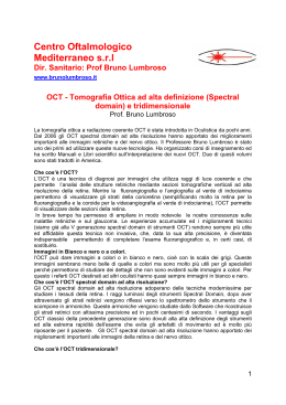
CASO CLINICO
Tomografia ottica a radiazione coerente in un
caso di Multiple Evanescent White Dot Syndrome
Optical Coherence Tomography in a case of Multiple
Evanescent White Dot Syndrome
M. Nicolò, M. Badino
Riassunto
La Multiple Evanescent White Dot Syndrome (MEWDS) è una patologia a eziologia sconosciuta che colpisce tipicamente donne tra la seconda e quinta decade di vita. L’esame oftalmoscopico mostra molteplici chiazzette color giallo
delle dimensioni di 100-200 μm situati negli strati retinici più profondi così
come una tipica sgranatura della regione foveale. La fluorangiografia (FAG) e gli
studi elettrofunzionali suggeriscono che la sede del processo patologico è nella
retina esterna e/o a livello dell’epitelio pigmentato retinico (EPR). All’angiografia
con verde indocianina (ICG), le lesioni della MEWDS appaiono ipofluorescenti
e scompaiono una volta che si è verificato il recupero clinico, suggerendo una
ipoperfusione della coroide o l’infiammazione dell’EPR come base patogenetica. Le alterazioni del campo visivo sono in correlazione con l’ICG. Recentemente alcuni autori hanno dimostrato che la risoluzione assiale di 3 μm che è
possibile con l’ultra-high resolution OCT (UHR-OCT) consente di visualizzare in
vivo la microstruttura della retina quasi come una sezione istologica. Sebbene
l’UHR-OCT offra un significativo miglioramento rispetto alla risoluzione di 10
μm dello Stratus OCT, riportiamo e correliamo il recupero dell’acuità visiva con
la stato della giunzione tra segmento esterno e interno dei fotorecettori, utilizzando lo Stratus OCT.
Dipartimento di Neuroscienze,
Oftalmologia e Genetica, Istituto di
Clinica Oculistica, Università di Genova
Summary
Multiple evanescent white dot syndrome (MEWDS) is a disease of unknown
aetiology that characteristically occurs in women in the second to fifth decades. Ophthalmoscopic examination typically reveals multiple, 100- to 200-μm
yellow-white dots deep on the retina as well as a unique foveal granularity.
Fluorescein angiography (FA) and electrophysiologic studies suggest that the
location of the disease process is in the outer retina and/or the retinal pigment
epithelium (RPE). Upon indocyanine green angiography (ICG), MEWDS lesions
are hypofluorescent and disappear with clinical recovery, suggesting pathologic
choroidal nonperfusion or RPE inflammation. Visual field alterations correlate
well with ICG findings. It has been recently reported that 3 μm axial resolution
of the ultrahigh resolution optical coherence tomography (UHR-OCT) offers the
possibility to visualize detailed retinal microstructure in vivo, with a resolution
approaching that of histology. Although UHR-OCT offers significant improvement over the 10 μm resolution available with a StratusOCT (Carl Zeiss Meditec,
Inc., Dublin, CA, USA), we report and correlate the recovery of visual acuity
with the status of the junction between inner and outer segments of the photoreceptor using StratusOCT.
Indirizzo per la corrispondenza:
Dott. M. Nicolò, Dipartimento di
Neuroscienze, Oftalmologia e Genetica,
Istituto di Clinica Oculistica, Università
di Genova Viale Benedetto XV, Genova.
E-mail: [email protected]
41
M. Nicolò, et al.
Descrizione del caso clinico
Donna di 33 anni con storia di repentino calo del
visus, scotoma paracentrale e annebbiamento visivo
nell’occhio di sinistra insorto da due giorni. La paziente è stata sottoposta a misurazione della acuità
visiva con migliore correzione, esame biomicroscopico del segmento anteriore, esame oftalmoscopico
indiretto, FA, ICG, campo visino e OCT, all’esordio e
alla fine delle manifestazioni cliniche. L’acuità visiva
era 10/10 in OD e 5/10 in OS. Il segmento anteriore era nella norma in entrambi gli occhi. L’esame
del fundus dell’occhio sinistro mostrava sgranatura
della regione maculare e molteplici lesioni giallobianche delle dimensioni di 100-200 μm situate in
profondità nella retina. Nelle fasi precoci della FA
le lesioni erano iperfluorescenti e si impregnavano
nelle fasi tardive dell’esame. La FA mostra inoltre
edema della papilla. All’ICG le lesioni apparivano
ipofluorescenti. L’esame del campo visivo mostrava
un allargamento della macchia cieca. L’esame OCT
mostrava attenuazione del segnale proveniente dalla giunzione tra segmento esterno e interno dei fotorecettori e l’impossibilità di individuare la sottile
linea che corrisponde a tale giunzione. Il segmento posteriore dell’occhio destro era normale. Due
settimane dopo l’acuità visiva nell’occhio sinistro
era 10/10. La FA e l’ICG mostravano la completa
regressione delle lesioni retiniche e la granularità
2
3
Figg. 2-3. La fluorangiografia mostra precoce
iperfluorescenza e impregnazione tardiva delle lesioni
– Fluorescein angiography showing early hyperfluorescence
and late staining of the lesions.
della macula era notevolmente regredita. L’esame
OCT mostrava chiaramente la linea di giunzione del
segmento esterno e interno dei fotorecettori.
Fig. 1. La fotografia a colori del fondo oculare mostra la
sgranatura della macula e le molteplici lesioni giallo-bianche
delle dimensioni di 100-200 μm situate in profondità
nella retina – Colour fund photograph showing macular
granularity and the yellow-white 100 to 200 μm lesions
deep on the retina.
42
Fig. 4. L’angiografia con verde indocianina mostra
ipofluorescenza della lesioni – Indocyanine green
angiography showing hypofluorescence of the lesions.
CASO CLINICO
Discussione
Nel 1984, Jampol et al. descrissero la MEWDS come
una malattia infiammatoria intraoculare idiopatica,
caratterizzata dalla formazione transitoria di lesioni
giallo-bianche, la cui localizzazione presunta era a
livello dello strato dei fotorecettori o dell’EPR 1. Le
lesioni sono meglio descritte come aree puntiformi
iperfluorescenti alla FA. Al contrario all’ICT tali lesioni appaiono ipofluorescenti e molto più numerose
rispetto alla FA. Precedenti studi hanno tentato di
localizzare e interpretare le lesioni della MEWDS sulla base dei dati oftalmoscopici e fluorangiografici.
Usando lo Stratus OCT siamo stati in grado di visualizzare la giunzione tra segmento interno ed esterno
dei fotorecettori sia prima che dopo il recupero della
malattia. L’OCT consente di visualizzare la microstrut-
tura della retina in corso di patologia. Studi basati sulla Fa e ICG suggeriscono che le anomalie dei
fotorecettori nella MEWDS siano conseguenza o di
ischemia della coroide o di infiammazione dell’EPR 2.
Tuttavia l’esame OCT evidenzia che la MEWDS è più
probabilmente associata a infiammazione dell’EPR.
Cellule dell’EPR rigonfie ma integre appaiono normali all’esame OCT. L’infiammazione dell’EPR potrebbe essere in grado di interrompere in modo acuto
l’orientamento del segmento esterno dei fotorecettori, causando l’attenuazione del segnale OCT che
corrisponde alla giunzione tra segmento esterno e
interno dei fotorecettori. Inoltre, dato che la sottile
linea di giunzione tra segmento interno ed esterno
dei fotorecettori ricompare dopo che la malattia è regredita, l’OCT può essere usato come metodo non
invasivo di follow-up in patologie di questo tipo.
Fig. 5. L’esame OCT mostra rarefazione della giunzione
tra segmento interno ed esterno dei fotorecettori – Optical
Coherence Tomography (OCT) showing the reduction of
the junction between the inner and outer segments of the
photoreceptor.
Fig. 6. L’esame OCT eseguito 2 settimane dopo mostra
la riformazione della giunzione tra segmento interno ed
esterno dei fotorecettori – Optical Coherence Tomography
(OCT) two weeks later showing the recovery of the junction
between the inner and outer segments of the photoreceptor.
Report of the clinical case
sibility to discern the thin reflection line that represents
that junction. The posterior segment of the right eye was
unremarkable. Two weeks later, BCVA in the left eye was
20/20. FA and ICG-A showed complete disappearance
of the spots, although there was still a minimal foveal
granularity. However, OCT showed a clearly discernible
reflection line between inner and outer photoreceptor
segments.
A 33-year-old woman presented with a rapid onset (2
days) history of photopsias, paracentral scotoma and
blurred vision in the left eye. Best-corrected visual acuity
exam (BCVA), complete slit lamp anterior segment examination, indirect ophtalmoscopic exam, FA, ICG-A, visual
field test and OCT were performed at the onset and at
the end of clinical manifestations. BCVA was 20/20 in
the right eye and 20/40 in the left eye. Anterior segment
exam was unremarkable in both eyes. In the left eye, fundus examination revealed foveal granularity and multiple,
yellow-white 100 to 200 μm lesions deep on the retina. FA showed typical early hyperfluorescence and late
staining of these lesions as well as disk leakage. ICG-A
showed hypofluorescence of the spots. Visual field testing showed an enlargement of blind spots. OCT imaging
demonstrated attenuation of the signal from the photoreceptors inner/outer segment junction and the impos-
Discussion
In 1984, Jampol et al. described MEWDS as an idiopathic intraocular inflammatory disorder, characterized by the development of transient small white-yellowish lesions, presumed to occur at the level of the
retinal photoreceptors or RPE 1. The lesions are best
characterized as wreathlike punctuate areas of early
hyperfluorescence on FA. In contrast, ICG-A in patients
43
M. Nicolò, et al.
with MEWDS reveals numerous hypofluorescent lesions
that significantly outnumber their counterparts seen by
clinical examination and FA. Previous studies have attempted to localize and interpret the lesions of MEWDS
on a clinical and angiographic basis. Using StratusOCT,
capable of 10 μm axial resolution, we were still able to
visualize the photoreceptor IS/OS junction of this patient
with MEWDS before and after recovery of the disease.
OCT offers a unique opportunity to visualize the retinal microstructure in the disease. FA and ICG-A reviews
suggest that photoreceptor abnormalities of MEWDS
are due to either choroidal ischaemia or RPE inflammation 2. However, OCT images suggest that MEWDS is
more likely associated with RPE inflammation. Swollen
but intact RPE cells would likely appear normal on OCT
imaging. Inflammation of RPE cells could interrupt the
orientation of photoreceptor outer segments acutely,
causing attenuation of OCT signal from the junction
between photoreceptors inner/outer segments, that becomes misaligned. Since there are no reports of other
conditions causing similar changes in the outer retina,
we believe that OCT represents a useful adjunct to diagnosis. Moreover, because the thin reflection line of junction between photoreceptors inner and outer segments
reappears at the disappearance of clinical picture, OCT
may be used as non-invasive exam during follow-up.
References
2
1
Jampol LM, Sieving PA, Pugh D, Fishman GA, Gilbert H.
Multiple evanescent white dot syndrome. I. Clinical findings. Arch Ophthalmol 1984;102:671-4.
44
Nguyen MH, Witkin AJ, Reichel E, Ko TH, Fujimoto
JG, Schuman JS, et al. Microstructural abnormalities in
MEWDS demonstrated by ultrahigh resolution optical
coherence tomography. Retina 2007;27:414-8.
Scaricare