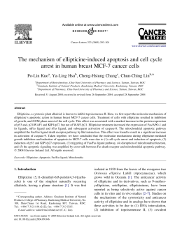
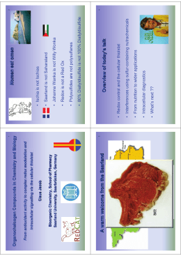
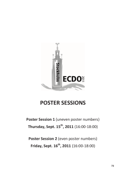
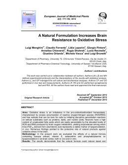
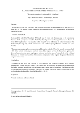
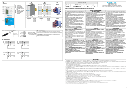
Toxicology Letters 131 (2002) 153– 159
www.elsevier.com/locate/toxlet
Altholactone, a novel styryl-lactone induces apoptosis via
oxidative stress in human HL-60 leukemia cells
Salmaan H. Inayat-Hussain a,*, Annuar Bin Osman b, Laily Bin Din b,
Naoyuki Taniguchi c
a
Department of Biomedical Science, Faculty of Allied Health Sciences, Uni6ersiti Kebangsaan Malaysia, Jalan Raja Muda Abdul
Aziz, 50300 Kuala Lumpur, Malaysia
b
Faculty of Science and Technology, Uni6ersiti Kebangsaan Malaysia, Bangi 43600, Selangor, Malaysia
c
Department of Biochemistry, Faculty of Medicine, Uni6ersity of Osaka, Suita, Japan
Received 9 October 2001; received in revised form 11 January 2002; accepted 14 January 2002
Abstract
Plant styryl-lactone derivatives isolated from Goniothalamus sp. are potential compounds for cancer chemotherapy.
In this study, we have examined the mechanisms of apoptosis induced by altholactone, a stryl-lactone isolated from
the Malaysian plant G. malayanus on human HL-60 promyelocytic leukemia cells. Flow cytometric analysis of the
externalization of phosphatidylserine (PS) using the annexin V/PI method on altholactone treated HL-60 cells showed
a concentration-dependent increase of apoptosis from concentrations ranging from 10.8 (2.5 mg/ml) to 172.4 mM (40
mg/ml). Pre-treatment with the antioxidant N-acetylcysteine (1 mM) completely abrogated apoptosis induced by
altholactone, suggesting for the involvement of oxidative stress. Further flow cytometric assessment of the level of
intracellular peroxides using the fluorescent probe 2%,7%-dichlorofluorescein diacetate (DCFH-DA) confirmed that
altholactone induced an increase in cellular oxidative stress in HL-60 cells which was suppressed by N-acetylcysteine.
In summary, our results demonstrate for the first time that altholactone induced apoptosis in HL-60 cells occurs via
oxidative stress. © 2002 Elsevier Science Ireland Ltd. All rights reserved.
Keywords: Apoptosis; Altholactone; Styryl-lactone; Oxidative stress; N-acetylcysteine
1. Introduction
Low molecular weight compounds especially
derived from natural sources such as plants are
* Corresponding author. Tel.: +60-3-40405-606; fax: + 603-26929-032.
E-mail address: [email protected] (S.H. Inayat-Hussain).
currently being investigated for their pharmaco
logical properties in regulating apoptosis, a cell
death program which is pivotal in the pathological process of tumor development (Kinloch et al.,
1999). In this respect, the styryl-pyrone derivatives
found abundantly in the genus Goniothalamus
species have also been investigated for cytotoxic
and antitumor properties (Ali et al., 1997; Cao et
al., 1998; Hawariah and Stanslas, 1998; Bermejo
et al., 1999).
0378-4274/02/$ - see front matter © 2002 Elsevier Science Ireland Ltd. All rights reserved.
PII: S 0 3 7 8 - 4 2 7 4 ( 0 2 ) 0 0 0 2 5 - 5
154
S.H. Inayat-Hussain et al. / Toxicology Letters 131 (2002) 153–159
2. Materials and methods
2.1. Reagents and cells
Fig. 1. Structure of altholactone (2-phenyl-3-hydroxy-6,7-dihydro-furano-pyrone).
Recent studies have demonstrated that goniothalamin, a plant styryl-lactone isolated from
Goniothalamus malayanus is a potential cytotoxic
compound especially by inducing apoptosis in a
variety of tumor cell lines (Ali et al., 1997;
Hawariah and Stanslas, 1998; Inayat-Hussain et
al., 1999). In addition, altholactone which is
also a styryl-lactone (Fig. 1) isolated from the
Goniothalamus sp. has been shown to possess
some cytotoxicity properties (Bermejo et al.,
1999). These earlier observations allude to the
potential of styryl-lactones to be developed as
anti-cancer agents.
A flurry of recent work has clearly demonstrated the importance of apoptosis as a mechanism of cell death in the treatment of cancer
(Jaffrezou et al., 1998; Debatin, 2000). It is currently known that chemotherapeutic agents can
induce apoptosis via multiple mechanisms including DNA topoisomerase inhibition, intercalation into DNA, cell membrane damage or
generation of reactive oxygen species (ROS)
(Barry et al., 1990; Chandra et al., 2000). Although the roles of ROS in apoptosis are controversial, many cytotoxic compounds such as
etoposide, adriamycin and methotrexate require
the involvement of ROS in signaling apoptotic
cell death (Verhaegen et al., 1995; McClain et
al., 1995; Davis et al., 2001). In this study, we
have investigated the mode of cell death induced
by altholactone and the possible role of ROS in
altholactone induced cytotoxicity in human
promyelocytic HL-60 leukemia cells. Our results
show for the first time that the mode of death
induced by altholactone is apoptosis, which occurs via oxidative stress.
The human promyelocytic HL-60 cells were
obtained from ATCC (Rockville, MD) and cultured as described previously (Inayat-Hussain et
al., 2000). Annexin V/FITC was purchased from
R&D Laboratories and 2%,7%-dichlorofluorescein
diacetate (DCFH-DA) from Molecular Probes.
Altholactone (MW. 232) was extracted from G.
malayanus as described previously (Zakaria et
al., 1998). All other reagents were from Sigma
Chemical (St. Louis, MO).
2.2. Flow cytometric assessment of apoptosis
using annexin V/PI assay
HL-60 cells were treated with altholactone
(10.8–172.4 mM) for 14 h prior to apoptosis assessment. In some experiments, the antioxidant
N-acetylcysteine (1 mM) was added 1 h prior to
addition of altholactone. The measurement of
phosphatidylserine (PS) exposure was carried
out using the annexin V assay as described previously (Inayat-Hussain et al., 2000). Briefly,
1× 10⁶ cells were collected and resuspended in 1
ml annexin V buffer containing 1.5 ml annexin V
and incubated for 8 min. Propidium iodide (2.5
mg/ml) was then added and followed by flow
cytometric analysis using FACS Calibur (Becton
Dickenson Laboratory).
2.3. Flow cytometric analysis of oxidati6e stress
using DCFH-DA
Oxidative stress was analyzed using the
fluorescent dye DCFH-DA as described previously (Kayanoki et al., 1996). For these experiments, altholactone (86.2 mM) was added to
HL-60 cells (1×10⁶) and further incubated for
3 h. In experiments employing NAC (1 mM),
this antioxidant was added 1 h prior to addition
of altholactone. This was followed by centrifuging the cells at 1000 rpm per 5 min and resuspending it in PBS. About 1 ml of DCFH-DA (5
mM in DMSO) was then added to the cell suspension and incubated in a waterbath (37 °C)
S.H. Inayat-Hussain et al. / Toxicology Letters 131 (2002) 153–159
for 15 min. Subsequently, the cells were kept on
ice and flow cytometry was immediately carried
out on the samples.
3. Results and discussion
Apoptosis has been an intensive research area,
which involves the study of compounds that trigger or inhibit this mode of death. Being an important process involved in many pathological
diseases including cancer, a number of low molecular weight compounds have been used to inhibit
or trigger this fundamental cellular process making apoptosis amenable to pharmacological intervention (Kinloch et al., 1999). In this study, we
report the potential apoptogenic activity of a styryl-lactone compound, altholactone isolated from
G. malayanus plant found abundantly in
Malaysia, on human promyelocytic HL-60
leukemia cells. Apoptosis induced by altholactone
in HL-60 cells was determined using the annexin
V/PI assay in conjunction with flow cytometry.
Annexin V is used to detect the externalization of
PS on the outer leaflet of plasma membrane during the apoptotic process (Fadok et al., 1992).
Cells were treated with a range of concentrations from 10.8 to 172.4 mM for 14 h. As shown in
the histogram (Fig. 2A), altholactone induced
apoptosis in a concentration dependent manner.
At 86.2 mM altholactone, 55.597.3% apoptosis
was observed in HL-60 cells and the highest concentration used in this study (172.4 mM) clearly
showed that this compound was able to induce
apoptosis culminating to 89.593.1% (Fig. 2B).
Although previous studies (Cao et al., 1998;
Bermejo et al., 1999) have demonstrated that altholactone is cytotoxic to a variety of human
tumor cell lines, no studies to date have elucidated
the mode of cell death induced by this compound.
It is important to note that apoptosis has always
been a preferred mode of cell death in the treatment of cancer as this death process unlike necrosis does not result in inflammatory reactions
(Fadok et al., 1992).
Recently, Peris et al. (2000) have reported that
styryl-lactone compounds such as altholactone,
3-acetylaltholactone and 5-acetoxyisogoniothala-
155
min oxide inhibit the mitochondrial respiratory
chain by studying the NADH oxidase activity of
beef heart submitochondrial particles. In addition,
inhibition of mitochondrial respiratory chain
complex I could affect the electron flow through
other complexes leading to release of cytochrome
c in an antioxidant sensitive pathway during
apoptosis (Higuchi et al., 1998). From these observations, it may be possible that styryl-lactones
disrupt the function of mitochondria, which may
lead to homeostasis imbalance leading to oxidative stress. Current models of apoptosis favor that
the loss of mitochondrial transmembrane potential occurs earlier during apoptotic death which
results in the release of cytochrome c and the
apoptotic inducing factors (Zamzami et al., 1995;
Liu et al., 1996; Kluck et al., 1997; Yang et al.,
1997). In agreement, goniothalamin, another styryl-lactone induces the loss of mitochondrial
transmembrane potential during apoptosis in HL60 cells further supporting the involvement of
mitochondria in this cell death process (data not
shown).
In order to investigate the possible role of
oxidative stress in altholactone induced apoptosis,
we pretreated the cells with the antioxidant NAC
as shown in Fig. 3. The cytogram in Fig. 3A
shows a bivariate annexin V/PI analysis of HL-60
cells. Viable cells were negative for both PI and
annexin V (lower left quadrant), apoptotic cells
were positive for annexin V and negative for PI
(early apoptosis, lower right quadrant) whereas
late apoptotic cells displayed both high annexin V
and PI labeling (upper right quadrant). Non-viable cells which underwent necrosis were positive
for PI and negative for annexin V (upper left
quadrant). Apoptosis induced by altholactone
(AL, 57.2%) was inhibited back to control levels
by pretreatment of the cells with NAC (AL+
NAC, 6.2%, Fig. 3A). It is clear from this cytogram that altholactone induced massive
apoptosis and most of the cells were already at
late apoptotic stage at 14 h. Nevertheless, pretreatment with NAC was effective in abrogating
apoptosis induced by altholactone resulting in a
cytogram with similar profile to control cells.
NAC alone did not induce any increase of apoptosis and essentially similar to control cells (Fig.
3B). These results are in agreement with previous
156
S.H. Inayat-Hussain et al. / Toxicology Letters 131 (2002) 153–159
Fig. 2. Concentration dependent increase in altholactone induced apoptosis in HL-60 cells. (A) Cells were incubated with either
alone (CON) or in the presence of various concentrations of altholactone (AL). The percentage of apoptosis was determined by
annexin V-FITC as shown in the histogram using flow cytometry. The M1 region represents normal viable cells and M2 region
represents apoptotic cells, which stained positive for annexin V. (B) Cumulative data represent the mean 9 S.E.M. of at least three
separate experiments.
studies where NAC inhibits oxidative stress and
apoptosis in HL-60 cells induced by certain compounds including actinomycin D, gallic acid and
ß-lapachone (Chau et al., 1998; Ikeda et al., 1999;
Inoue et al., 2000).
The results with NAC (Fig. 3) strongly suggest
that oxidative stress is a key feature of apoptosis
induced by altholactone. In order to confirm that
oxidative stress occurred in altholactone treated
cells, flow cytometric analysis was carried out to
S.H. Inayat-Hussain et al. / Toxicology Letters 131 (2002) 153–159
determine if DCFH-DA was oxidized to DCF, an
indicator for oxidative stress in cells (Burow and
Valet, 1987). In this experiment, the cells were
treated with altholactone for only 3 h before
addition of the dye. As shown in Fig. 4, there was
157
a marked increase in the fluorescence of DCFHDA in altholactone treated cells (AL) as compared with control cells (CON), further
confirming the presence of ROS. This marked
increase in the fluorescence of DCFH-DA in al-
Fig. 3. Flow cytommetry of N-acetylcysteine (NAC) inhibition of altholactone (AL) induced apoptosis. (A) cells treated as shown
in Fig. 2 were analyzed using annexin V/PI method. NAC (1 mM) was preincubated for 1 h prior to addition of AL (86.2 mm). The
x-axis FL1-H represents annexin V labeling while the y-axis (FL2-H) represents the PI staining. (B) The cumulative data obtained
from the cytogram which represents the means ( 9) S.E.M. of three separate experiments.
158
S.H. Inayat-Hussain et al. / Toxicology Letters 131 (2002) 153–159
ing that will initiate the apoptotic cell death execution program (Davis et al., 2001).
In summary, our results have demonstrated
that altholactone induces apoptosis in HL-60 cells
via the generation of oxidative stress. Future studies will be required to further understand the
molecular mechanisms of this potential anticancer
compound.
Acknowledgements
Fig. 4. Effects of N-acetylcysteine (NAC) on Altholactone
(AL) treated HL-60 cells as assessed by changes in DCFH
fluorescence using flow cytometry. Cells were treated either
alone (CON), with Al (86.2 mM) or Al in the presence of NAC
(AL+ NAC) for 3 h, followed by flow cytometric analysis as
described in the Section 2.
tholactone treated cells was reduced in the presence of NAC (Fig. 4, AL +NAC) resulting in a
similar pattern with the control treatment. Annexin V assay on altholactone treated cells for 3 h
showed that the treatment did not induce any
increase of apoptosis over control untreated cells
(data not shown). Therefore, any increase in
fluorescence seen in this study was not contributed by oxidative stress generated as a result
of cell death.
The redox status of a cell is influenced by the
balance between the levels of ROS and endogenous thiols such as gluthathione (Davis et al.,
2001). Recently, Chen et al. (2001) have demonstrated that NAC can reverse the depletion of
GSH which occurs during caffeic acid phenylethyl
ester induced apoptosis in HL-60 cells. In agreement, Anuradha et al. (2001) have shown that
sodium fluoride induces apoptosis in HL-60 cells
via an oxidative stress dependent pathway resulting in the loss of mitochondrial transmembrane
potential which can be blocked by the antioxidants NAC and GSH. It is tempting to speculate
that in our study, altholactone treatment may
result to a reduction of cellular GSH and subsequently leading to elevation of ROS. Such elevation as shown in Fig. 4, alters the redox status of
the cells, causing a sustained activation of signal-
Salmaan H. Inayat-Hussain is a recipient of the
International Fellowship of Dan Charitable Trust
Fund for Research in Biological Sciences and
would like to thank the Nippon Trust Bank for
this support.
References
Ali, A.M., Mackeen, M.M., Hamid, M., Aun, Q.B., Zauyah,
Y., Azimahtol, H.L., Kawazu, K., 1997. Cytotoxicity and
electron microscopy of cell death induced by goniothalamin. Planta Med. 63, 81 – 83.
Anuradha, C.D., Kanno, S., Hirano, S., 2001. Oxidative
damage to mitochondria is a preliminary step to caspase3 activation in fluoride-induced apoptosis in HL-60 cells.
Free Radic. Biol. Med. 31, 367 – 373.
Barry, M.A., Behnke, C.A., Eastman, A., 1990. Activation of
programmed cell death (apoptosis) by cisplatin, other anticancer drugs, toxins and hyperthermia. Biochem. Pharmacol. 40, 2353 – 2362.
Bermejo, A., Leonce, S., Cabedo, N., Andreu, I., Caignard,
D.H., Atassi, G., Cortes, D., 1999. Semisynthesis and
cytotoxicity of styryl-lactone derivatives. J. Nat. Prod. 62,
1106 – 1109.
Burow, S., Valet, G., 1987. Flow-cytometric characterization
of stimulation, free radical formation, peroxidase activity
and phagocytosis of human granulocytes with 2,7-dichlorofluorescein (DCF). Eur. J. Cell. Biol. 43, 128 – 133.
Cao, S.-G., Wu, X.-H., Sim, K.-Y., Tan, B.K.H., Pereira,
J.T., Goh, S.-H., 1998. Styryl-lactone derivatives and alkaloids from Goniothalamus borneensis (Annonaceae). Tetrahedron 54, 2143 – 2148.
Chandra, J., Samali, A., Orrenius, S., 2000. Triggering and
modulation of apoptosis by oxidative stress. Free Radic.
Biol. Med. 9 (3 – 4), 323 – 333.
Chau, Y.P., Shiah, S.G., Don, M.J., Kuo, M.L., 1998. Involvement of hydrogen peroxide in topoisomerase inhibitor
beta-lapachone-induced
apoptosis
and
differentiation in human leukemia cells. Free Radic. Biol.
Med. 24, 660 – 670.
S.H. Inayat-Hussain et al. / Toxicology Letters 131 (2002) 153–159
Chen, Y.J., Shiao, M.S., Wang, S.Y., 2001. The antioxidant
caffeic acid phenethyl ester induces apoptosis associated
with selective scavenging of hydrogen peroxide in human
leukemic HL-60 cells. Anticancer Drugs 12, 143 –149.
Davis, W. Jr, Ronai, Z., Tew, K.D., 2001. Cellular thiols and
reactive oxygen species in drug-induced apoptosis. J. Pharmacol. Exp. Ther. 296, 1 –6.
Debatin, K., 2000. Activation of apoptosis pathways by anticancer treatment. Toxicol. Lett. 112 –113, 41 – 48.
Fadok, V.A., Voelker, D.R., Campbell, P.A., Cohen, J.J.,
Bratton, D.L., Henson, P.M., 1992. Exposure of phosphatidylserine on the surface of apoptotic lymphocytes
triggers specific recognition and removal by macrophages.
J. Immunol. 148, 2207 –2216.
Hawariah, A., Stanslas, J., 1998. Antagonistic effects of
styrylpyrone derivative (SPD) on 7,12-dimethylbenzanthracene-induced rat mammary tumors. In Vivo 12, 403 –
410.
Higuchi, M., Proske, R.J., Yeh, E.T., 1998. Inhibition of
mitochondrial respiratory chain complex I by TNF results
in cytochrome c release, membrane permeability transition,
and apoptosis. Oncogene 17, 2515 –2524.
Ikeda, K., Kajiwara, K., Tanabe, E., Tokumaru, S., Kishida,
E., Masuzawa, Y., Kojo, S., 1999. Involvement of hydrogen peroxide and hydroxyl radical in chemically induced
apoptosis of HL-60 cells. Biochem. Pharmacol. 57, 1361 –
1365.
Inayat-Hussain, S.H., McGuinness, S.M., Johansson, R.,
Lundstrom, J., Ross, D., 2000. Caspase-dependent and
-independent mechanisms in apoptosis induced by hydroquinone and catechol metabolites of remoxipride in
HL-60 cells. Chem. Biol. Interact. 128, 51 –63.
Inayat-Hussain, S.H., Osman, A.B., Din, L.B., Ali, A.M.,
Snowden, R.T., MacFarlane, M., Cain, K., 1999. Caspases-3 and -7 are activated in goniothalamin-induced
apoptosis in human Jurkat T-cells. FEBS Lett. 456, 379 –
383.
Inoue, M., Sakaguchi, N., Isuzugawa, K., Tani, H., Ogihara,
Y., 2000. Role of reactive oxygen species in gallic acid-induced apoptosis. Biol. Pharm. Bull. 23, 1153 –1157.
159
Jaffrezou, J.P., Bettaieb, A., Levade, T., Laurent, G., 1998.
Antitumor agent-induced apoptosis in myeloid leukemia
cells: a controlled suicide. Leuk. Lymphoma 29, 453 – 463.
Kayanoki, Y., Fujii, J., Islam, K.N., Suzuki, K., Kawata, S.,
Matsuzawa, Y., Taniguchi, N., 1996. The protective role of
glutathione peroxidase in apoptosis induced by reactive
oxygen species. J. Biochem. (Tokyo) 119 (4), 817 – 822.
Kinloch, R.M., Treherne, J.M., Furness, L.M., Hajimohamadreza, I., 1999. The pharmacology of apoptosis.
Trends Pharm. Sci. 20, 35 – 42.
Kluck, R.M., Bossy-Wetzel, E., Green, D.R., Newmeyer,
D.D., 1997. The release of cytochrome c from mitochondria: a primary site for Bcl-2 regulation of apoptosis.
Science 275, 1132 – 1136.
Liu, X., Kim, C.N., Yang, J., Jemmerson, R., Wang, X., 1996.
Induction of apoptotic program in cell-free extracts: requirement for dATP and cytochrome c. Cell 12, 147 – 157.
McClain, D.E., Kalinich, J.F., Ramakrishnan, N., 1995.
Trolox inhibits apoptosis in irradiated MOLT-4
lymphocytes. FASEB J. 9, 1345 – 1354.
Peris, E., Estornell, E., Cabedo, N., Cortes, D., Bermejo, A.,
2000. 3-Acetylaltholactone and related styryl-lactones, mitochondrial respiratory chain inhibitors. Phytochemistry
54, 311 – 315.
Verhaegen, S., McGowan, A.J., Brophy, A.R., Fernandes,
R.S., Cotter, T.G., 1995. Inhibition of apoptosis by antioxidants in the human HL-60 leukemia cell line. Biochem.
Pharmacol. 50, 1021 – 1029.
Yang, J., Liu, X., Bhalla, K., Kim, C.N., Ibrado, A.M., Cai,
J., Peng, T.I., Jones, D.P., Wang, X., 1997. Prevention of
apoptosis by Bcl-2: release of cytochrome c from mitochondria blocked. Science 275, 1129 – 1132.
Zakaria, Z., Din, L.B., Said, I.M., Salleh, K.M., Latiff, A.,
1998. Components from Goniothalamus fasciculatus and G.
maiayanus. Sains Malaysiana 27, 135 – 138.
Zamzami, N., Marchetti, P., Castedo, M., Decaudin, D.,
Macho, A., Hirsch, T., Susin, S., Petit, P.X., Mignotte, B.,
Kroemer, G., 1995. Sequential reduction of mitochondrial
transmembrane potential and generation of reactive oxygen species in early programmed cell death. J. Exp. Med.
182, 367 – 377.
Scarica