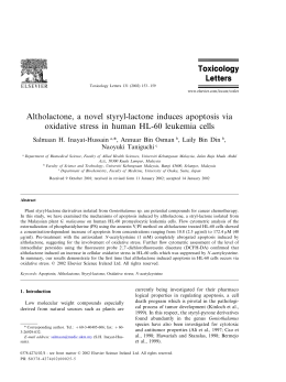
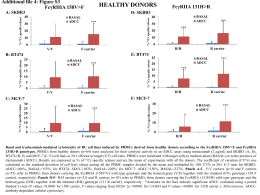
Cancer Letters 223 (2005) 293–301
www.elsevier.com/locate/canlet
The mechanism of ellipticine-induced apoptosis and cell cycle
arrest in human breast MCF-7 cancer cells
Po-Lin Kuoa, Ya-Ling Hsub, Cheng-Hsiung Changc, Chun-Ching Linb,*
a
Department of Biotechnology, Chia-Nan University of Pharmacy and Science, Tainan, Taiwan, ROC
b
Graduate Institute of Natural Products, Kaohsiung Medical University, Kaohsiung, Taiwan, ROC
c
Department of Pharmacy, Chia-Nan University of Pharmacy and Science, Tainan, Taiwan, ROC
Received 31 August 2004; received in revised form 26 September 2004; accepted 28 September 2004
Abstract
Ellipticine, a cytotoxic plant alkaloid, is known to inhibit topoisomerase II. Here, we first report the molecular mechanism of
ellipticine’s apoptotic action in human breast MCF-7 cancer cells. Treatment of cells with ellipticine resulted in inhibition
of growth, and G2/M phase arrest of the cell cycle. This effect was associated with a marked increase in the protein expression
of p53 and, p21/WAF1 and KIP1/p27, but not of WAF1/p21. Ellipticine treatment increased the expression of Fas/APO-1 and
its ligands, mFas ligand and sFas ligand, and subsequent activation of caspase-8. The mitochondrial apoptotic pathway
amplified the Fas/Fas ligand death receptor pathway by Bid interaction. This effect was found to result in a significant increase
in activation of caspase-9. Taken together, we have concluded that the molecular mechanisms during ellipticine-mediated
growth inhibition and induction of apoptosis in MCF-7 cells were due to (1) cell cycle arrest and induction of apoptosis, (2)
induction of p53 and KIP1/p27 expression, (3) triggering of Fas/Fas ligand pathway, (4) disruption of mitochondrial function,
and (5) the apoptotic signaling was amplified by cross-talk between Fas death receptor and mitochondrial apoptotic pathway.
q 2004 Elsevier Ireland Ltd. All rights reserved.
Keywords: Ellipticine; Apoptosis; Fas/Fas ligand; Mitochondria
1. Introduction
Ellipticine (5,11-dimethyl-6H-pyrido[4,3-b]carbazole) is one of the simplest naturally occurring
alkaloids, having a planar structure [1]. It was first
* Corresponding author. Address: Graduate Institute of Natural
Products, College of Pharmacy, Kaohsiung Medical University, No.
100, Shin-Chuan 1st Road, Kaohsiung 807, Taiwan, ROC.
Tel.: C886 7 312 1101x2122; fax: C886 7 313 5215.
E-mail address: [email protected] (C.-C. Lin).
isolated in 1959 from the leaves of the evergreen tree
Ochrosia elliptica Labill (Apocynaceae), which
grows wild in Oceania [1]. The anticancer activity
of ellipticine and its derivatives, such as 9-methoxyellipticine, retelliptine, ellipticiniums, have been
reported as being selectively active against cancer
cells in in vitro and in vivo studies [2–7]. Studies on
the mechanisms of the cytotoxicity and anticancer
activity of ellipticine and its analogs have shown that
these activities to be due to (1) DNA intercalation,
(2) inhibition of topoisomerase II, (3) covalent
0304-3835/$ - see front matter q 2004 Elsevier Ireland Ltd. All rights reserved.
doi:10.1016/j.canlet.2004.09.046
294
P.-L. Kuo et al. / Cancer Letters 223 (2005) 293–301
alkylation of macromolecules, and (4) induction of
endoplasmic reticulum stress [2–9].
Apoptosis signaling converges at the activation of
initiator caspases (i.e. caspase-8 and caspase-9),
which leads to the proteolytic activation of effectors
caspase (i.e. caspase-3) that then cleaves the cellular
substrate, resulting in cell death [10,11]. The
death receptor pathway is triggered by members of
the death receptor superfamily, such as Fas/APO-1.
Ligation of Fas/APO-1 by agonistic antibody or its
mature ligand (Fas ligand) induces receptor oligomerization and formation of death-inducing signaling
complex (DISC), followed by activation of caspase-8.
The mitochondrial apoptotic pathway is controlled by
Bcl-2 family protein, including the proapoptotic Bax
and antiapoptotic Bcl-2 and Bcl-XL [12,13]. Death
stimuli induce the release of cytochrome c, procaspase-9 and other proapoptotic factors from the
mitochondria into the cytoplasm, thereby activating
downstream effector caspases such as caspase-3.
Cross-talk between death receptor and mitochondrial
pathway is provided by Bid (a member of Bcl-2
family). Caspase-8-mediated cleavage of Bid greatly
increases its pro-death activity and results in its
translocation to the mitochondria, where it promotes
cytochrome c exit [14].
In this study, to establish the anticancer mechanism
of ellipticine in MCF-7 cells, we assayed the death
receptor and mitochondrial apoptotic pathway-related
molecules, including Fas/APO-1, Fas ligand, caspase8, cytochrome c, caspase-9, and Bcl-2 family proteins,
which are strongly associated with the signal
transduction pathway of apoptosis and affect the
chemosensitivity of tumor cells to anticancer agents.
Nucleosome ELISA, WAF1 ELISA, Fas Ligand,
Fas/APO-1 ELISA, and caspase-8, caspase-9 activity
assay kits, caspase-8 inhibitor (Z-IETD-FMK) and
caspase-9 inhibitor (LEDH-CHO) were purchased
from Calbiochem (Cambridge, MA). The antibodies
to KIP/p27, Bax, Bcl-2, and Bcl-XL were obtained
from Santa Cruz Biotechnology (Santa Cruz, CA).
The antibodies to Bid and cytochrome c were obtained
from Cell Signaling Technology (Beverly, MA).
2.2. Cell line and culture
MCF-7 (American Type Culture Collection [ATCC]
HB8065) was maintained in a monolayer culture at 37 8C
and 5% CO2 in DMEM supplemented with 10% FCS,
10 U/mL of penicillin, 10 mg/mL of streptomycin, and
0.25 mg/mL of amphotericin B and 5 mg/mL of insulin.
2.3. Cell proliferation assay
Inhibition of cell proliferation by ellipticine was
measured by XTT (sodium 3 0 -[1-(phenylamino-carbonyl)-3,4-tetrazolium]-bis(4-methoxy-6-nitro)benzene-sulfonic acid hydrate) assay. Briefly, cells were
plated in 96-well culture plates (1!104 cells/well).
After 24 h incubation, the cells were treated with
ellipticine (0.5, 1, 2, and 3 mM) for 48 h. Fifty
microliters of XTT test solution, which was prepared
by mixing 5 mL of XTT-labeling reagent with 100 mL
of electron coupling reagent, was then added to each
well. After 4 h incubation, the absorbance was
measured on an ELISA reader (Multiskan EX,
Labsystems) at a test wavelength of 492 nm and a
reference wavelength of 690 nm.
2.4. Cell cycle analysis
2. Materials and methods
2.1. Materials
Fetal calf serum (FCS), penicillin G, streptomycin,
amphotericin B, Dulbecco’s modified Eagle’s medium (DMEM) and insulin were obtained from GIBCO
BRL (Gaithersburg, MD). Ellipticine, dimethyl sulfoxide (DMSO), ribonuclease (RNase), and propidium
iodide (PI) were purchased from Sigma Chemical Co.
(St Louis, MO). XTT and p53 pan ELISA kits were
obtained from Roche Diagnostics GmbH (Germany).
To determine cell cycle distribution analysis,
5!105 cells were plated in 60-mm dishes and treated
with ellipticine (0, 1.5, and 3 mM) for 6 h. After
treatment, the cells were collected by trypsinization,
fixed in 70% ethanol, washed in PBS, resuspended in
1 mL of PBS containing 1 mg/mL RNase and
50 mg/mL propidium iodide, incubated in the dark
for 30 min at room temperature, and analyzed by
EPICS flow cytometer. The data were analyzed using
the Multicycle software (Phoenix Flow Systems, San
Diego, CA).
P.-L. Kuo et al. / Cancer Letters 223 (2005) 293–301
2.5. Apoptosis assay
Cells (1!106) were treated with vehicle alone
(0.1% DMSO) and various concentrations of ellipticine for 48 h, and then collected by centrifugation.
Pellets were lysed by DNA lysis buffer (10 mM Tris,
pH 7.5, 400 mM EDTA, and 1% Triton X-100) and
then centrifuged. The supernatant obtained was
incubated overnight with proteinase K (0.1 mg/mL)
and then with RNase (0.2 mg/mL) for 2 h at 37 8C.
After extraction with phenol–chloroform (1:1), the
DNA was separated in 2% agarose gel and visualized
by UV after staining with ethidium bromide.
Quantitative assessment of oligonucleosomal DNA
fragmentation was assayed using the Nucleosome
ELISA kit. Briefly, the cells were treated with vehicle
alone (0.1% DMSO) and ellipticine (1.5 and 3 mM)
for the indicated times. The induction of apoptosis
was evaluated by assessing the enrichment of
nucleosome in cytoplasm, and determined exactly as
described in the manufacturer’s protocol.
2.6. Assaying the levels of p53, p21/WAF1, Fas ligand
(mFasL and sFasL) and Fas/APO-1
p53 pan ELISA, WAF1 ELISA, Fas Ligand and
Fas/APO-1 ELISA kits were used for the detection of
p53, p21/WAF1, Fas ligand (mFasL and sFasL) and
Fas/APO-1 receptor. Briefly, cells were treated with
vehicle alone (0.1% DMSO) or ellipticine (1.5 and
3 mM) for the indicated times. Samples of cell lysate
were placed in 96 well (1!106 per well) microtiter
plates that were coated with monoclonal detective
antibodies, and incubated for 1 h (Fas/APO-1), 2 h
(p53 or p21/WAF1) or 3 h (FasL) at room temperature. The soluble Fas ligand in cell culture supernatant
also needed to be determined by using a Fas Ligand
ELISA kit. Upon removing unbound material by
washing with buffer, horseradish peroxidase conjugated streptavidin was added to bind to the antibodies.
Horseradish peroxidase catalyzed the conversion of a
chromogenic substrate (tetramethylbenzidine) to a
colored solution with a color intensity proportional to
the amount of protein present in the sample. The
absorbance of each well was measured at 450 nm,
and concentrations of p53, p21/WAF1, FasL and
Fas/APO-1 were determined by interpolating from
295
standard curves obtained with known concentrations
of standard proteins [15,16].
2.7. Assay for caspase activity
The assay is based on the ability of the active
enzyme to cleave the chromophore from the enzyme
substrate, Ac-IETD-pNA (for caspase-8), and LEHDpNA (for caspase-9). The cell lysates were incubated
with peptide substrate in assay buffer (100 mM NaCl,
50 mM HEPES, 10 mM dithiothreitol, 1 mM EDTA,
10% glycerol, 0.1% CHAPS, pH 7.4) for 2 h at 37 8C.
The release of p-nitroaniline was monitored at
405 nm. Results are represented as the percent change
of the activity compared to the untreated control.
2.8. Western blotting
Cells (8!106/dish) were seeded in a 10 cm dish.
After 24 h of incubation, the cells were treated with
3 mM ellipticine for the indicated times. Mitochondrial and cytoplasmic fractions were separated using
Cytochrome c Releasing Apoptosis Assay Kit (BioVision, California, USA). Total cell extracts were
prepared in lysis buffer (50 mM Tris–HCl, 150 mM
NaCl, 1 mM EGTA, 1 mM EDTA, 20 mM NaF,
100 mM Na3VO4, 0.5% NP-40, 1% Triton X-100,
1 mM PMSF, 5 mg/mL Aprotinin, 5 mg/mL Leupetin).
Equivalent amounts of protein were resolved by
SDS-PAGE (10–12%) and transferred to PVDF
membranes. After the membrane was blocked in
Tris–buffer saline (TBST) containing 0.05% Tween
20 and 5% nonfat powdered milk, the membranes
were incubated with primary antibodies specific to
Bax, Bid (full and cleaved length), Bcl-2, Bcl-XL, and
cytochrome c at 4 8C for 16 h. After washing three
times with TBST for 10 min each, the membrane was
incubated with horseradish peroxidase-labeled secondary antibody for 1 h. The membranes were washed
again, and detection was performed using the
enhanced chemiluminescence blotting detection system (Amersham, USA).
2.9. Statistical analysis
Data were expressed as meansGSD. Statistical
comparisons of the results were made using analysis
of variance (ANOVA). Significant differences
296
P.-L. Kuo et al. / Cancer Letters 223 (2005) 293–301
Fig. 1. Effect of ellipticine on inhibiting the growth of MCF-7 cells.
Cells were seeded into 96-well plates (104 cells/well) and allowed to
adhere overnight. The next day, the cells were incubated with
vehicle (0.1% DMSO) and different concentrations of ellipticine for
48 h. Cell proliferation was determined by XTT assay. Results are
expressed as the percentage of cell proliferation relative to the
proliferation of control. Each value is the meanGSD of three
determinations.
(P!0.05) between the means of control and ellipticine-treated cells were analyzed by Dunnett’s test.
3. Results
3.1. Effect of ellipticine on MCF-7 cell
growth inhibition
We first determined the effect of ellipticine on
growth of MCF-7 cells cultured for 48 h, either in the
presence or absence of ellipticine, using XTT assay.
As shown in Fig. 1, the growth of these MCF-7 cell
lines was inhibited by ellipticine in a dose-dependent
manner. The IC50 value was 1.52 mM.
the treatment. The treatment caused an arrest of
30.3% cells in the G2/M phase of the cell cycle at
1.5 mM concentration that further 39.1% at 3 mM in
MCF-7 cells (Fig. 2).
We next assessed the effect of ellipticine on the
induction of apoptosis in MCF-7 cells by DNA
fragmentation assay. The results showed that ellipticine treatment results in the formation of DNA
fragments in MCF-7 cells, as assessed by agarose
gel electrophoresis at 48 h (Fig. 3A). Additionally, a
quantitative evaluation was then sought using an
ELISA to detect histone-associated oligonucleosome
DNA fragments. Compared with vehicle-treated cells,
1.5 mM ellipticine induced 3.5 and 5.29-fold of
oligonucleosome in MCF cells at 24 and 48 h. This
effect was more pronounced with 3 mM ellipticine
treatment (Fig. 3B).
3.3. Ellipticine increases the expression of p53 and
KIP1/p27, but not WAF1/p21 proteins in MCF-7 cells
To determine whether the cell growth inhibition
effects of ellipticine are mediated through changes in
several key cell cycle regulatory factors, we examined
the effects of treatment on the cell cycle regulatory
proteins p53, WAF1/p21, and KIP1/p27 in MCF-7
cells (Fig. 4A–C). Treatment with ellipticine resulted
in an increase of p53 and KIP1/p27 that was observed
at 3 h and persisted until 24 h. However, treatment of
MCF-7 cells with 1.5 or 3 mM ellipticine failed
to show any significant effects on the levels of
WAF1/p21 proteins at any examined times point.
These data suggest that one potential mechanism of
action of the observed anticancer effects of ellipticine
3.2. Ellipticine-induced cell cycle arrest
and apoptosis in MCF-7 cells
To examine the mechanism responsible for ellipticine-mediated cell growth inhibition, cell cycle
distribution was evaluated using flow cytometric
analysis. Compared with the vehicle-treated control,
ellipticine treatment resulted in an appreciable arrest
of cells in G2/M phase of cell cycle after 6 h of
Fig. 2. Effect of ellipticine on cell cycle distribution and apoptosis
induction in MCF-7 cells. Cells were treated with vehicle and
ellipticine for 6 h, and cell cycle distribution was assessed by flow
cytometry. The asterisk indicates a significant difference between
control and ellipticine-treated cells, *P!0.05.
P.-L. Kuo et al. / Cancer Letters 223 (2005) 293–301
297
of exposure to the drugs. Compared to the control,
3 mM ellipticine increased 2.9, 4.2, and 3.1-fold of
Fas/APO-1 at 6, 12, and 24 h, respectively. Results
on Fas ligand assay indicate that FasL, mFasL, and
Fig. 3. The induction and quantification of apoptosis in ellipticinetreated MCF-7 cells. The DNA fragmentation was assessed by gel
electrophoresis (A) and quantitated by Nucleosome assay kit. For
(A), cells were treated with vehicle and ellipticine for 48 h, and then
the fragmentation of DNA was assessed by agarose gel electrophoresis. For (B), the cytoplasmic oligonucleosome of ellipticine
treated cells at the indicated times was estimated by Nucleosome
ELISA kit. Each value is the meanGSD of three determinations.
The asterisk indicates a significant difference between control and
ellipticine-treated cells, *P!0.05.
may be through the up-regulation of p53 and
the cell cycle regulatory proteins KIP1/p27, but not
WAF/p21.
3.4. Fas/FasL apoptotic system might be a possible
pathway of ellipticine-mediated apoptosis
Fig. 5A shows that treatment of MCF-7 cells
with ellipticine increased Fas/APO-1 protein levels;
maximum enhancement was observed within 12 h
Fig. 4. Effect of ellipticine on the expression of p53, WAF1/p21 and
KIP1/p27 of MCF-7 cells. (A) The level of p53. (B) The amount of
WAF1/p21. (C) The level of KIP1/p27 in MCF-7 cells. Cells were
treated with vehicle (0.1% DMSO), 1.5, and 3 mM ellipticine for the
indicated times. The levels of p53 and WAF1/p21 protein were
measured by p53 pan and WAF1 ELISA kit. Each value is the
meanGSD of three determinations. The asterisk indicates a
significant difference between control and ellipticine-treated cells,
*P!0.05. The KIP1/p27 expression level of 3 mM ellipticinetreated MCF-7 cells was determined by Western blotting.
298
P.-L. Kuo et al. / Cancer Letters 223 (2005) 293–301
Fig. 5. The Fas/FasL system was involved in ellipticine-mediated apotopsis. (A) The amount of Fas/APO-1 receptor. (B) The amount of mFasL.
(C) The amount of sFasL. (D) The activation of caspase-8 in MCF-7 cells. (E) The effect of caspase-8 inhibitor on ellipticine-mediated
antiproliferation. (F) The effect of caspase-8 inhibitor on ellipticine-induced apoptosis. MCF-7 cells were incubated with ellipticine for the
indicated times. The amounts of Fas/APO-1 and Fas ligand were determined by Fas/APO-1 and Fas ligand ELISA kit. For blocking
experiments, cells were preincubated with Z-IETD-FMK (10 mM) for 1 h before the addition of 3 mM ellipticine. After 48 h of treatment, cell
viability and induction of apoptosis was measured by XTT and Nucleosome ELISA kit. Each value is the meanGSD of three determinations.
The asterisk indicates a significant difference between control and ellipticine-treated cells, *P!0.05.
sFasL increased in a dose-dependent manner (Fig.
5B and C). The accumulation of mFasL was
observed at 6 h after ellipticine treatment, and
increased progressively for up to 12 h (Fig. 5B). A
similar result was observed for sFasL (Fig. 5C).
However, the amount of mFasL by ellipticine was
more than sFasL at all time points.
We next measured the initiator caspase-8 of the
Fas/FasL apoptotic system. The results showed that
caspase-8 activity increased at 6 h, and reached
maximum induction at 12 h in ellipticine treated
MCF-7 cells (Fig. 5D). Furthermore, our results
showed that the ellipticine’s cell growth inhibition
and apoptosis induction decreased significantly in
P.-L. Kuo et al. / Cancer Letters 223 (2005) 293–301
the presence of caspase-8 inhibitor (Z-IETD-FMK)
(Fig. 5E and F).
3.5. Mitochondrial apoptotic pathway is involved
in ellipticine-mediated apoptosis
299
near that of the untreated control cells (Fig. 7).
However, pre-treatment with the specific caspase-9
inhibitor LEHD-CHO did not prevent the disappearance of native Bid protein (Fig. 7). These results
To investigate the mitochondrial apoptotic events
involved in ellipticine-induced apoptosis, we first
analyzed the changes in the levels of pro-apoptotic
protein Bax and anti-apoptotic proteins Bcl-2 and BclXL. Western blot analysis showed that treatment of
MCF-7 cells with ellipticine increased Bax protein
levels (Fig. 6A). In contrast, ellipticine markedly
decreased Bcl-2 levels, which led to an increase in the
Bax/Bcl-2 ratio (Fig. 5A). In addition, ellipticine
also decreased the expression of Bcl-XL. These effects
of ellipticine on Bcl-2 family proteins led to the
release of mitochondrial cytochrome c content into
the cytosol (Fig. 6A). Next, we investigated
the implication of initiator caspases and effector
caspases in ellipticine-induced apoptosis. Biochemical analyses showed that treatment with ellipticine
increased caspase-9 activity in MCF-7 cells, consistent with the release of cytochrome c into the cytosol
(Fig. 6B).
3.6. The activation of the mitochondrial pathway
was through caspase-8-mediated Bid cleavage
Since ellipticine-mediated apoptosis involved
initiation of Fas/Fas ligand death receptor and
mitochondrial signaling, it is possible that ellipticine
activates the mitochondrial apoptotic pathway
through caspase-8-mediated Bid cleavage, which
then results in cytochrome c release and caspase-9
activation. To test this idea, we checked the status of
Bid protein during ellipticine-induced apoptosis.
Fig. 5D shows that full size Bid (22 kDa) protein
was cleaved to yield a 15 kDa fragment after
treatment of cells with ellipticine, and this closely
matched the appearance of caspase-8 activation. To
assess whether proteolytic cleavage by caspase-8 is
really responsible for the truncation of Bid, we
investigated the effects of various caspase inhibitors
on Bid cleavage. When cells were pre-treated with the
specific caspase-8 inhibitor Z-IETD-fmk before
ellipticine treatment, the decreased level of Bid
protein recovered in ellipticine-treated cells was
Fig. 6. Ellipticine-induced apoptosis through the initiation of the
mitochondrial pathway. (A) The release of cytochrome c and the
expression level of Bcl-2 family proteins in ellipticine treated MCF7 cells. (B) The activation of caspase-9 in ellipticine treated MCF-7
cells. (C) The cleavage of Bid in ellipticine treated MCF-7 cells. For
(A), cells were treated with 3 mM ellipticine for the indicated times.
Cytoplasm and mitochondria were separated from the cell pellets by
lysis buffer and centrifugation. Western blotting analysis assessed
protein expressions. For (B), the activity of caspase-9 was assessed
by a caspase-9 activity assay kit. Each value is the meanGSD of
three determinations. The cleavage of Bid was assessed by Western
blotting analysis.
300
P.-L. Kuo et al. / Cancer Letters 223 (2005) 293–301
Fig. 7. The inhibitory effects of caspase-8 and caspase-9 on
ellipticine-mediated cleavage of Bid. For blocking experiments,
cells were preincubated with Z-IETD-FMK (10 mM) or LEHDCHO (20 mM) for 1 h before the addition of 3 mM ellipticine for
another 12 h. The cleavage of Bid was assessed by Western blotting
analysis.
indicate that Bid protein is cleaved by caspase-8,
which is activated by ellipticine treatment, and that
the activation of caspase-9 may be a downstream
event of this caspase-8-mediated Bid cleavage.
4. Discussion
Ellipticine is a potent anti-neoplastic agent whose
mechanism of action is considered to be based mainly
on DNA intercalation and/or inhibition of topoisomerase II [2–9]. However, the molecular mechanism for
its apoptotic effect as an anticancer agent has not yet
been clarified. Our results demonstrate that ellipticine
inhibits the growth of human breast MCF-7 cancer
cells. Treatment of MCF-7 cells with ellipticine
accumulated at the G2/M phase of cell cycle and
underwent apoptosis. We found that ellipticine
increased the expression of p53 and KIP1/p27, but
did not affect the expression of WAF1/p21,
suggesting that up-regulated of p53 and KIP1/p27
might contribute to ellipticine’s blockade effect on
cell cycle progression at the G2/M phase.
Numerous studies have reported that the involvement of p53 in ellipticine-mediated cytotoxic effect
[17–19]. Ellipticine has been found to restore the
transcription function of mutant p53 (175 H, 248W,
249S, 273 H, 281G, 194F, 233L, 241F, and 273C
mutants), and this property may contribute to resisting
tumor cell lines expressing mutant p53 [17]. 9-Hydroxyellipticine (9-HE) treatment caused induction of
apoptosis in the G1 phase of the cell cycle in mutant
p53 (p53ala143, p53his175, orp53his273) transfected
Saos-2 cells, but not in p53-deficient parental Saos-2
cells. Similar induction of apoptosis was observed
24–48 h after treatment with 9-HE in mutant p53-containing SW480, SK-BR-3 and MKN-1, but not in
p53-deficient KATO cells [18]. Ellipticine and 9-HE
caused selective inhibition of p53 protein phosphorylation in Lewis lung carcinoma and SW480 (human
colon cancer cell line) [19]. In our study, we found
that the expression of p53 increased in ellipticine
treated MCF-7 cells. Two downstream molecules of
p53, Fas/APO-1 and Bax, were accumulated after p53
increased by treatment with ellipticine. However, the
expression of WAF1/p21 known to be regulated by
p53 dose not increase in ellipticine-treated MCF-7
cells. Therefore, the actual role of p53 in ellipticinemediated apoptosis in MCF-7 cells needs additional
investigation in further studies.
In our ongoing efforts to determine the apoptotic
mechanism of ellipticine against MCF-7 cells, we
studied the involvement of Fas/Fas ligand death
receptor and the mitochondrial pathway. Our study
indicates that the Fas ligands mFasL and sFasL
increase in ellipticine-treated MCF-7 cells. Fas/APO1 levels and caspase-8 activity are simultaneously
enhanced in FasL-upexpressing MCF-7 cells. Furthermore, cell growth inhibition and apoptotic induction of ellipticine decreases in MCF-7 cells treated
with caspase-8 inhibitor. On the other hand, we found
that ellipticine increased the expression of Bax and
decreased the expression of Bcl-2 and Bcl-XL in
MCF-7 cells, and subsequently induced the release of
cytochrome c from the mitochondria into the
cytoplasm, then activated caspase-9. Furthermore,
we have also demonstrated that cross-talk between
Fas/Fas ligand and the mitochondrial pathway is
provided by Bid in ellipticine-treated MCF-7 cells.
The cleavage of Bid by caspase-8 supports the
hypothesis that the proteolysis event of Bid is
suppressed by caspase-8 inhibitor pre-treatment, but
not in caspase-9 inhibitor pre-treated MCF-7 cells.
In summary, the overall goal of our studies is to
characterize the signaling pathways producing ellipticine-mediated cell growth inhibition, cell cycle arrest
and apoptosis. The members of the ellipticine family
might be adopted clinically as useful anticancer drugs
if their mechanisms of action and related activities in
tumors with particular molecular characteristics are
better understood. Our studies prove that induction of
p53, Fas/Fas ligand death receptor, and mitochondrial
P.-L. Kuo et al. / Cancer Letters 223 (2005) 293–301
proapoptotic pathways are involved in ellipticinemediated cell growth inhibition in MCF-7 cells.
[9]
References
[10]
[1] S. Goodwin, A.F. Smith, E.C. Horning, Alkaloids of
Ochrosia elliptica Labill, J Am. Chem. Soc. 81 (1959)
1903–1908.
[2] Z. Djuric, C.K. Everett, F.A. Valeriote, DNA damage
cytotoxicity in L1210 cells by ellipticine and a structural
analogue,
N-2-(diethylaminoethyl)-9-hydroxyellipticinium
chloride, Cancer Res. 52 (1992) 1515–1519.
[3] M. Monnot, O. Mauffret, V. Simon, E. Lescot, B. Psaume,
J.M. Saucier, et al., DNA-drug recognition and effects on
topoisomerase II-mediated cytotoxicity. A three-mode binding
model for ellipticine derivatives, J. Biol. Chem. 266 (1991)
1820–1829.
[4] L.M. Shi, T.G. Myers, Y. Fan, P.M. O’Connor, K.D. Paull,
S.H. Friend, J.N. Weinstein, Mining the National Cancer
Institute Anticancer Drug Discovery Database: cluster analysis of ellipticine analogs with p53-inverse and central
nervous system-selective patterns of activity, Mol. Pharmacol.
53 (1998) 241–251.
[5] R. Devraj, J.F. Barrett, J.A. Fernandez, J.A. Katzenellenbogen,
M. Cushman, Design, synthesis, and biological evaluation of
ellipticine-estradiol conjugates, J. Med. Chem. 39 (1996)
3367–3374.
[6] S. Kenney, D.T. Vistica, H. Linden, M.R. Boyd, Uptake and
cytotoxicity of 9-methoxy-N2-methylellipticinium acetate in
human brain and non-brain tumor cell lines, Biochem.
Pharmacol. 49 (1995) 23–32.
[7] W.K. Anderson, A. Gopalsamy, P.S. Reddy, Design, synthesis, and study of 9-substituted ellipticine and 2-methylellipticinium analogues as potential CNS-selective antitumor
agents, J. Med. Chem. 37 (1994) 1955–1963.
[8] J. Kattan, M. Durand, J.P. Droz, M. Mahjoubi, J.P. Marino,
M. Azab, Phase I study of retelliptine dihydrochloride
[11]
[12]
[13]
[14]
[15]
[16]
[17]
[18]
[19]
301
(SR 95325 B) using a single two-hour intravenous infusion
schedule, Am. J. Clin. Oncol. 17 (1994) 242–245.
M. Hagg, M. Berndtsson, A. Mandic, R. Zhou, M.C. Shoshan,
S. Linder, Induction of endoplasmic reticulum stress
by ellipticine plant alkaloids, Mol. Cancer Ther. 3 (2004)
489–497.
M.O. Hengartner, The biochemistry of apoptosis, Nature 407
(2000) 770–776.
T. Suda, T. Takahashi, P. Golstein, S. Nagata, Molecular
cloning and expression of the Fas ligand, a novel member of
the tumor necrosis factor family, Cell 75 (1993) 1169–1178.
Y. Pommier, O. Sordet, S. Antony, R.L. Hayward,
K.W. Kohn, Apoptosis defects and chemotherapy resistance:
molecular interaction maps and networks, Oncogene 23
(2004) 2934–2949.
I. Herr, K.M. Debatin, Cellular stress response and apoptosis
in cancer therapy, Blood 98 (2001) 2603–2614.
C. Gajate, F. An, F. Mollinedo, Rapid and selective apoptosis
in human leukemic cells induced by Aplidine through a
Fas/CD95- and mitochondrial-mediated mechanism, Clin.
Cancer Res. 9 (2003) 1535–1545.
M.D. Mediavilla, S. Cos, E.J. Sanchez-Barcelo, Melatonin
increases p53 and p21WAF1 expression in MCF-7 human
breast cancer cells in vitro, Life Sci. 65 (1999) 415–420.
F. Castaneda, R.K. Kinne, Apoptosis induced in HepG2 cells
by short exposure to millimolar concentrations of ethanol
involves the Fas-receptor pathway, J. Cancer Res. Clin. Oncol.
127 (2001) 418–424.
Y. Peng, C. Li, L. Chen, S. Sebti, J. Chen, Rescue of mutant
p53 transcription function by ellipticine, Oncogene 22 (2003)
4478–4487.
E. Sugikawa, T. Hosoi, N. Yazaki, M. Gamanuma,
N. Nakanishi, M. Ohashi, Mutant p53 mediated induction of
cell cycle arrest and apoptosis at G1 phase by 9-hydroxyellipticine, Anticancer Res. 19 (1999) 3099–3108.
M. Ohashi, E. Sugikawa, N. Nakanishi, Inhibition of p53
protein phosphorylation by 9-hydroxyellipticine: a possible
anticancer mechanism, Jpn. J. Cancer Res. 86 (1995) 819–827.
Scaricare