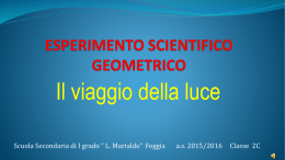
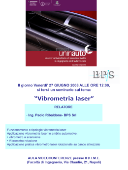
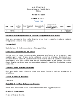
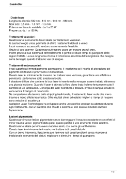
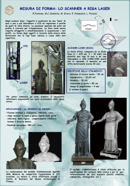
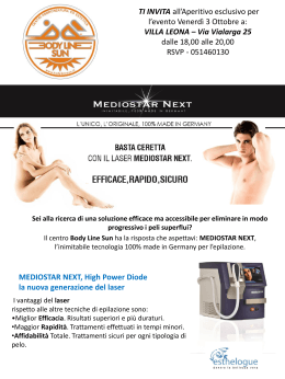
J. Appl. Cosmetol. 26, 29-34 (Jonuory/Morch 2008)
THE EFFECTS ON THE SKIN OF A NEW
FRACTIONAL LASER. MACROSCOPICAL ANO
HISTOLOGICAL EXAMINATION
Paolo Mezzana', M.D. Giuseppe Coppola', M.D.
' Plastic. Reconstructive Surgery Specialist. Rome - ltaly; R&D lnternational Society of Cosmetic
Dermatology. Rome - ltaly
2
Director of the Division of Pathology of the S.Glovanni Addolorata Hospital of Rome - ltaly
Received: Ju/y, 2007
Key words: Froctionol resurfocing;
Photoogeing:
Froctionol
photothermolysis;
Skin
rejuvenotion:
Summary
Fractional resurfacing is an exciting new technology th at fi lls a uniq ue niche in any skin care rejuvenati on protocol. " Fractional Photothermolys is" seeks to onl y damage certai n zones within the selected target area, (producing tiny dot, or pi xel-like treated areas on the skin) , leaving the othe r zones
within it perfec tly intact; hence only causing fractional damage through the heat of the light source.
T his allows the skin to heal much fas ter than if the whole area was treated , as the ' healthy' untreated
tissue surrounding the treated zones helps to fili in the damaged area with new cells .
T he Authors examine the effects of this new procedure on the skin , w ith the aim of analyze the treatment, exploring the c utaneous reaction to various laser pulse durations a nd energies by means of
the macroscopic reaction and histo logical appraisal.
Riassunto
Il res urfacing fraz ionale è una nuova tecno logia che colma una nicchia unica nei protocoll i di ring iovanimento c uta neo . "La Fototermolisi fraz ionale" riesce a danneggiare solo parzialmente il tessuto
bersaglio, producendo piccole colonne di coagulazione, lasc iando le altre zone tra di esse pe rfettamente intatte. Provoca, quindi, esclusivamente un danno frazionale grazie al calore della luce laser,
e le zone risparmjate dal trauma fungono da serbato i di rigenerazione per la produzione delle nuove
cellule .
Gli A utori esaminano gli effetti di q uesta nuova procedu ra sulla pelle, con lo scopo d i a nalizzare il
tratta mento attraverso le reazioni cutanee da un punto di vista macroscopico ed isto logico a d iverse
durate dell'impulso e a diverse energie.
29
The Effects on the Skin of a New Fractional Laser. Macroscopica/ snd Histological Examiriation
INTRQDUCTIQN
durations of the laser (fig. 2) and the " punch
biopsy" has been done 24 hours after the laser
The Fractional Skin Resurfacing is a new laser
technique that is becorning day by day more
popular in the aesthetic surgery and dermatology
sector. 1• 3 It is used for the treatment and improvement of many symptoms of the skin ageing, fo r
some traumatic lesions and acne scars2-' . The aim
of this study is to analyze the treatment of
Fractional Skin Laser Resurfacing, exploring the
cutaneous reaction to various laser pulse durations and energies by means of the macroscopic
reaction and histological appraisal.
exposure.
MATERIALS ANO METHODS
• Skin portion of the retro-auricular region irradiateci imrnediately after the surgical excision.
Biopsy immediately after laser irradiation (fig.
l).
• Skin of the forearm region irradiated at four
d ifferent pulse/energy configurations (Area A,
B, C, D) and "punch biopsy" of 3mm, 24 hours
after the laser exposure (fig. 2).
•Coloration with Haematoxylin-Eosin.
• Macroscopic pictures and images captured
with digitai polarized camera.
• Histology with Zeiss microscope and JVC
camera.
• Fractional Skin Resurfacing Laser System
1540 nm (Matissetm , Quanta System S.p.a.,
Varese, Italy).
A
e
e
o
000
I hour
A
e
e
o
24 hours
RESULTS
First series of tests (fig. 1), has been treated with
laser, with energies equivalent to those of the
second series (fig.2) and after the histology there
are no visible alterations to the laser treatment.
No signs of carbonization, vaporization or ablation of the cutaneous tissues. The second series
has been treated with various energies and pulse
30
A5ms
5 rnliUTS
B 5ms
8.UUTS
C7mo
ltnUUTS
D oma
BmJIUTS
OOQ,
P. Mezzana, G . Coppola
ces, other times by rough mass . In some
"columns" the superficial small blood vessels
show coagulati ve necrosis of the wall with pyknosis of the endothelial nucleus. At the lower
lirnit of the "column" often small blood vessels
are observed that seem to represent the deep
limit of the derma! alteration (fig. 5).
Fig. 3 Area A- 5ms, 5111}/MTS. Small vacuo/ization of a
group of keratinocytes, with lysis of the correspo11dent basai membrane .
;
Fig. 5 Area C-7ms, l lmJ/MTS.
.;'
' .fig. 4
Fig. 4 Area B-5ms, 8mJIMTS. Coagulative necrosis and of
a group of keratinocytes with gaps for111atio11 in the
medium assizes of the 111alpighia11; it is accompcmied
with severa/ alterations of rhe basai membrane:
delami11atio11 or histolysis, with small enlargement
of the collagen, or disi11tegratio11 of the same collagen.
Conservation , like always, of the stratum corneum, inconstant conservation of the granular
Jayer, coagulative necrosis of a group of keratinocytes generally close to a dense eosinophil
mass, other times they are disintegrated in
grains, with intra-epidermal gap formation: The
basai membrane is no longer recognizable, the
derma! collagen in a "column" is changed in
small dense bands, eosinophil with a light blue
tonality, the bands are separated by empty spa-
Fig. 6 Area D-9ms, 9mJIMTS. Skin a/rerations simi/ar ro
the case of 5ms, 8mJ/ MTS.
The development of the erythema
A moderate erythema was produced by al1 the
pulse/energy laser configurations
More detailed:
A. This configuration produces a very light erythema which disappears within an hour.
31
The Effects on the Skin of a New Fractional Laser. Macroscopica/ snd Histological Examination
B. The erythema is more stable, 24 hours later
has almost completely disappeared.
C.One hour later the erythema is stili evident
but moderate. Within 24 hours it is in regression but stili visible.
D. Similar to the B configuration.
Images in dermatoscopy, 24 hours after the
laser treatment
fig.9
Fig. 9: C. Homoge11011s p1111ctifor111 alterations of the cutaneous pigmelllation. The retic11!11111 of "microscab"
is completely similar 10 the theoretical print of the
ji-actio11al /e11s array.
fig.
Fig. 7: A. No 111acroscopic visible alrerarions.
Fig.10: D. Similar 10 the A conjiguration.
DISCUSSION
Fig. 8: B. No ho111oge11ous pu11c1ifor111 alrerarions of the
c111a11eo11s pigmemation. Jr is possible to identify a
reticulum of "microscabs" simi/ar to the theoretical
print of the fractional lens array of the laser.
32
From the comparison between the first series
and the second one, probably the alterations of
the keratinocytes and the dermis after the laser
irradiation , is a "vita!" phenomenon that has
developed within the 24 hours of observation
and is consequent to a thermal insult concentrated inside the microspots and caused by the laser
beam.
P.Mezzana, G. Coppola
In ali the cases, damage to leve! of the corne um
layer does not exist and it is interesting to notice
the presence of free melan in in the epidermis .
Lowe r times of exposure and fl uence (es . 5ms,
5mJ/MTS) determi ne damage to the leve! of the
basai membrane and an overheating of the
superficial dermis without coagulation.
The basai membrane carries out a fundamental
ro le of support and regulation of the cellular prolife ration (sees improvement of the texture), it is
a structural support and al lows fo r the nourishment and the refuse to diffuse . It acts as a fi lter for macromolecules, a nd also a zone of polarization of the cells, in the regeneration process
as it works a "freeway" fo r the cellular migrati on. Its alteration therefore, that happens also at
re latively low parameters, can already justify the
positive observations on the improvement of the
c utaneous texture at low laser parameters .
The max imum depth of coagulation in the test is
600 m icron using 11 mJ/MTS (fig. 5). The
syste m can reach 20 mJ/MTS and the coagulation columns can reach in the dermis at approximately 900 micron.
33
The Effects on the Skin of a New Fractiona/ Laser. Macroscopica/ snd Histological Examination
References
1) Manstein D, Herron GS, Sink RK, et al. (2004) Fractional photothermolysis: A new concept
for c utaneous remodeli ng using microscopie patterns of thermal injury. Lasers Surg Med. 34:
363-7.
2) Tanzi EL, Alster TS, Williams CM. (2003) Treatment of facial rhytides with a nonablative,
1450 nm diode laser: A controlled clinica] and histologic study. Dermatol Surg. 29 (2): 124-8.
3) Bass LS. (2005) Reju venation of the aging face using Fraxel laser treatment. Aesthet Surg
Journal 25: 307-309.
4) Rokhsar CK, Fitzpatrick RE. (2005) The treatment of melasma with fractional photothermolys is: A pilot study. Dermatol Surg. 31: 1645-1650.
Author Address:
Paolo Mezzana, M.D.
Via Merulana 6 1I A
00185 Rome ltaly
[email protected]
34
Scaricare