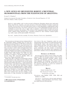
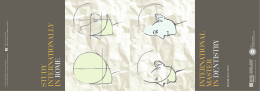
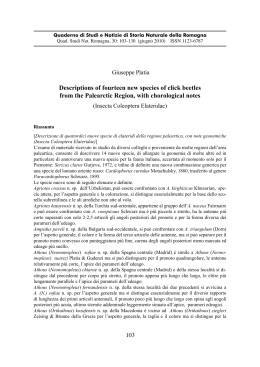
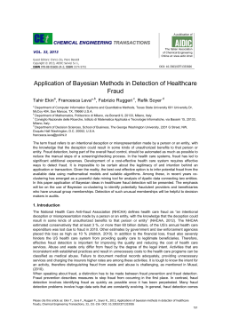
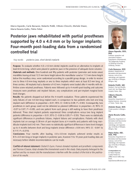
AMERICAN
MUSEUM
Novitates
PUBLISHED BY THE AMERICAN MUSEUM OF NATURAL HISTORY
CENTRAL PARK WEST AT 79TH STREET, NEW YORK, N.Y. 10024
Number 2730, pp. 1-23, figs. 1-1 3, tables 1, 2
June 7, 1982
Results of the Archbold Expeditions. No. 107.
A New Genus of Arboreal Rat from Luzon Island
in the Philippines
GUY G. MUSSER'
ABSTRACT
The taxon latidens Sanborn, 1952 originally
nar-like occlusal surfaces are not characteristic of
named and described as a species of Rattus is taken
out of that genus and placed in Abditomys, new
genus. Some of its distinctive features, such as
nails instead of claws on the halluces, wide upper
incisors, and large molars with simple and lami-
Rattus. The species is known by two specimens
from the island of Luzon in the Philippines. The
relationship of latidens to other Murinae with nails
on the halluces is discussed. Aspects of its natural
history are presented.
INTRODUCTION
the fauna will be analyzed in a separate reI propose a new genus for a native Philippine rat originally described as Rattus la- port.
tidens (Sanborn, 1952), endemic to the island
There is an impression among mammalof Luzon in the Philippines. This report is ogists and biogeographers that the native
one of several already published (Musser,
Philippine rats are well known, especially
1977a, 1977b, 1979, 1981a; Musser and that spectacular segment of the fauna that
Freeman, 1981; Musser and Gordon, 1981) lives on the western highlands in northern
Luzon (Thomas, 1898). This is not so. We
or in manuscript dealing with the taxonomy
and geographic distribution of Philippine know less about the Philippine rats than most
murids. With this and future papers, I intend other murid groups in the Far East. The natto document the morphological limits of cerural history of nearly all the native species
tain species and provide information about is either unknown or encapsulated in sketchy
their insular distributions and natural his- descriptions made by collectors. Many of the
tories. Possible phylogenetic relationships of native genera were thought to be restricted
I
Archbold Curator, Department of Mammalogy, American Museum of Natural History.
Copyright ©) American Museum of Natural History 1982
ISSN 0003-0082 / Price $1.90
2
1
AMERICAN MUSEUM NOVITATES
bis
a-lab
alc
pd-
NO. 2730
for their work with the Scanning Electron
Microscope and the resulting micrographs.
Mrs. Frances A. Hufty, of Archbold Expeditions, Inc., contributed part ofthe financial
support for my work.
plc
hda-lab-
pdplc-
7-
hd-
-labPd-
hd-
FIG. 1. Nomenclature of dental structures using right upper and lower molars of Lenothrix
canus. Upper molars: cusps are numbered according to Miller's (1912) scheme and are referred to
in the text with the prefix t; pc, posterior cingulum.
Lower molars: a-cen, anterocentral cusp; a-lab,
anterolabial cusp; a-ling, anterolingual cusp; pd,
protoconid; hd, hypoconid; md, metaconid; ed,
entoconid; pc, posterior cingulum; alc, anterior
labial cusplet; plc, posterior labial cusplet.
to Luzon or Mindanao but actually have
more extensive distributions over the archipelago. The diversity ofthe fauna, at the level
of genera and species, is greater than generally
realized. There are specimens hidden under
misidentifications in museum collections
that represent new taxa, there are samples of
new species obtained from various islands
during recent exploration, and there are
many species now placed in Rattus that really
do not belong there. The species, latidens, is
one of these. To separate it from Rattus, to
contrast it to other species that have nails
instead of claws on their halluces, and to provide notes and speculations about its natural
history are my intentions here.
I am grateful to the curators who allowed
me access to collections under their care; to
Ms. Fran Stiles, Ms. Marjorie Shepatin, and
Mr. Helmut Sommer for the artwork; to
Messrs. Arthur Singer, Peter Goldberg, and
Jim Coxe for the photographs; and to Mr.
Robert J. Koestler and Mr. Richard Sheryll
ABBREVIATIONS AND METHODS
Specimens referred to here are in collections of the American Museum of Natual History, New York (AMNH); the British Museum (Natural History), London
(BMNH); the Field Museum of Natural History, Chicago (FMNH); the Rijksmuseum
van Natuurlijke Historie, Leiden (RMNH);
and the National Museum of Natural History
of the Smithsonian Institution, Washington,
D.C. (USNM).
Values for lengths of head and body, tail,
hind foot, and ear are from the labels attached
to study skins. Cranial and dental measurements were taken with dial calipers graduated
to tenths of millimeters; limits of those measurements are explained in Musser (1970,
1979). The nomenclature I use for cusps and
cusplets on the molars is diagrammed in figure 1.
DIAGNOSIS AND DESCRIPTION
Named and described by Sanborn (1952)
as a species of Rattus, the characteristics of
latidens are known from an adult female (the
holotype) and an adult male, both from the
island of Luzon in the Philippines. Sanborn
(1952, p. 125) diagnosed latidens as "A large
dark species with tail slightly longer than
head and body; skull short, with thick rostrum, large bullae and short palate ending
between last molars; molar pattern like that
of Rattus, but incisors nearly twice the width
found in most species of Rattus and wider
than in the largest species." Those wide incisors should have prompted Sanbom to look
closer at the holotype. The configuration of
the cranium and the occlusal patterns of the
molars are unlike species of Rattus; furthermore, each hallux has a nail instead of a claw,
a specialization not found in any Rattus. The
two specimens of latidens represent a species
outside of the morphological radiation characterizing not only Rattus but all other known
1982
MUSSER: LUZON ISLAND RAT
genera of murids and requires its own genus,
which is named and diagnosed below.
ABDITOMYS, NEW GENUS
TYPE SPECIES: Rattus latidens Sanbom
(1952, p. 125); based on the holotype, an
adult female (FMNH 62347), collected April
29, 1946, from Mount Data, 7500 feet,
Mountain Province, Luzon, the Philippines.
An adult male (USNM 357244) obtained on
March 14, 1967 from Los Baflos, Laguna
Province, Luzon (Barbehenn, Sumangil, and
Libay, 1972-1973) is the only other known
specimen of the species.
KNowN DISTRIBUTION: The island of Luzon in the Philippine Islands (fig. 2).
ETYMOLOGY: Abditomys is formed by combining the Latin, abditus, meaning hidden or
concealed, with the Greek, mys, for mouse
or rat. I allude to the past concealment of
latidens-so distinctive in features of skin,
skull, and teeth-within the morphological
confines of the genus Rattus, to which it is
not closely related.
DIAGNOSIS: A genus of arboreal murid distinguished from all other species by the following combination of features: large body
size, long, monocolored tail; four pairs of
mammae; a large nail instead of a claw on
each hallux; squamosal roots of the zygomatic arches situated high on sides of the
braincase; squarish interparietal, its anterior
one-third between the parietals, the posterior
two-thirds roofing the occipital bulge; no
strut of alisphenoid bone covering lateral part
of the alisphenoid canal; masticatory-buccinator foramina merged with foramen ovale
accessorius; incisive foramina long and thin;
palatal bridge short, ending well before backs
of third molars; deep mesopterygoid and
pterygoid fossae; sphenopalatine and sphenopterygoid vacuities large; bullae large and
inflated; upper incisors very wide, absolutely
and relative to breadth of rostrum; molars
long and wide, gleaming white, the uppers
large relative to area of palatal bridge; high
cusps that slant back with appreciable overlap between first and second molars, and between second and third, especially in the upper molars; first upper molars five-rooted,
3
first lower molars four-rooted; each third upper molar squarish in occlusal outline, only
a little smaller than the second; upper molars
with simple occlusal surfaces, no cusps t7, no
posterior cingula, no cusps t3 on second and
third molars; cusp t9 broadly merged with
cusp t8 on first and second molars; front two
rows of cusps form thin and gently arcuate
laminae on each first upper molar and on the
front row of each second and third upper
molar; last row of cusps on each upper molar
laminar, nearly transverse; occlusal surfaces
of lower molars consisting of transverse
laminae formed from two elongate cusps;
posterior cingulum a wide flat-topped cone
in center of posterior margin on first and second lower molars; small anterolabial cusps
on second and third molars; no anterocentral
cusp at front of first lower molar; no labial
cusplets on any lower molars.
DESCRIPTION: Abditomys latidens is a largebodied rat with a tail longer than the combined lengths of head and body (table 1, fig.
3). The upperparts of the head and body are
brown with tawny highlights. The underparts
are buffy yellow with gray suffused through
the base of the coat. Color of the venter is
indistinctly demarcated from that of the dorsum. The pelage covering the head and body
is long (30-3 5 mm. over the back) and coarse,
but not spinous. Long (up to 50 mm.) and
conspicuous guard hairs emerge from the
coat everywhere over the top and sick.s Z-f the
body, and are especially prominent over the
rump. The ears are large and brown. The tail
is brown everywhere, appears naked, but is
covered with short, stiff, inconspicuous hairs.
The feet are dark or pale brown over the entire dorsal surfaces down to the base of the
claws. The claws on the front feet are sharp
and small, especially relative to the large digital pads all textured with transverse and
semicircular striations. There is a prominent
pointed nail on each hallux; the other digits
terminate in large, elongate, sharp claws (fig.
4). The female has four pairs of mammae: a
pectoral pair, postaxillary pair, and two inguinal pairs. The male (testes were abdominal) has a prominent midventral cutaneous
glandular area, 70 mm. long and 6 mm.
4
AMERICAN MUSEUM NOVITATES
NO. 2730
LI
0-1500 ft.
EZ
1500 - 5000 ft.
E
5000 - 8000 ft.
E
8000 + ft.
.
.
*0
;.
C%@:' .
0
FIG. 2. Map of Luzon Island in the Philippines. Specimens of Abditomys latidens are from Mount
Data and Los Bafios.
MUSSER: LUZON ISLAND RAT
1982
5
TABLE 1
Measurements (in Millimeters) of Adult Abditomys latidens from Luzon Island in the Philippines
w
i.
A.
.t
.*.
4
*.t.
,-
'-:
I
.v
O.,
I
FIG. 3. Abditomys latidens. Dorsal and ventral
views of USNM 357244, the adult male from Los
Bafios. Its measurements are listed in table 1.
across
its widest place (partially
seen
in fig.
3).
The cranium of A. latidens is large, high,
stocky, and roughly rectangular from a dorsal
view (table 1, fig. 5; Sanborn, 1952, fig. 10).
Measurement
Length of head and body
Length of tail
Scale rows/cm on tail
Length of hind foot
Length of ear
Weight (g)
Greatest length of skull
Zygomatic breadth
Interorbital breadth
Length of nasals
Length of rostrum
Breadth of rostrum
Breadth of braincase
Height of braincase
Breadth of incisor tips
Breadth of zygomatic plate
Depth of zygomatic notch
Length of diastema
Palatilar length
Palatal length
Postpalatal length
Length of incisive foramina
Breadth of incisive foramina
Length of palatal bridge
Breadth of bridge at M'
Breadth of bridge at M3
Breadth of mesopterygoid fossa
Length of bulla
Height of bulla
Alveolar length of M'-3
Breadth of M'
Breadth of M2
Breadth of M3
Actual length of M,_3
Breadth of Ml
Breadth of M2
Breadth of M3
a
Holotype.
FMNH
62347a
female
232
242
7
45
21
50.6
25.9
6.0
19.4
16.6
9.6
18.7
14.1
5.4
6.0
3.1
13.4
22.5
26.3
19.4
9.0
2.9
9.2
3.4
4.7
2.7
9.0
7.8
9.7
3.4
3.5
2.6
10.4
2.9
3.2
3.0
USNM
357244
male
216
271
9
47
24
269
49.5
24.2
5.7
18.9
15.3
8.6
18.0
13.5
3.9
5.4
3.1
13.0
22.1
25.3
18.3
9.3
2.8
9.1
3.4
3.9
3.1
9.5
8.2
9.9
3.1
3.0
2.6
9.8
2.9
3.0
2.8
Strong and nearly parallel zygomatic arches,
together with a short and broad rostrum and
squarish braincase provide the rectangular
appearance. Nasals, which are narrow proximally but expand to become wide distally,
enhance the broadness ofa squarish rostrum.
6
AMERICAN MUSEUM NOVITATES
NO. 2730
IN
--!- 'N
-
-'ov
<.
I
--
O.ViWAF,--
FIG. 4. The hind foot of Abditomys latidens. Drawn from USNM 357244. Note the nail on the
hallux.
The nasals are nearly flat for about threefourths of their lengths, which gives the rostrum a broad, flat dorsal surface. The interorbital region is pinched in, narrower than
the rostral breadth. High, thick ridges define
the dorsolateral margins of the interorbital
region and extend back along the postorbital
area to the dorsolateral margins of the braincase where they become reduced in size and
prominence and nearly disappear near the
occiput. The interparietal is about as long as
it is wide; the front one-third is between the
parietals, the posterior two-thirds forms the
top of the deep occipital region. High, thin
ridges mark the sides of the braincase where
the occipital and squamosal bones join. The
walls of the braincase above the squamosal
roots of the zygomatic arches slope toward
the midline of the cranium.
In side view, the zygomatic plates are wide,
appear robust, and their anterior borders
project well in front of the dorsal roots of the
zygomatic arches. The squamosal roots of the
zygomatic arches are set high on the braincase.
The configuration of each alisphenoid region at the base of the braincase just above
each pterygoid ridge is like that in Rattus
(Musser, 1981a). A strutlike wall of alisphenoid bone, present in many murids
(Musser, 1981 a), is gone from the lateral side
of the alisphenoid canal, which is now an
open channel. Above the channel is a large
foramen ovale and anterior to that a small
opening where the contents ofthe canal enter
the sphenoidal fissure (fig. 7A). When a strut
of alisphenoid bone is present, it separates
the masticatory-buccinator foramina, anterior to the strut, from the foramen ovale accessorius, posterior to the strut (as in Crateromys, for example; see Musser and
Gordon, 1981). With the strut of bone missing, the masticatory-buccinator foramina
have merged with the foramen ovale accessorius and appear functionally absent. The
foramen ovale and opening of the alisphenoid canal into the sphenoidal fissure are now
seen where they would be hidden if a strut
were present (fig. 7A).
The inflated bullae, large absolutely and
relative to size of the cranium, are evident
in side view. Each bulla is separated from the
squamosal bone by a wide postglenoid vacuity. Above each bulla, the squamosal bone
is complete, not perforated or notched by a
squamoso-mastoid vacuity that would separate the bone into a dorsal part and a ventral
tympanic hook (fig. 7A).
In ventral view (fig. 5, fig. 7B), the incisive
foramina are narrow, long, and slightly constricted posteriorly; their back margins penetrate between the anterior margins of the
first molars by 0.5 mm. in FMNH 62347 and
0.4 mm. in USNM 357244. The palatal
bridge is narrow, thick, scored by deep palatine grooves, and short; its posterior edge
is situated 1.0 mm. before the ends of the
toothrows in FMNH and 0.8 mm. in USNM
1982
MUSSER: LUZON ISLAND RAT
7
a
A..
b
* #~ ~ ~ ~ ~ ~ .
c
'-.
FIG. 6. Dentaries from the same specimens
shown in figure 5: a, labial view of R. r. diardii;
b, labial view of A. latidens; c, lingual view of A.
latidens. Approximately X 1.
IC, X I -t.
.I
a
b
a
b
FIG. 5. Views of adult crania: a, Abditomys
latidens (USNM 357244) is contrasted with b, an
example of Rattus rattus diardii from West Java
(AMNH 250104). Approximately xl.
357244. The mesopterygoid fossa is deep and
conspicuously narrower than the palatal
bridge; its walls are breached by spacious
sphenopalatine vacuities that extend to the
back of the orbits (fig. 8). Each pterygoid fossa
is deep, its floor slanted down toward the
midline of the cranium and its anterior twothirds perforated by a large sphenopterygoid
vacuity. The posterolateral edge of the pterygoid platform behind each foramen ovale
is a definite, high ridge. In ventral view the
bullae are large and inflated; the eustachian
tubes are short and inconspicuous.
Shapes and positions of some foramina in
each orbit are of interest. The sphenopalatine
foramen is very large and conspicuous. It is
set well anterior to the much smaller and
nearly hidden dorsal palatine foramen (fig.
8). The optic foramen is also large. It is separated from the anterior end of the large and
elongate sphenopalatine vacuity by a narrow
bony strut.
Each dentary is large and stocky (fig. 6).
That part of the ramus anterior to the toothrow is short and deep. The coronoid process
is large. A deep concavity forms the back
margin of the dentary. The masseteric ridge
is thick and prominent. On the inner side of
the mandible, a shelf extends from the back
of the toothrow diagonally past the mandibular foramen onto the bottom of the condyloid process.
8
~ ~ bs
AMERICAN MUSEUM NOVITATES
aIs'~
NO. 2730
~ ~ p
q
ab
max
=
-
>
~~~~~ptr--
trc
mif
*~~~~~
ab
ptb
AL,.~~~~~~~~~~~~~~~~~'t,
FIG. 7. The back part of the cranium in Abditomys latidens (USNM 357244): A, lateral view; B,
ventral view. Abbreviations: a, anterior opening of the alisphenoid, where it empties into the sphenoidal
fissure; ab, auditory bulla; alc, alisphenoid canal, which is an open channel; als, alisphenoid bone; bo,
basioccipital bone; bs, basisphenoid bone; et, bony eustachian tube; fo, foramen ovale; mas, mastoid
portion of the petromastoid; max, maxillary bone; mlf, middle lacerate foramen; oc, occiput; pa, parietal
bone; pgl, postglenoid vacuity; ps, presphenoid bone; pt, periotic portion of the petrosal; ptb, pterygoid
bridge, which is a definite ridge; ptf, pterygoid fossa; ptr, pterygoid ridge; sf, sphenoidal fissure; spt,
sphenopterygoid vacuity; sq, squamosal bone; srza, squamosal root of the zygomatic arch; sv, sphenopalatine vacuity; trc, transverse canal. Hill (1935), Wahlert (1974), and Musser (1981a) discuss the
foramina and significance of some configurations.
MUSSER: LUZON ISLAND RAT
1982
9
fr
r
IC
1.
I
max
W ..- -W !,
9
.1
*.c
li.'
.",,
,WI
I&
',
FIG. 8. The orbital and postorbital region in Abditomys latidens (USNM 357244). Abbreviations:
dpl, dorsal palatine foramen; fr, frontal bone; max, maxillary bone; ml, first upper molar; op, optic
foramen; spl, sphenopalatine foramen; sq, squamosal bone; sv, sphenopalatine vacuity. Note the slant
to the high cusps and the way one molar overlaps another.
The incisors of A. latidens are large and
smooth, the enamel layers of the uppers are
orange, those of the lowers yellowish orange.
The uppers emerge from the rostrum at a
right angle (opisthodont) and are very wide
(56% of the rostral breadth in FMNH 62347
and 45% in USNM 357244).
Abditomys latidens has big, chunky hyposodont molars that are ivory-colored, glisten,
and without the brown or blackish tarry encrustations often found in other murids. Each
first upper molar is anchored by five roots
(large anterior and posterior, two lingual, and
a labial), the second by four roots, and the
third by three. Each first lower molar has four
roots (large anterior and posterior, small labial, and small lingual), each second and third
lower molar is anchored by three roots (two
small anterior and a large posterior).
The upper molars form wide, prominent
rows in the bony palate. The second upper
molar is about as wide as the first, and the
third, which is squarish in occlusal outline,
is only a little smaller than the second. The
high cusps slant back so that the first molar
appreciably overlaps the front part of the second and the second overlaps the third (figs.
8 and 9). The occlusal surfaces of the uppers
are simple in configuration: no cusp tlbis,
t2bis, t7, accessory labial cusp behind cusp
t6, or posterior cingulum occurs in any of the
teeth; and cusp t3 is absent from the second
and third molars. The cusps on all the teeth
form either gently arcuate rows-such as the
first and second laminae on the first molar,
and the first lamina of the second and third
molars-or nearly transverse laminae, those
at the back of each tooth. Cusp tl on all
three teeth, and cusp t4 on each first and
second molar are round and discrete-especially cusp t4 on the first molar, which is
nearly separate from cusp t5 -but the labial
cusps are elongate and merged with the central cusps to form most of the surface of each
10
AMERICAN MUSEUM NOVITATES
+/
s
tA ^ vV
7)j
./
;i4,
"ql
i
.1
J"/
FIG. 9. Occlusal views of right molar rows of
Abditomys latidens (USNM 357244). M13 is
the left, Ml3
on
the right. See table
1
for
on
mea-
surements.
lamina. Cusp t9, for example, is large but
poorly defined because it is broadly merged
with cusp t8 on the first and second molars.
The stocky lower molars also have high,
slanting cusps, and are simple in occlusal topography (fig. 9). There are three transverse
rows of cusps (two in each row) on the first
molar and two rows on each second and third
molar. The laminae are separated from each
other and lean forward. A large and discrete
posterior cingulum sits at the back of each
first and second molar, distinctly separate
from the lamina in front of it. The first molar
is oblong in occlusal outline, the second and
third squarish. The front lamina on each first
lower molar is formed of a large anterolingual
cusp and a smaller anterolabial cusp merged
to form a lamina distinctly set apart from the
one behind; there is no anterocentral cusp.
No anterior or posterior cusplets occur along
NO. 2730
the labial margins of the first molars, and no
posterior labial cusplets occur on either the
second or third molars in the two specimens
I examined. There is a small anterolabial cusp
on each second and third molar.
COMPARISONS WITH RATTUS
I compare Abditomys latidens with Rattus
because latidens was originally described as
a species in that genus. The Asian forms of
R. rattus, R. r. diardii (Java) and R. r. flavipectus (China), are the examples I use; specimens of them are compared with that of A.
latidens in figure 5 (crania), figure 6 (dentaries), and figure 10 (molar rows). Abditomys
latidens possesses the following characteristics that contrast with those of Rattus.
1. A nail (or scalelike claw) is on each hallux. Every species of Rattus has claws on the
halluces.
2. The dorsal surfaces of the nasals are
nearly flat for most of their lengths and only
gently convex near their tips. In Rattus, the
nasals are not flat but rounded on top along
their lengths.
3. Squamosal roots of the zygomatic arches
are set higher on sides of the braincase.
4. The sides of the braincase slope outward
from the temporal ridges to the squamosal
roots. In Rattus (certainly R. rattus), the sides
of the braincase are vertical or nearly so.
5. The interparietal is narrow, the anterior
one-third sits between the parietals, the posterior two-thirds roofs part of the occipital
region. Rattus has a short, wide interparietal
that is farther posteriorly, forming nearly the
entire roof of the occipital region.
6. Each zygomatic plate is taller, joining
the dorsal root of the zygomatic arch nearly
at a level with the top of the cranium. The
plate is relatively shorter in Rattus, meeting
the dorsal root of the zygomatic plate about
one-third of the way down from the top of
the cranium.
7. The bullae are much more inflated and
appreciably larger relative to size of the cranium.
8. The cranium is relatively deeper, especially between the interorbital area and the
bony palate.
9. The palatal bridge sits well above the
floor ofthe basisphenoid (as seen from a ven-
1982
MUSSER: LUZON ISLAND RAT
tral view), is deeply scored by palatine
grooves, is narrow, and ends anterior to the
posterior margins of the toothrows. In Rattus, the bridge is much lower relative to the
basisphenoid, very wide, with either shallow
or faint palatine grooves, and projects beyond
the posterior margins of the toothrows to
form a wide and long shelf.
10. The mesopterygoid and pterygoid fossae are relatively much deeper, a reflection
of the raised bony palate.
11. The upper incisors are orthodont and
very wide, about half the width of the rostrum. The uppers are either orthodont or opisthodont and much narrower, about a third
the width of the rostrum (an average of 33%
of the rostral breadth in a series of 20 R. r.
diardii from Java).
12. The upper molars are big and chunky,
the toothrows occupy about two-thirds of the
bony palate. Rattus has small molars, the
toothrows are short and narrow relative to
the wide and long expanse of bony palate.
13. Each third upper molar is squarish and
only a little smaller than the second. Each
third molar is round or oblong in Rattus and
much smaller than the second.
14. Occlusal surfaces of the uppers consist
of wide and gently bowed laminae in which
only some of the lingual cusps are still discrete. The lamina at the back of each molar
is transverse or nearly so, a configuration due
to an elongate cusp t9 merged on the same
axis with an elongate cusp t8. The laminae
in Rattus are arcuate and conspicuously cuspidate. Cusp t9 on each first and second molar is discrete and directed anterolabially;
those posterior laminae are not transverse.
15. No cusp t3 occurs on either the second
or third upper molars. Cusp t3 is often present, although small, on the second and third
molars of Rattus.
16. Each dentary is more robust and
higher; the portion anterior to the molar row
is relatively shorter and deeper.
17. The lower molars are large and chunky,
especially relative to the dentary. Rattus has
much smaller molars.
18. Each third lower molar is squarish in
occlusal outline and nearly as large as the
second molar. Each third molar is triangular
in outline in Rattus.
11
19. Occlusal surfaces of the lower molars
consist of wide, transverse laminae, each
composed of two elongate cusps. The laminae are set apart from each other. The laminae are close together in Rattus, most are
chevron-shaped rather than transverse.
20. The front lamina of each first lower
molar is slightly angled to the posterolateral
side, distinctly separated from the lamina
behind, and consists ofdiscrete anterolingual
and anterolabial cusps set on about the same
axis. In Rattus, the front lamina is formed
of a large anterolingual cusp and a smaller
anterolabial cusp set at nearly a right angle
to the other; the backs of the cusps are pressed
tightly against the anterior surface of the second lamina.
21. The lamina at the back of each third
lower molar is nearly as wide as the front
lamina and transverse. In Rattus, the posterior lamina is narrower relative to the one
in front, and slanted anterolabially.
22. Each posterior cingulum on the first
and second lower molars is round or slightly
elliptical in cross-section and distinctly separated from the lamina in front of it. The
posterior cingula are elliptical in Rattus and
set closer to the laminae, often merging with
them at places.
23. There are no anterior or posterior labial
cusplets on the first lower molars, and no
posterior labial cusplets on the second molars. There is an anterior labial cusplet on
each first molar in some examples of Rattus;
posterior labial cusplets occur on both the
first and second molars in most specimens.
COMPARISONS WITH MURINES
HAVING NAILS
In having nails on its halluces, Abditomys
latidens is unique among rats native to the
Philippine Islands. The character is rare even
within muroid rodents and is found in only
two groups. One consists of Dendromus,
Megadendromus, and Prionomys, which are
in the Dendromurinae, a small assemblage
of species native to Africa.
The other group is the Murinae. Among
the hundreds of species in that group, only
the following seven genera and 14 species
have nails on halluces.
12
AMERICAN MUSEUM NOVITATES
NO. 2730
p
4
f
I
'
"..
1
I.
1.
x 1,p
!,--
.s~~~~~~
sr
t-.
.I
I
_iwt~~~~~~~~~~~~~~~~~~~~~~~~~~~~~~~1
i~'
FIG. 10. Occlusal views contrasting right molar rows of Abditomys and Rattus. A and D: MI-3 and
M, 3, respectively of A. latidens (USNM 357244). B and C: M'-3 and M,-3, respectively of R. rattus
flavipectus from China (AMNH 84677). Approximately x10.
1. Hapalomys: H. longicaudatus and H. delacouri; the mainland of Southeast Asia
and the Malay Peninsula (Musser, 1972).
2. Chiropodomys: C. gliroides, C. muroides,
C. calamianensis, C. major, and C. karlkoopmani; Southeast Asia, the Sunda
Shelf, and the Mentawai Islands (Musser,
1979).
3. Vandeleuria: V. oleracae and V. nolthenii;
India, Ceylon, and Southeast Asia (Musser, 1979).
4. Pithecheir: P. melanurus and P. parvus;
the Malay Peninsula, Sumatra, and Java
(Ellerman, 1941).
5. Chiromyscus: C. chiropus; Southeast Asia
(Musser, 1981a).
6. Kadarsanomys: K. sodyi; Java (Musser,
1981c).
7. Abditomys: A. latidens; Luzon, the Philippine Islands.
Because nails on halluces is a specialization
and one that occurs infrequently among muroid rodents, especially among the Murinae,
1982
MUSSER: LUZON ISLAND RAT
13
TABLE 2
Distribution of Primitive (P) or Derived (+) Expressions of 26 Traits among 7 Genera of
Murine Rodents
Chiro- Hapalo- VandeChiro- Kadar- Abditopodomys mys
leuria Pithecheir myscus sanomys mys
1. Dorsolateral margins of braincase
P
+
+
+
+
+
+
2. Sides of braincase
+
P
P
+
P
+
P
3. Squamx.sal roots of zygomatic arches
P
P
+
+
+
P
+
4. Alisphenoid complex
+
+
P
+
P
+
+
5. Size of bulla
P
+
P
+
P
+
+
6. Attachment of bulla
+
+
+
P
P
+
+
7. Incisive foramina
P
P
P
P
+
+
+
8. Mesopterygoid fossa
P
P
+
P
P
P
+
9. Pterygoidfossa
P
P
P
P
+
+
+
10. Pterygoid platform
P
+
P
+
P
+
+
11. Roots of M'
P
P
P
P
P
+
+
12. Roots of M,
P
P
P
P
+
+
+
13. Height of cusps
P
P
P
P
P
P
+
14. Sizeofmolars
P
P
P
P
+
P
P
15. Overlap of molars
P
P
P
+
+
+
+
16. Cuspstlbisandt2bis
P
+
P
+
P
P
P
17. Cuspt7
+
+
+
+
P
P
P
18. Posterior cingulum
P
+
P
P
+
P
+
19. Cuspt9
P
P
P
P
+
P
+
20. Cuspt.,
P
P
P
P
+
P
+
21. Unionofcusps
P
P
P
P
+
P
+
22. Anterolabial and anterolingual cusps
P
P
P
P
+
P
P
23. Anterolabial cusp
P
P
+
P
P
P
P
24. Fifth digit
P
P
+
P
P
P
P
25. Lamina
P
P
P
P
P
+
P
26. Rows of cusps
P
+
P
P
P
P
P
Total derivedtraits
11
5
8
5
13
11
17
a Numbers correspond to
numbered descriptions of the traits in the text. See Musser (198 la) for more information
about some of the features.
I studied. species in the seven genera listed
above to determine if they were closely related because they shared this derived trait.
Elsewhere (Musser, 1981a), I presented hypotheses ofderived and primitive conditions
in 29 external, cranial, and dental features of
several genera from the Indo-Australian region that were once part of Rattus to note
their possible phylogenetic relationships relative to Rattus. I examined 23 of those same
characteristics, as well as three new ones, in
the seven genera. These traits are briefly presented below: the primitive condition is described first, followed by the derivation in
parentheses. Distributions of the primitive
or derived states in the 26 traits are listed in
table 2.
1. Dorso lateral margins of braincase: Slight
beading al(ong dorsolateral margins of the interorbital aand postorbital regions; the braincase is eith er smooth all over or has low beading along the dorsolateral margins of the
anterior hailf and is smooth over the posterior
half. (Medilium to high ridges or shelves outline the iInterorbital area and sweep back
along the (dorsolateral margins of the braincase, all tthe way to the occiput in some
species.)
2. Sides of braincase: Sides ofthe braincase
slope outwiard from the dorsolateral margins
of the cranium to the squamosal roots of the
zygomatic arches. (The sides are vertical or
nearly so.)
3. Squaimosal roots of zygomatic arches:
14
AMERICAN MUSEUM NOVITATES
The roots originate high on the upper half of
the braincase, often close to the temporal
ridges and well above the bullae. (The roots
are set halfway down the braincase or lower,
just above the bullae.)
4. Alisphenoid complex: A strut of alisphenoid bone covers the lateral part of the
alisphenoid canal and separates the foramen
ovale accessorius from the masticatory-buccinator foramina. (The strut is gone, the alisphenoid canal is an open channel and the
foramina have merged-they appear to be
functionally absent.)
5. Size of bullae: The bullae are moderately
to very small relative to size of the cranium.
(The bullae are large relative to cranial size,
sometimes highly inflated.)
6. Attachment of bullae: Each bulla is
tightly attached to the squamosal bone. The
postglenoid vacuity is small and usually confined to the top of the bulla between the periotic and squamosal. (A wide, spacious vacuity separates the dorsal and anterior margins
of the periotic and bulla from the squamosal.)
7. Incisive foramina: The incisive foramina are short, their posterior edges terminating well anterior to the maxillary molar rows.
(The foramina are long, the posterior margins
ending at the fronts of the first upper molars
or beyond them.)
8. Mesopterygoid fossa: The fossa is nearly
as wide as the back part of the palatal bridge.
Its walls are breached by thin, short sphenopalatine vacuities or slits. (The fossa is onethird to one-half the width of the palatal
bridge. The sphenopalatine vacuities are
huge so that the presphenoid and anterior
part of the basisphenoid appear suspended
in air.)
9. Pterygoid fossae: Each fossa is nearly flat
or shallowly excavated, its anterior twothirds entire and not perforated by a large
sphenopterygoid opening. (Each fossa is excavated or tilted toward the midline of the
skull and perforated by a large sphenopterygoid vacuity.)
10. Pterygoid platform: The posterolateral
portion of each pterygoid platform behind
the foramen ovale is either nearly flat, a low
mound, or a wide and smooth low ridge. (The
edge is thrown up into a definite high and
thin ridge.)
NO. 2730
11. Roots of first upper molars: Each first
upper molar is three-rooted (anterior, lingual,
and posterior) or four-rooted (a labial root
in addition to the three primary roots or a
divided lingual root along with only an anterior and posterior root). (Each first upper
molar is five-rooted -anterior, posterior, two
lingual, and one labial.)
12. Roots of first lower molars: Each first
lower molar is two-rooted (an anterior and
posterior root) or three-rooted (either a labial
or a lingual rootlet in addition to the two
primary roots). (Each lower molar is fourrooted-anterior, posterior, labial, and lingual.)
13. Height of cusps: The molars have low
to moderately high cusps, but definitely
brachyodont. (The molars have high cusps,
clearly hypsodont.)
14. Size of molars: The upper molars gradually decrease in size from the first to the
third. (Each second upper molar is smaller
than the first, but the third is reduced in size
and much smaller relative to the second.)
15. Overlap of molars: Cusps on the upper
molars are only slightly slanted and there is
little or no overlap among the three teeth in
a toothrow, each essentially abutting against
the other. (The cusps slant conspicuously
back, so that the first molar overlaps the second, and the second overlaps the third.)
16. Cusps tlbis and t2bis: These cusps do
not occur on the first and second upper molars of most specimens. (Either tlbis, t2bis,
or both are present on first upper molars in
most specimens, sometimes the second molars.)
17. Cusp t7: No cusp t7 is on any of the
upper molars. (A cusp t7 occurs on the first
molars, usually on the second molars, and
sometimes on the third.)
18. Posterior cingulum: There is a posterior
cingulum at the back of each first upper molar, usually the second, and sometimes the
third. (No posterior cingulum occurs on any
ofthe upper molars, or ifit occurs is indicated
only by a triangular bump at the back of each
first upper molar.)
19. Cusp t9: This cusp is large and discrete
on each first and second upper molar. (Cusp
t9 is nearly incorporated into the much larger
cusp t8 and inconspicuous after wear.)
1982
MUSSER: LUZON ISLAND RAT
20. Cusp t3: In all or most of the samples,
cusp t3 is present on the second and often on
the third upper molars. (Cusp t3 is absent
from the second and third molars in all or
most specimens.)
21. Union of cusps: Cusps on the upper
and lower molars are weakly connected so
the occlusal patterns appear strongly cuspidate. (All or most cusps are strongly joined,
some merged to the point where they nearly
lose their identities in several species.)
22. Anterolabial and anterolingual cusps:
The anterolabial and anterolingual cusps on
the first lower molar are large and discrete,
forming a lamina nearly as wide as the rest
of the tooth. (The two cusps are smaller,
pressed against each other to form a lamina
narrower than the rest of the tooth. In young
rats they may be separate but soon merge into
an oblong lamina.)
23. Anterolabial cusp: The anterolabial
cusp on each second lower molar, and often
on each third molar, is present in all or most
of the samples. (Anterolabial cusps are absent
from the second and third molars in all or
most of the samples.)
24. Fifth digit: The fifth digit on each hind
foot bears a claw. (A nail is embedded in each
fifth digit.)
25. Laminae: The rows of cusps on the
upper and lower molars are arcuate or chevron-shaped. (The cusps form nearly straight,
laminar-like rows.)
26. Rows of cusps: Cusps are weakly joined
or strongly joined but form uneven rows on
the molars. (Large and discrete cusps form
two or three straight rows on each molar.)
Except for having nails on the halluces,
Abditomys is unlike Chiropodomys, Hapalomys, Vandeleuria, Pithecheir, Chiromyscus, or Kadarsanomys in external, cranial, or
dental characteristics. I explain below and in
figure 11, where crania of the seven genera
are compared, and in figures 12 and 13, which
contrast toothrows.
Vandeleuria is arboreal, small-bodied, and
long-tailed. The tail is scantily haired. Digits
on the front and hind feet are long and slender. Both the fifth digit and hallux of each
hind foot have nails. A nail instead of a claw
on the fifth digit is unique among the Mu-
15
rinae. The structural specializations of Van-
deleuria are those associated with climbing
among slender branches or high grass. Among
living rodents, Vandeleuria has been allied
with Micromys, Vernaya, and Chiropodomys
(Ellerman, 1941); based on dental characters,
Misonne (1969) regarded Vandeleuria to be
a close relative of Chiropodomys.
Species of Chiropodomys are also smallboddieLand long-tailed, but the tails are penicillate. The fifth digit of each hind foot bears
a claw-only the halluces have nails. The
crania of Vandeleuria and Chiropodomys differ strikingly from that of Abditomys in size
and configuration (fig. 11). The dentitions of
the two genera are unlike those of A. latidens.
The molars of both Vandeleuria and Chiropodomys are anchored by fewer roots than
are those ofAbditomys (upper first molars are
three-rooted, lowers are two-rooted in Chiropodomys; upper first molars are fourrooted, the lowers two-rooted in Vandeleuria); the upper molars either abut against one
another without overlapping (Chiropodomys)
or overlap slightly ( Vandeleuria), as opposed
to the extensive overlap among the teeth of
Abditomys; and the molars of Chiropodomys
and Vandeleuria have complex and highly
cuspidate occlusal surfaces, unlike the simple, laminar-like chewing areas ofA. latidens.
Complexity of the occlusal surface in the upper molars is partly due to large cusps t7 and
posterior cingula on all or most of the teeth,
and to large cusps t3 on the second molars.
Large anterocentral cusps, wide anterolabial
cusps on the second and third molars, and
prominent labial cusplets are features of the
lower molars contributing to the complex
chewing surfaces. Such structures are not
present in the simple molars of Abditomys.
Among the traits listed in table 2, Abditomys has many more derived features (17)
than does either Vandeleuria (5) or Chiropodomys (5). No single derivation, other than
nails on halluces, occurs in all three genera.
Whether Vandeleuria and Chiropodomys are
as closely related as some taxonomists claim
remains to be tested but neither one seems
phylogenetically close to Abditomys.
Species of Hapalomys are arboreal and at
least one of them, H. longicaudatus, is specialized for living in bamboo and feeding on
i
.,I
V.
".4,
14viO
,--
-
1*.
r.
_:
-,d4j
._
01
"'Aor-.-L
dowIr
00
S"i
itwh.,;
'%A?fr: /4b,
A-,
IA
,%KU
4'.
o -
,,
-
-
Nt
*s,
IY
..~~~~~~~~~~~~~~~~~~~~~~~~~~~~~~~~~~~~~~~~~~~~~~~~~t -
,
"*
C)
-
_-
-0
0
c0
a°
CU3
a,~~~0
0U
3
-_
.a.. r4
Fr
--.
AN.I.
-* otak
r
-A' A
S
~~~-r
co
n
k
A.
CU*
::
C's
.r
Ad
*4. ~ ~ ~ ~:~
*f *Q i
co
o
I~~~~W
a>>^
>
_
.eCm
1982
MUSSER: LUZON ISLAND RAT
it (Medway, 1964). In addition to being
smaller in body size than A. latidens, and
differing in coloration and texture of the fur,
and in basic shape of the cranium (especially
the interorbital region and braincase), the
species of Hapalomys have different occlusal
patterns on the molars. Straight rows of discrete gleaming cusps form the chewing surfaces of the upper and lower molars, a configuration not found in any of the other genera
discussed here or in the rest of the Murinae
in general (figs. 12 and 13). The upper molars
in Hapalomys abut against one another without overlap. Four roots anchor the first upper
molars, the first lower molars are two-rooted.
The simple, laminar-like occlusal topography, strong overlap among the molars, and
additional roots in Abditomys are unlike the
complex, cuspidate occlusal surfaces in Hapalomys. Except for nails on the halluces, no
other derived trait occurs only in Hapalomys
and Abditomys. Misonne (1969) suggested
that Chiropodomys may be the closest relative of Hapalomys. Abditomys is certainly
not.
Both species of Pithecheir are arboreal,
have prehensile tails, and the most grasping
type of hind foot in any of the arboreal rats
and mice noted here (Musser, 1979). Both
the tail and feet are specialized for climbing
on slender supports such as twigs, narrow
branches, and palm and fern fronds (Bartels,
1937b). The configuration of the craniumparticularly the wide interorbital and postorbital regions bordered by wide shelves, the
interparietal enclosed on both sides by the
parietals, lack of occipital bulge at the back
of the braincase, and the large and highly
inflated bullae-is unlike that in Abditomys
(fig. 11). The upper molars are cuspidate
rather than laminar-like and have more complex occlusal surfaces due to cusps t lbis at
the front of the first molars, large cusps t7 on
the first and second molars, and prominent
cusps t3 on the second and third teeth. The
lower molars are also cuspidate: the rows of
cusps are chevron-shaped rather than nearly
transverse, separated farther from one
another, and the anterocentral cusp (absent
from Abditomys) is bifurcate and large.
Judged from characteristics of skin, skull, and
teeth Pithecheir has no close morphological
17
relationship to Abditomys, and no derived
traits are common to only those two genera,
at least in the characteristics surveyed here.
Although it possesses some highly specialized
structures, Pithecheir has far fewer (8) derived traits than does Abditomys (17), may
be part of an older group of murines, and
possibly more closely related to Lenothrix
than to anything else (Misonne, 1969). There
appears -to be no close phylogenetic tie between Pithecheir and Abditomys.
Little is known about the habits of Chiromyscus chiropus. The rat is specialized for
arboreal living (Musser, 1981 a) and was
thought to occur in tropical deciduous forest
(Marshall, 1977). Recently, Joe T. Marshall
wrote me that he has seen a new specimen
of Chiromyscus collected in Thailand from
a ravine in which bamboo densely filled the
bottom and broad-leaf tropical forest covered the upper slopes. Possibly Chiromyscus-like Chiropodomys, Hapalomys, and
Kadarsanomys-may be ecologically closely
tied to bamboo. A small and low cranium,
narrow zygomatic plates, wide interorbital
region, postorbital margins and dorsolateral
sides of braincase defined by shelves, wide
and flat- braincase, a large interparietal roofing most of the occiput, tiny bullae, small and
low-crowned molars, third molars much
smaller relative to others in the rows, chevron-shaped laminae, no anterolabial cusps on
the second and third lower molars, and first
upper molars with four roots are some of the
cranial and dental features of Chiromyscus
that contrast with those of Abditomys (figs.
1 1-13). Three derived traits (cusp t9 broadly
merged with cusp t8 on the first and second
upper molars, no cusp t3 on the second and
third molars, and all cusps strongly connected) occur in Chiromyscus and Abditomys
but not in any of the other genera in which
the halluces bear nails. Judged from my own
studies, all three traits have developed at different times and are widely distributed
among African, Indian, and Indo-Australian
murines. The three features reflect simple
occlusal surfaces, simpler than those found
in Chiropodomys or Vandeleuria (figs. 12 and
13), and do not indicate close phylogenetic
ties between Abditomys and Chiromyscus.
The latter is actually closely related to Nivi-
~zlZ
-
q
0q
0af
v,
-
-..1%
so
!~~~C
~0
Cd
18
0Z0
~
_
LL
os
L
v
liii
.d
b4;
,.,
A"
I
At.
": 4
2. 7_-7
-
-17,
cD~
.14 va
.;,
.4
19
ZS
20
AMERICAN MUSEUM NOVITATES
venter of Southeast Asia and the Sunda Shelf
(Musser, 1981 a).
Kadarsanomys sodyi is of medium body
size, arboreal, and associated with bamboo
in mountain forests on West Java (Bartels,
1937a; Musser, 1981c). The tail is long,
brown, and monocolored, as is the tail of A.
latidens, but the upperparts of the head and
body are brown, the underparts white, and
the fur short, with very short guard hairs. All
these features are unlike the coloration and
length of pelage in Abditomys. In configuration of the skull and in some dental features,
A. latidens is more like K. sodyi than any of
the other species discussed here. In both, the
cranium is high, the interparietal is small, the
incisive foramina are long, the palatal bridge
is short, the pterygoid fossae and alisphenoid
region are similar in shape, the bullae are
large relative to size of the braincase, the upper molars are large relative to the bony palate, each third molar is only a little smaller
than the second, the first upper molars are
five-rooted, and the first lowers are fourrooted. The two species, however, differ in
conspicuous ways: K. sodyi has a shorter and
relatively broader rostrum with relatively
narrower nasals that are convex on top rather
than flat; a wider interorbital region; wider
and more prominent shelves along the postorbital margins; a more expansive braincase
with straighter sides; relatively narrower zygomatic plates with much shorter anterior
spines; squamosal roots of the zygomatic
arches situated lower on sides of the braincase; shallower mesopterygoid fossa with
much smaller sphenopalatine vacuities, molars with low cusps; less overlap among the
upper and lower molars, and occlusal surfaces that are complex and cuspidate rather
than simple and laminar-like (figs. 1 1-13).
Abditomys and Kadarsanomys have fiverooted first upper molars, a derived trait not
found in any of the other five genera with
nails instead of claws on the halluces. The
character is significant because it is found in
Rattus and many genera distantly and closely
related to Rattus (Arvicanthis is an example)
and usually occurs in combination with other
specializations, such as the derived conditions of the alisphenoid region, incisive foramina, size of bullae, and configurations of
NO. 2730
the mesopterygoid and pterygoid fossae
(Musser, 198 la and 198 lb). Five-rooted first
upper molars indicate only that A. latidens
and K. sodyi are more closely related to each
other than to the species of Chiropodomys,
Vandeleuria, Hapalomys, Pithecheir, and
Chiromyscus. The character has no information that can be used to determine whether
Abditomys is phylogenetically closer to Kadarsanomys than to other genera, such as
Rattus, which have five-rooted first upper
molars. That trait occurs widely throughout
other murines from Africa, India, and the
Indo-Australian region, genera with clawed
halluces. Both A. latidens and K. sodyi must
be compared with those forms to determine
whether the two are closely related or whether
they are actually more closely allied to a genus in which the halluces are clawed.
The configuration of the nail bears further
study because it differs among the species and
finer resolution of its structure may provide
additional information with which to test
hypotheses of relationships among the seven
genera. In Hapalomys, Chiropodomys, Vandeleuria, and Chiromyscus each nail is small,
nearly flat (slightly convex), with a straight
or gently arcuate distal margin, and embedded in the top of the digital pad. The configuration is similar to that illustrated for Chiropodomys in Musser (1979, fig. 14). The nail
of Kadarsanomys is very small relative to the
size of the large digital pad and embedded in
it, has a highly convex dorsal surface, is short,
and has a truncate distal margin (Musser,
1981 c, pl. 1). In Abditomys, the base of each
nail is embedded in the top of the digital pad
but the triangular distal margin is not, and
the dorsal surface is moderately convex (fig.
4). It is a longer structure relative to size of
the digital pad compared with the nails in the
other genera, except for Pithecheir.
The nails in Pithecheir are essentially very
short claws (as I called them, Musser, 1979)
or scalelike claws (in Medway's, 1969, terminology) and range in size among the specimens. On some rats, each structure is short,
sits on top of the digital pad, and appears to
be a pointed nail with a highly convex surface. On other specimens, it is larger, overhangs the pad and resembles a much reduced
claw. The configuration is unlike that in any
1982
MUSSER: LUZON ISLAND RAT
of the other genera and represents the most
primitive state since it is the most clawlike.
To me, the structures on the halluces of the
other genera are good nails (Medway, 1964,
described that in Hapalomys as a flat, naillike scale). The nails in Chiropodomys, Hapalomys, Vandeleuria, and Chiromyscus are
similar in shape and size relative to the digital
pad and represent the most derived configuration. The small, highly convex, and truncate nail of Kadarsanomys is distinctive,
highly derived, and set apart from the others.
The long, pointed nail of Abditomys is
unique. Based only on configurations of the
nails, Pithecheir is isolated from the other
genera, Abditomys and Kadarsanomys each
stands by itself, and Chiropodomys, Hapalomys, Vandeleuria, and Chiromyscus cluster. That the nails of the halluces in Abditomys represent not close phylogenetic affinity
with the other six genera but rather independent derivation seems to be the best hypothesis.
NATURAL HISTORY
Little is known about the natural history
of Abditomys latidens. The male from Los
Bafios "was collected from the electric fence
at the International Rice Research Institutean area of intensive rice cultivation at an elevation no higher than 200 feet!" (Barbehenn, Sumangil, and Libay, 1972-1973). The
holotype from Mount Data was obtained
during the Philippine Zoological Expendition
of 1946-1947. According to Rabor (1955, p.
197), a member of that expedition, the collection on Mount Data ". . . was made mostly
on the western slope ... at elevations ranging
from 2000 meters at the base, up to 2310 or
more meters at the very top of the plateau.
The locality was never worked previously by
any collector, because Whitehead ... made
his collections on the eastern and northern
slopes of the mountain ...."
On Mount Data, Rabor (1 9 5 5, pp.
195-196) noted that "The vegetation is
mainly pine forest, consisting of pure stands
of Benguet pine (Pinus insularis Endl.), on
the sides; and mossy forest at the top, above
the pine belt, consisting mainly of oaks
(Quercus spp.) and mountain yew (Taxus
wallichiana Zucc.). The trunks and branches
21
of the trees in the mossy forests are so heavily
coated with moss that the observer is easily
deceived as to the actual diameters of the tree
trunks.
"The banks of the deep gullies, that have
been cut by the descending mountain streams,
are also densely vegetated with almost the
same species as those found on the top; but
never by pine trees. In fact, these gullies are
easily traced even at a distance, because the
vegetation growing in them contrasts markedly with the surrounding pine forests."
The holotype was caught in the following
manner (Rabor, 1955, p. 205): "A member
of the collecting party went out with a net to
collect insects. Instead, he caught a big female
rat in his net. He was chasing insects under
the pine trees close to the bank of a wellvegetated deep gully, at about 2000 meters
elevation, when he saw a large rat apparently
dozing on its haunches, close to a decaying
pine stump. The animal did not make any
attempt to escape even when the collector
was already close to it so that it was easily
caught. We found out later that the animal's
forefoot was badly swollen due to a bad
wound. Doubtless it must have been caught
in one of our traps set in the vicinity but had
managed to escape with a hurt foot. The bad
foot prevented it from going about its normal
feeding activities, and it was very emaciated
when caught; its stomach was empty when
opened.
"The palage was stained with intense yellow all over when it was freshly caught, but
this yellow color faded fast after the animal
was skinned, especially so when the skin was
thoroughly dried."
Because Abditomys latidens is likely arboreal and may have a special diet, it is apparently rarely encountered by collectors and
unattracted to usual baits. Several external
features of A. latidens indicate arboreal habits: a tail that is much longer than the combined lengths of head and body; long vibrissae and long guard hairs over most of the
upperparts; large, fleshy, striated pads covering the palmar and much of the plantar
surfaces; large digital pads; long claws with
sharp, recurved tips; long and wide hind feet;
a nail partially embedded in a large, grooved
digital pad on each hallux; and probably
22
AMERICAN MUSEUM NOVITATES
some pseudo-opposability of the hallux and
fifth digit. The relative length of the tail and
structure of the hind feet are adaptive for
moving about on surfaces above the ground;
the long tail would provide counterbalance
to the body motion; the fleshy and striated
palmar, plantar, and digital pads would provide good adhesion to smooth surfaces; the
sharp claws would dig into the substrate and
help maintain balance and position; a short,
stout hallux terminating in a large fleshy pad
and nail would provide the adhesion and
strength to enhance the forces involved in
gripping the substrate; long vibrissae and long
guard hairs would help detect and distinguish
among objects and surfaces in the arboreal
habitat, and also gauge diameters of nest
holes.
The arboreal adaptations ofbody, tail, and
feet of A. latidens are complemented by the
structure of the skull and teeth, configurations possibly reflecting adaptations to dealing with fibrous and siliceous substances.
The cranium of A. latidens is compact,
stocky, and robust. The upper incisors are
wide, emerge from the rostrum at a right angle, and appear strong. The rostrum is short
and probably stout enough to absorb the
force generated as the wide incisors are used
to chip away at hard substances, such as the
edges around entrance holes into tree cavities, the internodes of large bamboo stems,
or through the partitions between the internodes inside the stem. The high ridges
bounding the frontal bones and portions of
the parietals offer additional surface for the
origin of temporal musculature. The high,
wide, gleaming molars provide large laminarlike surface areas that suggest a predominantly herbaceous diet-leaves, buds, flowers, young twigs, for example.
Some arboreal rats that have nails instead
of claws on the halluces are associated with
bamboo, as either a nesting site or source of
food. Species in Hapalomys, Chiropodomys,
and Kadarsanomys are wholly or partly ecologically tied to bamboo (Medway, 1964;
Rudd, 1 979; Musser, 1 979, 198 1c). The morphological adaptations in rats and mice utilizing bamboo are special because, as I noted
elsewhere (Musser, 1981c, p. 28), "a dense
stand of smooth bamboo stems is not easily
NO. 2730
negotiated. Some large dead and decaying
stems will have fallen on the ground, but
most of them are still erect, supported by the
living stems at different angles. To move over
the smooth surfaces of the stems, and to run
up and down through the hollow internodes,
requires feet with good adhesive qualities. To
climb about in the slim twigs at the end of
bamboo stems, on the other hand requires
abilities to grasp and negotiate very slender
supports. A nail imbedded in a very large
distal pad on the hallux apparently is one of
the specializations of the hind feet associated
with grasping and moving over smooth
round surfaces."
Abditomys latidens is probably highly arboreal and it may live in bamboo. Its morphological traits are consistent with that kind
of habitat. The arboreal adaptations of A.
latidens are similar to those ofKadarsanomys
sodyi, which is definitely associated with
bamboo, at least as a nesting site (Musser,
1981c). Neither of the two specimens of A.
latidens were taken in bamboo but the scanty
notes associated with each simply indicates
they were caught where the collectors happened to find them. One was found wounded
on the ground at the base of a stump, the
other was picked up against an electric fence
in a ricefield. Future collectors should look
for the species in bamboo, as well as in conifer
and broadleaf forests.
LITERATURE CITED
Barbehenn, K. R., J. P. Sumangil, and J. L. Libay
1972-1973. Rodents of the Philippine croplands. The Philippine Agriculturist, vol.
LVI, nos. 7 and 8, pp. 217-242, 1 fig.
Bartels, M., Jr.
1937a. A new rat from Java. Treubia, vol. 16,
pp. 45-46, 1 fig.
1937b. Zur Kenntnis der Verbreitung und der
Lebensweise Javanischer Saugetiere.
Treubia, vol. 16, pp. 149-164.
Ellerman, J. R.
1941. The families and genera of living rodents. London, British Museum (Nat.
Hist.), vol. 2, Family Muridae, pp. i-xii,
1-690, figs. 1-50.
Hill, J. E
1935. The cranial foramina in rodents. Jour.
Mammal., vol. 16, pp. 121-129, figs.
1-3.
MUSSER: LUZON ISLAND RAT
1982
Marshall, Joe T., Jr.
1977. Family Muridae: rats and mice. Pp.
396-487. Reprinted in Mammals of
Thailand (Boonsong Lekagul and J. A.
McNeely). Assoc. Conserv. of Wildlife,
Bangkok, Thailand.
Medway, Lord
1964. The marmoset rat, Hapalomys longicaudatus Blyth. Malayan Nat. Jour., vol.
18, nos. 2 and 3, pp. 104-110, pls.
XIII-XvIII.
1969. The wild mammals of Malaya and offshore islands including Singapore. Kuala
Lumpur and Singapore, Oxford Univ.
Press, xix + 127 pp., figs. 1-11, pls.
1-15.
Miller, Gerrit S., Jr.
1912. Catalogue of the mammals of Western
Europe (Europe exclusive of Russia) in
the collection of the British Museum.
London, British Museum (Nat. Hist.),
pp. i-xv, 1-10 19, figs. 1-213.
Misonne, Xavier
1969. African and Indo-Australian Muridae.
Evolutionary trends. Mus. Roy. l'Afrique
Cent., Tervuren, Zool., no. 172, pp.
1-219, figs. A-K, pls. 1-27.
Musser, Guy G.
1970. Species-limits of Rattus brahma, a murid rodent of northeastern India and
northern Burma. Amer. Mus. Novitates,
no. 2406, pp. 1-27, figs. 1-6.
1972. The species of Hapalomys (Rodentia,
Muridae). Ibid., no. 2503, pp. 1-27, figs.
1-12.
1977a. Epimys benguetensis, a composite, and
one zoogeographic view of rat and
mouse faunas in the Philippines and
Celebes. Ibid., no. 2624, pp. 1-15, figs.
1-4.
1977b. Results of the Archbold Expeditions.
No. 100. Notes on the Philippine rat,
Limnomys, and the identity of Limnomys picinus, a composite. Ibid., no.
2636, pp. 1-14, 1 fig.
1979. Results of the Archbold Expeditions.
No. 102. The species of Chiropodomys,
arboreal mice of Indochina and the Malay Archipelago. Bull. Amer. Mus. Nat.
23
Hist., vol. 162, art. 6, pp. 377-445, figs.
1-16.
1981a. Results of the Archbold Expeditions.
No. 105. Notes on systematics of IndoMalayan murid rodents, and descriptions of new genera and species from
Ceylon, Sulawesi, and the Philippines.
Ibid., vol. 168, art. 3, pp. 225-334, figs.
1-51.
198 lb. The giant rat of Flores and its relatives
east of Borneo and Bali. Ibid., vol. 169,
art. 2, pp. 67-176, figs. 1-40.
1981 c. A new genus of arboreal rat from West
Java, Indonesia. Zoologische Verhandelingen, no. 189, pp. 1-40, pls. 1-4.
Musser, Guy G., and Patricia W. Freeman
1981. A new species ofRhynchomys (Muridae)
from the Philippines. Jour. Mammal.,
vol. 62, no. 1, pp. 154-159, figs. 1-2.
Musser, Guy G., and Linda K. Gordon
1981. A new species of Crateromys (Muridae)
from the Philippines. Ibid., vol. 62, no.
3, pp. 513-525, figs. 1-6.
Rabor, D. S.
1955. Notes on mammals and birds ofthe central northern Luzon highlands, Philippines. Part I: notes on mammals. Silliman Journal, vol. 2, no. 3, pp. 193-218.
Rudd, R. L.
1979. Niche dimension in the bamboo mouse,
Chiropodomys gliroides (Rodentia: Muridae). Malay. Nat. Jour., vol. 32, pp.
347-349.
Sanborn, Colin Campbell
1952. Philippine Zoological Expedition
1946-1947. Fieldiana: Zoology, vol. 33,
no. 2, pp. 89-158, figs. 8-22.
Thomas, Oldfield
1898. VIII. On the mammals obtained by Mr.
John Whitehead during his recent expedition to the Philippines. Trans. Zool.
Soc. London, vol. 14, part 6, no. 1, pp.
377-414, pls. 30-36.
Wahlert, John A.
1974. The cranial foramina of protrogomorphous rodents; an anatomical and phylogenetic study. Bull. Mus. Comp. Zool.,
vol. 146, pp. 363-410, figs. 1-13.
Scarica