☰
Esplorare
registrati
Iscriviti
Caricare
×
Scaricare
senza categoria
Orali - Società Italiana di Cardiologia
Il piede diabetico - formazionesostenibile.it
Nessun titolo diapositiva
2.6 Mb Ppt file
Presentazione di PowerPoint - Registro Italiano Trombocitemie
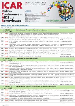
Elenco Poster Discussion Selezionate
Annoni - Parco Maraini
Diapositiva 1 - Pneumologia Veneto
Mosconi