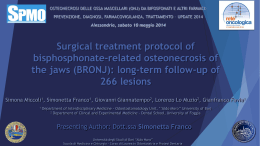
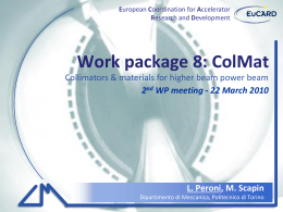
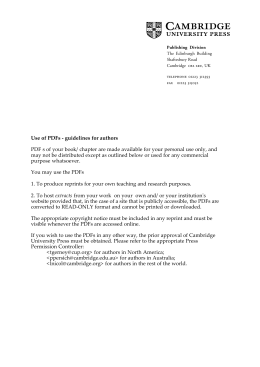
ACTA otorhinolaryngologica italica 2015;35:350-354; doi: 10.14639/0392-100X-546
Clinical techniques and technology
3D computed tomographic evaluation of the upper
airway space of patients undergoing mandibular
distraction osteogenesis for micrognathia
Valutazione mediante tomografia assiale computerizzata 3D delle vie aeree superiori
di pazienti sottoposti a distrazione osteogenetica mandibolare per micrognazia
A. Bianchi, E. Betti, G. Badiali, F. Ricotta, C. Marchetti, A. Tarsitano
Maxillofacial Surgery Unit, Policlinico “S. Orsola-Malpighi”, University of Bologna, Italy
Summary
Mandibular distraction osteogenesis (MDO) is currently an accepted method of treatment for patients requiring reconstruction of hypoplastic mandibles. To date one of the unsolved problems is how to assess the quantitative increase of mandible length needed to achieve a
significant change in the volume of the posterior airway space (PAS) in children with mandibular micrognathia following distraction osteogenesis. The purpose of this study is to present quantitative volumetric evaluation of PAS in young patients having distraction osteogenesis
for micrognathia using 3D-CT data sets and compare it with pre-operative situation. In this observational retrospective study, we report our
experience in five consecutive patients who underwent MDO in an attempt to relieve severe upper airway obstruction. Each patient was
evaluated before treatment (T0) and at the end of distraction procedure (T1) with computer tomography (CT) in axial, coronal, and sagittal
planes and three-dimensional CT of the facial bones and upper airway. Using parameters to extract only data within anatomic constraints,
a digital set of the edited upper airway volume was obtained. The volume determination was used for volumetric qualification of upper
airway. The computed tomographic digital data were used to evaluate the upper airway volumes both pre-distraction and post-distraction.
The mean length of distraction was 23 mm. Quantitative assessment of upper airway volume before and after distraction demonstrated
increased volumes ranging from 84% to 3,087% with a mean of 536%. In conclusion, our study seems to show that DO can significantly
increase the volume of the PAS in patients with upper airway obstruction following micrognathia, by an average of 5 times. Furthermore,
the worse is the starting volume, the greater the increase in PAS to equal distraction.
key words: 3D Computed tomographic evaluation • PAS • Mandibular distraction osteogenesis • Micrognathia
Riassunto
La distrazione osteogenetica mandibolare rappresenta oggi un metodo di trattamento consolidato per i pazienti affetti da ipoplasia mandibolare. Ad oggi, un problema insoluto, nei bambini affetti da micrognazia, è la modalità di valutazione del guadagno di lunghezza mandibolare necessario ad ottenere un miglioramento significativo a livello del volume dello spazio aereo posteriore (PAS). La proposta di
questo studio è la valutazione volumetrica quantitativa del PAS in giovani pazienti sottoposti a distrazione osteogenetica mandibolare per
severa micrognazia, attraverso l’analisi di ‘data-set’ di TC pre-trattamento in comparazione ai medesimi dati post-trattamento. In questo
studio retrospettivo osservazionale riportiamo la nostra esperienza relativa a cinque pazienti sottoposti a distrazione osteogenetica mandibolare. Per ciascuno dei pazienti in esame, è stata valutata la TC pre-trattamento (T0) ed al termine del trattamento (T1) nei piani assiale,
coronale, sagittale e 3D a livello del PAS. Utilizzando parametri di estrazione dei dati anatomici, è stato ottenuto un modello di analisi
dello spazio aereo posteriore, utilizzato per comparare le differenze volumetriche quantitative a T0 e T1. La lunghezza media di distrazione
ottenuta è stata di 23 mm. L’analisi volumetrica quantitativa del PAS ha mostrato un incremento di volume, al termine del trattamento,
variabile dal 84% sino 3,087% (media 536%) rispetto alla situazione pre-trattamento. Concludendo, il presente studio sembra confermare
che la distrazione osteogenetica incrementi in maniera significativa il volume del PAS in pazienti con ostruzione delle vie aeree dovuta
alla micrognazia. La quantificazione di tale incremento appare lineare con il guadagno ottenuto grazie alla distrazione. Nella suddetta
popolazione di studio, tale guadagno è stato, in media, di 5 volte rispetto al volume di partenza. Il dato da sottolineare è che tanto minore
è il volume del PAS al T0, tanto maggiore risulta il guadagno volumetrico al T1.
parole chiave: Tomografia computerizzata 3D • PAS • Distrazione osteogenetica mandibolare • Micrognazia
Acta Otorhinolaryngol Ital 2015;35:350-354
Introduction
Upper airway obstruction occurs most commonly in individuals with craniofacial anomalies associated with
350
micrognathia such as Pierre Robin syndrome, hemifacial
microsomia, Treacher Collins and Nager syndromes 1.
In these disorders, the reduced size of mandible and its
Volumetric 3D assessment of PAS in mandibular distraction osteogenesis
retruded position cause retro-displacement of the tongue
and concomitant reduction of the oropharyngeal airway
that may lead to upper airway obstruction.
Patients have symptoms of obstructive sleep apnoea
(OSA) that in severe cases require tracheal intubation and
tracheostomy 2. More than 60% of children with craniofacial abnormalities require airway intervention as part of
their overall treatment 2.
Tracheostomy is traditionally the safest and most effective treatment option in patients with micrognathia
in Pierre Robin sequence and severe upper airway obstruction and is performed in as many as 12% of cases 2.
Tracheostomy, however, is associated with high cost,
frequent morbidity, and occasional mortality, with an
average age of 3.1 years at decannulation 2. Therefore,
the development of other effective methods of airway
management is desirable.
Mandibular distraction osteogenesis (MDO) has become
an accepted method of treatment for patients requiring reconstruction of hypoplastic mandibles and may
achieve mandibular lengthening without need for bone
graft 2. During the past few years mandibular reconstruction by distraction osteogenesis has been demonstrated
to be effective in resolving upper airway obstruction and
need for tracheostomy decannulation. Mandibular distraction has also been used in respiratory-distressed neonates and infants to avoid tracheostomy 2.
To date one of the unsolved problems is how to assess
the quantitative increase of mandible length needed to
achieve a significant change in the volume of posterior
airway space (PAS) in children with mandibular micrognathia following distraction osteogenesis.
The purpose of this study is to present the quantitative
volumetric evaluation of PAS in young patients having
distraction osteogenesis for micrognathia using 3D-CT
data sets and compared it with pre-operative situation.
Description of clinical techniques
and technology
In this observational retrospective study, we report our experience in five consecutive patients (1 with Treacher Collins syndrome, 1 with Nager syndrome, 2 with bilateral
hemifacial mycrosomia and 1 with severe micrognathia)
Fig. 1. Anatomic limits of upper airway in sagittal and 3D reconstructed CT scans.
who underwent mandibular DO in an attempt to relieve
severe upper airway obstruction between March 2008 and
May 2011 at the Maxillofacial Surgery Unit of “S. Orsola-Malpighi” University Hospital of Bologna, Italy.
Inclusion criteria were the presence of syndromic or nonsyndromic mandibular hypoplasia and respiratory distress
with episodes of severe desaturation (oxygen saturation
below 70%).
Exclusion criteria included central apnoea, apnoea that
was dependent on other levels of airway impairment, such
as laryngomalacia/tracheomalacia, and previous surgical
procedures.
Mandibular distraction was planned bilaterally in order
to advance the mandible and to increase upper airway
volume. Unidirectional extra-oral (n = 4) and intra-oral
(n = 1) distraction devices were used.
Each patient was evaluated before treatment (T0) and
three months after the end of the distraction procedure
(T1) with computed tomography (CT) in axial, coronal
and sagittal planes and three-dimensional CT of the facial
bones and upper airway.
Regarding CT scans, each child received the same protocol for three-dimensional CT of whole head, with 1 mm
continuous axial slices, parallel to the Frankfurt horizontal 3. The CT data were converted into DICOM (Digital
Imaging and Communication in Medicine) format, after
which the images were reconstructed for a 3D-model with
Table I. Reference points and planes.
Area
Definition Explanation
AREA 1 (the area between the CV1
and CV2 planes)
AREA 2 (The area between the CV2
and CV3 planes)
AREA 3 (The area between the CV3
and CV4 planes)
CV1
Horizontal plane passing for the most anterior point of the anterior arch of the atlas
CV2
Horizontal plane passing for the most anterior inferior point of the body of the 2nd cervical vertebra
CV3
Horizontal plane passing for the most anterior inferior point of the body of the 3rd cervical vertebra
CV4
Horizontal plane passing for the most anterior inferior point of the body of the 4th cervical vertebra
351
A. Bianchi et al.
Fig. 2. 3D reconstructed CT scan volumetric assessment of
the upper airway.
Simplant O&O (Materialise Dental, Leuven, Belgium).
The automatically fixed threshold value was manually increased for each dataset until the nasopharyngeal airway
was adequately depicted. The maximum value established
as the reference threshold for this study of all patients thus
defined was -306 and the minimum value was -1024.
Preparatory to upper airway analysis, the relevant reference planes and points were determined.
The upper airway was divided into three regions relative
to the reference planes: area 1 (between the CV1 and CV2
plane); area 2 (between the CV2 plane and CV3 plane); area
3 (between the CV3 and the CV4 plane) (Fig. 1a, b; Table I)
Using parameters to extract only the data within anatomic
constraints, a digital set of the edited upper airway volume was obtained (Fig. 2). The volume determination was
used for volumetric qualification of the upper airway. The
digital CT data were used to evaluate upper airway volumes both pre-distraction and post-distraction (Fig. 3).
Table II. Clinical and volumetric data.
Patient
Age
Length of DO
Results
Five infants (1 boy and 4 girls) with micrognathia, glossoptosis and severe upper airway obstruction underwent
bilateral mandibular DO. The mean length of distraction
was 23 mm (range, 13-35 mm) (Table II). Information on
patient age, type of DO performed and the exact length of
distraction for each patient is detailed in Table II.
The lateral, axial, coronal and 3D CT at the end of distraction revealed forward lengthening of the mandible and hyoid bone as exemplified in Figure 3 for all patients. These
resulted in forward traction of the tongue and increased
Pre-DO upper airway
volume (mm3)
Post-DO upper airway
volume (mm3)
∆ Volume (mm3)
∆ Volume (%)
A1: 113
A2: 330
A3: 546
A1: 0
A2: 148
A3: 251
A1: 811
A2: 635
A3: 351
A1: 3106
A2: 2700
A3: 2670
A1: 3808
A2: 1485
A3: 2425
A1: 1500
A2: 2194
A3: 2017
A1: 3528
A2: 4717
A3: 2527
A1: 3500
A2: 1695
A3: 1781
A1: 7405
A2: 5582
A3: 7023
A1: 7035
A2: 3116
A3: 6098
A1: +1387
A2: +1864
A3: +1471
A1: +3528
A2: +4569
A3: +2276
A1: +2689
A2: +1060
A3: +1430
A1: +4299
A2: +2882
A3: +4350
A1: +3227
A2: +1631
A3: +3673
A1: +1227%
A2: +564%
A3: +269%
A1: +352%
A2: +3087%
A3: +906%
A1: +331%
A2: +166%
A3: +407%
A1: +138%
A2: +106%
A3: +162%
A1: +84%
A2: +109%
A3: +151%
3
20 mm
oblique
#2
4
35 mm
oblique
#3
6
22 mm
oblique
#4
5
13 mm
oblique
#5
5
25 mm
horizontal
352
The present study is retrospective in nature. The study
was conducted in accordance with the tenets of the WMA
Declaration of Helsinki in the context of Ethical Principles for Medical Research Involving Human Subjects and
was granted exemption by the local IRB of our Institution.
Vector of DO
#1
Legend: A1: area 1; A2: area 2; A3: area 3.
Fig. 3. 3D reconstructed CT scan showing pre-treatment airway volume (left side) and post-treatment increased volume
(right side).
Volumetric 3D assessment of PAS in mandibular distraction osteogenesis
pharyngeal space. 3D-CT demonstrated improved airway in all patients. The three-dimensional digital image
demonstrated the increased airway in all areas (Table I).
Area 1 volume increased between 1.387 mm3 (pt. #1) and
4.299 mm3 (pt. #3). Area 2 volume increased between
1.060 mm3 (pt. #3) and 4.569 mm3 (pt. #2). Area 3 volume
increased between 1.430 mm3 (pt. #3) and 4.350 mm3
(pt. #4). The mean increased volume for all areas was
3.379 mm3 (Table II).
Quantitative assessment of the upper airway volume
before and after distraction demonstrated increased
volume ranging from 84% to 3.087% with a mean of
536%. All variations in terms of volumetric upper airway changes between pre and post-DO situation are detailed in Table II.
Qualitative increase of upper airway volume was assessed considering the decannulation rate at the end of
the DO procedure in patients undergoing tracheostomy.
In fact, all patients subjected to tracheostomy before
DO underwent decannulation after the end of the described procedure. This confirms the efficacy of DO in
terms of airway volume increase in patients with severe
micrognathia.
During the 24-36 months of follow-up none of the patients developed symptoms of OSA and none needed
CPAP treatment.
Discussion
Micrognathia is characterised by a small and retrusive
mandible. Children with craniofacial anomalies associated with micrognathia, retruded position of the mandible and glossoptosis often have compromised upper
airways, a condition with potential for morbidity and
mortality. The degree of respiratory difficulty is dependent on the severity of the micrognathia and glossoptosis. These patients usually fail to thrive, present
feeding problems, have insufficient weight gain associated with malnutrition, higher pulmonary morbidity
and long-term hospitalisation 1. There are a variety of
options available for airway management in the micrognathic child. Certainly, it is reasonable to begin with
the most conservative measures. This includes prone
positioning and placement of a nasal pharyngeal airway. The use of positive pressure mask ventilation
through the nasal pharyngeal airway will provide some
additional benefit. Other options include glossopexy
procedures, or subperiosteal release of the floor of the
mouth combined with glossopexy. For the most severe
cases, tracheotomy is traditionally considered to be
the definitive technique in securing a stable airway for
these children 2. Long-standing tracheostomies are associated with high morbidity such as tracheomalacia,
chronic bronchitis, laryngeal stenosis and risk of death
due to the mucus plug or dislodgement of tracheostomy
tube. Patients who undergo tracheostomy require complex nursing care.
The method of DO should have a substantial advantage
over all of the above mentioned techniques. It enables
gradual forward advancement of the mandible and tongue
that increase the pharyngeal space. The apnoea index and
O2 saturation should be markedly improved following distraction.
Most studies report improved airway and respiratory
status in patients after DO based on polysomnography,
cephalometry, decannulation rates and imaging studies.
It is known from the literature that posterior airway volume can be accurately measured three-dimensionally using CT 4-6. Nevertheless, few studies have measured the
extent of the increase in volume of PAS as a result of DO
and no study has related the extent of distraction with the
increase in volume itself. Herein, we present our quantitative assessment of increase in volume of PAS using
a three-dimensional method based on CT. Our results
show that the increase in volume can be much more extensive than previously assessed by other studies with
similar quantities of distraction. An average increase of
536% (ranging from 84 to 3,087%) is a surprisingly positive result which gives, according to the authors, a much
more important value to DO in the treatment of upper
airway obstruction in mandibular micrognathia. Positive discordance with previous studies could be further
investigated and can be attributed to slight differences
in the evaluation method, but our analysis appears to be
robust and reliable.
Furthermore, there seems to be no relation between the
entity of distraction and the increase in volume. In fact,
the greatest distraction (35 mm) caused the best increase
in PAS (356-3087%), but the other results show no proportion with the quantity of DO. Obviously, this is only
an underlying trend, because five cases are to few to
evaluate a statistically significant result. Nevertheless,
the slight inverse proportion between the pre-operative volume and the volume increase should be noted.
This, according to our previous results in adult OSAS
patients 5, seems to demonstrate that those with greater
preoperative PAS volumes can obtain lower increase in
volume, independently of the entity of DO, while patients with smaller preoperative PAS volume can obtain
a greater increase in volume, also independently from
the entity of DO. This could mean that the possibility to
augment the PAS volume varies according to the starting
volume in a non-linear manner.
In conclusion, our study seems to show that DO can
significantly increase the volume of the PAS in patients with upper airway obstruction following micrognathia, by an average of 5 times; the worse the starting volume, the greater the increase in PAS to equal
distraction.
353
A. Bianchi et al.
References
1
Cosman B, Crikelair GF. Mandibular hypoplasia and the late
development of glossopharyngeal airway obstruction. Plast
Reconstr Surg 1972;50:573-9.
2
Perkins JA, Sie KC, Milczuk H, et al. Airway management in
children with craniofacial anomalies. Cleft Palate Craniofac
J 1997;34:135-40.
3
Knapp RH, Vannier MW, Marsh JL. Generation of three dimensional images from CT scans: technological perspective.
Radiol Technol 1985;56:391-8.
4
Bianchi A, Betti E, Tarsitano A, et al. Volumetric three-dimensional computed tomographic evaluation of the upper
airway in patients with obstructive sleep apnoea syndrome
treated by maxillomandibular advancement. Br J Oral Maxillofac Surg 2014;52:831-7.
5
Tarsitano A, Marchetti C. Unusual presentation of obstructive sleep apnoea syndrome due to a giant mandible osteoma: case report and literature review. Acta Otorhinolaryngol
Ital 2013;33:63-6.
6
Saccomanno S, Greco F, D’Alatri L, et al. Role of 3D-CT
for orthodontic and ENT evaluation in Goldenhar syndrome.
Acta Otorhinolaryngol Ital 2014;34:283-7.
Received: December 24, 2014 - Accepted: March 8, 2015
Address for correspondence: Achille Tarsitano, Maxillofacial Surgery Unit, “S. Orsola” University Hospital, via G. Massarenti 9,
40100 Bologna, Italy. Tel. +39 051 6364197. E-mail: [email protected]
354
Scaricare