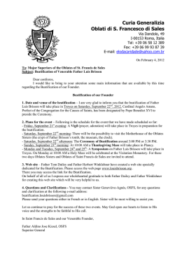
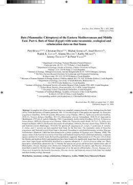
Acta Zoologica Academiae Scientiarum Hungaricae 59(1), pp. 41–59, 2013
THE SUBSPECIES OF MYOTIS MONTIVAGUS –
TAXONOMIC REVISION AND SPECIES LIMITS
(MAMMALIA: CHIROPTERA: VESPERTILIONIDAE)
Görföl, T.1,3, Estók, P.2 and Csorba, G.3
1
Institute for Veterinary Medical Research, Centre for Agricultural Research
Hungarian Academy of Sciences, H-1143 Budapest, Hungária krt. 21, Hungary
E-mail: [email protected]
2
Department of Zoology, Eszterházy Károly College, H-3300 Eger, Eszterházy tér 1, Hungary
E-mail: [email protected]
3
Department of Zoology, Hungarian Natural History Museum
H-1088 Budapest, Baross u. 13, Hungary; E-mail: [email protected]
A morphological evaluation of different Myotis montivagus subspecies and M. annectans is
presented. Using evidence provided by cranial and dental features and multivariate statistical analyses, we raise the four montivagus subspecies to species level, recognizing M.
borneoensis, M. federatus, M. montivagus and M. peytoni as distinct species. Diagnoses, distribution and ecological data are given for each of these species and the morphologically
very similar M. annectans. The conservation status of M. montivagus sensu lato should be reconsidered as our elevation of these subspecies to species results in a significantly smaller
distribution range for each taxon.
Key words: Indomalayan Region, Myotis annectans, species identification, taxonomy.
INTRODUCTION
Bats are a highly diverse group of mammals with 1259 species presently
known (Fenton 2012), a number which continues to grow as new taxa are
described every year (e.g. Velazco et al. 2010, Csorba et al. 2011, Francis &
Eger 2012). These discoveries are based not only on specimens collected during contemporary field expeditions but also on studies of museum specimens
sometimes over a hundred years old. Correct recognition and classification of
taxonomic units is essential for studies in biogeography, ecology and conservation. Conservation mainly focuses on species level taxonomic units; hence
failure to recognize these correctly could lead to vulnerable species being
overlooked. Re-evaluation of different taxonomic units, investigation of type
material and description of cryptic taxa based on morphology and morphometrics are consequently essential even in the age of genetics.
Myotis is the most speciose genus of bats and includes a large number of
morphologically similar species. M. montivagus (as Vespertilio montivagus) was
described by Dobson (1874) from Yunnan, China and is characterised by its
relatively small size (forearm 39.2–41.5 mm), small foot, and displaced middle
Hungarian Natural History Museum, Budapest
42
GÖRFÖL, T., ESTÓK, P. & CSORBA, G.
upper premolar (P3) and lower middle premolar (p3) in the toothrow. Four
subspecies are currently recognised. Wroughton and Ryley (1913) described
M. peytoni from Kanara, India as a new species of the “small-footed section” of
Myotis on account of its much larger size compared to mystacinus and nipalensis. Thomas (1916) distinguished a Malaysian bat as M. peytoni federatus by its
smaller forearm, metacarpals and hind legs. Based on dental characters, both
peytoni and federatus were regarded as closely allied to and synonymised with
M. montivagus by Hill (1962), the latter being regarded by him as a member
of the Selysius subgenus. Another subspecies, M. montivagus borneoensis, was
proposed by Hill and Francis (1984). These authors postulated that this form
is cranially the largest of the species whereas the nominotypical subspecies
from south China and north Myanmar is the smallest.
M. annectans was originally described (Dobson 1871) as a species of Pipistrellus due to its missing upper and lower middle premolars but was later
transferred to Myotis by Topál (1970) who (after investigating the type specimen of M. annectans held in the collection of Zoological Survey of India, Kolkata) also viewed M. primula Thomas, 1920 as a synonym of M. annectans. The
dentition of the type specimen of M. primula is complete with three premolars
in the upper and lower toothrows but the presence or absence of the minute
middle premolars within the species is variable. This is demonstrated, for example, by the presence of P3 and absence of p3 in a specimen from Cambodia (HNHM 2005.82.8.). Hill and Thonglongya (1972) also provided a detailed description of a M. annectans specimen (reportedly housed in the Thai
National Reference Collection) and compared it with the type of M. primula.
The specific distinctiveness of M. annectans and M. montivagus was, however,
questioned by Borisenko and Kruskop (2003: 125) in view of “the wide intraspecific variation of M. montivagus… and that both species are known from
very few specimens”.
M. montivagus, peytoni, federatus and primula (annectans) were analysed
using numerical taxonomic methods by Findley (1972). He placed primula
in the muricola-group of the Selysius subgenus; montivagus in the montivagusgroup and peytoni together with federatus in the peytoni-group. The two latter
groups were placed in the subgenus Leuconoe. He regarded the montivagus
and peytoni groups as comprising seemingly quite distinct taxa and further
noted that “the collocation of peytoni with montivagus, as suggested by Hill
(1962) is not strongly supported”.
Hill (1962) and Corbet and Hill (1992) distinguished montivagus and
annectans by the widely or narrowly separated anteorbital foramen but neither defined the measuring points or provided metric data. The anterior point
of the “anteorbital bridge” (AOB, the distance by which the anteorbital foramen is separated from the orbit) is evident but the posterior end is weakly
Acta zool. hung. 59, 2013
REVISION OF MYOTIS MONTIVAGUS SUBSPECIES
43
determined. In fact, two foramens (their visibility varies) open from the orbit,
namely the foramen lachrymale and the caudal opening of the infraorbital
canal. The anterior rim of the zygoma can extend to either of these and, depending on the taxon and age of the individual, can be developed to a different extent. The posterior point of AOB therefore requires precise definition. In
this paper, we consequently define the foramen infraorbitale and the foramen
lachrymale as landmark measurement points for AOB (Fig. 1).
We critically evaluate the morphological characters of all these taxa and
provide skull diagrams of their type specimens, in addition to craniodental
measurements and statistical comparisons. The four montivagus subspecies
are consequently raised to species level. An amended diagnosis of M. annectans (morphologically the closest species to the montivagus-group) and a
character matrix to compare and discriminate these five species are also given.
MATERIALS AND METHODS
Specimens used for comparisons and statistical analyses are listed under the corresponding taxa in the results section.
Institutional abbreviations include BM(NH): Natural History Museum, London,
United Kingdom, formerly British Museum (Natural History); HNHM: Hungarian Natural History Museum, Budapest, Hungary; HZM: Harrison Institute, Sevenoaks, United
Kingdom, formerly Harrison Zoological Museum.
Forearm length data were compiled from the literature (Hill 1962, Topál 1970, Hill
& Francis 1984, Das 1987, Heller & Volleth 1988, Mandal et al. 2000, Lunde et al. 2003,
Bates et al. 2005, Wilson et al. 2006, Suyanto & Struebig 2007) and taken by the authors from
alcohol-preserved museum specimens to the nearest 0.1 mm. Among the montivagus material from Malaysia listed by Heller and Volleth (1988), two specimens were excluded from
statistical analyses as they exhibit excessively large forearm measurements which cannot be
explained by intraspecific variability and most probably represent a different taxon.
Craniodental measurements were taken by the authors to the nearest 0.01 mm using digital calipers and a stereomicroscope. Measurements include only those taken from
adults. Abbreviations and definitions for craniodental measurements are GTL: greatest
length of skull – from the front of the 1st upper incisor to the most projecting point of the
occipital region; CCL: condylo-canine length – from the exoccipital condyle to the most
anterior part of the canine; C1C1W: width across the upper canines – greatest width across
the outer borders of the upper canines; M3M3W: width across the upper molars – greatest
width across the outer crowns of the last upper molars; ZYW: zygomatic width – greatest
width of the skull across the zygomatic arches; MAW: mastoid width – greatest distance
across the mastoid region; IOW: interorbital width – least width of the interorbital constriction; BCW: braincase width – greatest width of the braincase; BCH: braincase height – from
the basisphenoid at the level of the hamular processes to the highest part of the skull, including the sagittal crest (if present); AOB: anteorbital width – the distance by which the
anteorbital foramen is separated from orbit, measured from the foramen infraorbitale to
the foramen lachrymale; CM3L: maxillary toothrow length – from the front of the upper
canine to the back of the crown of the third molar; CP4L: upper canine–premolar length
Acta zool. hung. 59, 2013
44
GÖRFÖL, T., ESTÓK, P. & CSORBA, G.
– from the front of the upper canine to the back of the crown of the last premolar; ML:
mandible length – from the anterior rim of the alveolus of the 1st lower incisor to the most
posterior part of the condyle; cm3L: mandibular toothrow length – from the front of the
lower canine to the back of the crown of the 3rd lower molar; and CPH: least height of the
coronoid process – from the tip of the coronoid process to the apex of the indentation on
the inferior surface of the ramus adjacent to the angular process. Absolute crown height
was used in all height comparisons for individual teeth (e.g. C versus P4).
Statistical analyses were carried out with R (R Core Team 2012). Measurements were
compared using Welch two sample t-tests. All tests were two-tailed. Principal Component
Analysis (PCA) on the correlation matrix was used for multivariate comparisons. The data
were also analysed using conditional inference trees, which estimate regression relationships by binary recursive partitioning in a conditional inference framework (Hothorn et
al. 2006). We treated species as the response variable and morphological measurements
as explanatory variables. Due to incomplete datasets, external (forearm) and craniodental
data were analysed separately.
RESULTS
Morphology and morphometrics
The skull profile of M. annectans is characterised by a weak rostral depression; the skull is more domed in M. montivagus sensu lato although to a
variable extent among the subspecies (Fig. 2). There are significant differences in AOB values (Welch two-sample t-test, t= -19.81, p < 0.0001) which
are considerably smaller in M. annectans and greater in all subspecies of M.
montivagus (Table 1, Fig. 3). The second upper premolar is often lacking or
excessively reduced in M. annectans whereas in all M. montivagus subspecies it
is always present and usually larger in relation to P4 (Fig. 4).
Within montivagus, subspecies can be separated by a combination of their
external and craniodental features. The subspecies borneoensis (from Borneo)
and peytoni (from south and east-central India) have the largest forearm while
Fig. 1. Measuring points of the anteorbital bridge (AOB).
Acta zool. hung. 59, 2013
REVISION OF MYOTIS MONTIVAGUS SUBSPECIES
45
montivagus (from south China, north Myanmar) and federatus (from Peninsular
Malaysia) are smaller externally. Cranially, Bornean specimens are the largest,
whereas bats from south China and north Myanmar are the smallest. Cranial
dimensions overlap in Peninsular Malaysian and Indian bats. Bornean specimens have the highest sagittal and lambdoid crests; in other subspecies these
are poorly developed. The upper middle premolars (P3) of Bornean and Peninsular Malaysian specimens are much smaller than those of specimens from
south China, north Myanmar or south and east-central India, hence P2 and P4
are fully or nearly in contact with each other in Bornean and Peninsular Malaysian bats. The upper premolars stand loosely in Indian specimens whereas
in Peninsular Malaysian specimens these are crowded and P2 and P4 are in
full contact. The lower middle premolar of Bornean and Peninsular Malaysian
bats is fully intruded from the toothrow but remains within the toothrow in
south China, north Myanmar and south and east-central India specimens.
Multivariate statistical methods were employed alongside classical morphological comparisons to broadly elucidate relationships within montivagus.
Fig. 2. Lateral view of skulls: a = M. annectans BM(NH) 78.2355 from Thailand, b = M.
borneoensis BM(NH) 83.349 (holotype), c = M. federatus BM(NH) 16.4.20.5 (holotype), d =
M. montivagus BM(NH) 76.3.10.5 (holotype), e = M. peytoni BM(NH) 12.8.25.1 (holotype).
Scale = 5 mm.
Acta zool. hung. 59, 2013
46
GÖRFÖL, T., ESTÓK, P. & CSORBA, G.
Table 1. Selected external and craniodental measurements (in mm) of Myotis annectans, borneoensis,
federatus, montivagus and peytoni. Values are given as mean, ±SD (n ≥ 2), min-max, (n). Acronyms and
definitions for measurements are given in the text.
character
annectans
borneoensis
federatus
montivagus
peytoni
46.11±1.17
44.9–48.4 (8)
43.6±1.07
42.1–44.8 (8)
41.01±0.99
39.5–42.5 (9)
40.28±0.78
39.2–41.5 (6)
45.50±0.71
45.0–47.0 (7)
GTL
17.41±0.34
16.96–17.77 (6)
18.03±0.28
17.65–18.57 (7)
16.97
16.79–17.14 (2)
15.69
15.62–15.76 (2)
17.09±0.41
16.19–17.60 (11)
CCL
15.31±0.27
14.86–15.65 (7)
15.76±0.25
15.43–16.10 (7)
15.02
15.01–15.03 (2)
14.09
14.07–14.10 (2)
15.16±0.30
14.50–15.52 (9)
C1C1W
4.74±0.16
4.45–4.91 (7)
4.74±0.11
4.63–4.93 (7)
4.62
4.41–4.82 (2)
4.45±0.06
4.39–4.51 (3)
4.68±0.19
4.24–4.92 (11)
M3M3W
7.31±0.19
7.02–7.61 (7)
7.54±0.12
7.37–7.69 (7)
7.10±0.15
6.99–7.20 (2)
6.67±0.15
6.50–6.79 (3)
7.18±0.17
6.73–7.34 (11)
ZYW
11.41
11.30–11.51 (2)
11.98±0.09
11.82–12.09 (6)
–
–
11.45 (1)
MAW
8.55
8.53–8.57 (2)
8.70±0.13
8.48–8.88 (7)
8.49 (1)
7.97
7.93–8.01 (2)
8.49±0.19
8.12–8.78 (9)
IOW
4.27±0.15
4.01–4.46 (7)
4.02±0.10
3.85–4.15 (7)
3.97
3.96–3.98 (2)
3.81
3.74–3.88 (2)
4.08±0.16
3.78–4.37 (11)
BCW
8.11
7.86–8.36 (2)
7.77±0.11
7.62–7.91 (7)
7.64 (1)
7.17
7.15–7.19 (2)
7.80±0.21
7.54–8.10 (9)
5.78 (2)
6.73±0.17
6.51–6.98 (7)
5.95 (1)
5.75
5.73–5.76 (2)
5.91±0.15
5.65–6.09 (8)
AOB
0.56±0.07
0.47–0.67 (7)
1.42±0.12
1.25–1.65 (7)
1.38
1.35–1.40 (2)
1.06±0.03
1.03–1.08 (3)
1.41±0.08
1.24–1.49 (11)
CM3L
6.76±0.10
6.60–6.88 (7)
6.95±0.12
6.71–7.10 (7)
6.61
6.44–6.78 (2)
6.14±0.04
6.09–6.17 (3)
6.68±0.11
6.50–6.90 (11)
CP4L
3.21
3.09–3.32 (2)
3.15±0.06
3.07–3.24 (7)
3.04 (1)
2.91±0.03
2.89–2.94 (3)
3.10±0.08
2.98–3.22 (9)
12.81±0.23
12.60–13.21 (7)
13.57±0.22
13.33–14.03 (7)
12.62
12.41–12.82 (2)
11.66±0.19
11.48–11.85 (3)
12.72±0.32
12.16–13.16 (11)
cm3L
7.25±0.13
7.04–7.44 (7)
7.32±0.11
7.15–7.46 (7)
7.03
6.93–7.12 (2)
6.43±0.07
6.35–6.47 (3)
7.13±0.13
6.91–7.35 (11)
CPH
4.04
3.76–4.31 (2)
4.76±0.16
4.49–4.92 (7)
4.14 (1)
3.76±0.08
3.67–3.83 (3)
4.10±0.13
3.85–4.24 (9)
FA
BCH
ML
Principal Component Analysis (based on craniodental characters listed in Table 1) resulted in good separation of two groups representing specimens from
Borneo and from south China and north Myanmar, respectively (Fig. 5 and
Table 2). The third group contained specimens from south and east-central
India and Peninsular Malaysia. The latter bats differ, however, in the length of
their forearm: individuals from India have significantly longer forearms than
bats from Peninsular Malaysia (Welch two-sample t-test, t= -10.54, p < 0.0001).
Acta zool. hung. 59, 2013
REVISION OF MYOTIS MONTIVAGUS SUBSPECIES
47
The full cranial and dental metric datasets were also analysed using conditional interference trees. Fig. 6 provides statistical support for four groups:
M. annectans, south Chinese and north Myanmar montivagus sensu stricto, borneoensis, and a fourth comprising federatus and peytoni. However, individuals
within the latter group can be readily separated by their dental characters and
forearm measurements (see above).
Systematic descriptions
The presence of morphologically distinct and geographically separate
taxa within M. montivagus sensu lato is supported by morphometric statistical
analyses. These can be assigned to the four named forms of M. montivagus
Fig. 3. Detail of the rostral part of skulls: a = M. annectans BM(NH) 78.2355 from Thailand,
b = M. borneoensis BM(NH) 83.349 (holotype), c = M. federatus BM(NH) 16.4.20.5 (holotype),
d = M. montivagus BM(NH) 76.3.10.5 (holotype), e = M. peytoni BM(NH) 12.8.25.1 (holotype). Scale = 3 mm.
Acta zool. hung. 59, 2013
48
GÖRFÖL, T., ESTÓK, P. & CSORBA, G.
(previously generally regarded as subspecies), and based on the differences
above, are recognized here as valid species.
Widely accepted species of Myotis from the region are also separated from
each other by similar quantitative features and magnitude of mensural differences (Corbet & Hill 1992, Borisenko & Kruskop 2003). Recently described
species e.g. M. gomantongensis (Francis et Hill, 1998), M. annamiticus (Kruskop et
Tsytsulina, 2001), M. phanluongi (Borisenko et al., 2008) also exhibit similar differences compared to their morphologically closest relatives and once analysed
Fig. 4. Occlusal view of left upper premolar rows: a = M. annectans BM(NH) 78.2355 from
Thailand, b = M. annectans BM(NH) 16.3.25.30 (holotype of M. primula), c = M. borneoensis
BM(NH) 83.349 (holotype), d = M. federatus BM(NH) 16.4.20.5 (holotype), e = M. montivagus
BM(NH) 76.3.10.5 (holotype), f = M. peytoni BM(NH) 12.8.25.1 (holotype). Scale = 3 mm.
Acta zool. hung. 59, 2013
REVISION OF MYOTIS MONTIVAGUS SUBSPECIES
49
Fig. 5. Principal Component Analysis based on 15 craniodental characters of M. borneoensis
(black circles), M. federatus (black squares), M. montivagus (empty squares) and M. peytoni
(empty circles) specimens.
Fig. 6. Conditional interference tree based on 15 cranial characters of 29 specimens (a =
Myotis annectans, b = M. borneoensis, c = M. federatus, d = M. montivagus and e = M. peytoni).
Acta zool. hung. 59, 2013
50
GÖRFÖL, T., ESTÓK, P. & CSORBA, G.
Table 2. Factor loadings of craniodental
characters of M. montivagus-group obtained
by the PCA.
Character
Dim.1
GTL
0.9079
CCL
0.9054
UCCW
0.5712
UM3M3W
0.9095
Dim.2
IOW
0.5312
ZYW
–0.6942
MAW
0.8613
BCW
0.6508
0.6337
BCH
0.7101
–0.4605
AOB
0.6885
UCM3L
0.9424
UCP4L
0.8135
MANL
0.9478
LCM3L
0.9277
PCH
0.8802
Eigenvalue
9.1483
1.7133
Percentage of variance
60.9883
11.4218
genetically turn out to be quite distinct (Borisenko et al. 2008, Francis
et al. 2010).
Diagnoses, distribution (Fig. 7)
and ecological data for these species
and M. annectans are given below.
Myotis annectans (Dobson, 1871)
Hairy-faced Myotis
Pipistrellus annectans Dobson, 1871: 213
Myotis primula Thomas, 1920: 248
Pipistrellus annectens (sic!): Tate 1942: 251
Myotis annectans: Topál 1970: 374
(first use of current name combination)
Myotis annectans: Corbet et Hill 1992: 124
Myotis annectans: Koopman 1994: 104
Material investigated: INDIA (West
Bengal) – BM(NH) 16.3.25.30 (primula
holotype), BM(NH) 20.7.27.2, 20.7.27.3;
CAMBODIA – HZM 1.32758; HNHM
2005.82.8; THAILAND – BM(NH)
78.2355; VIETNAM – HNHM 2008.23.10.
Table 3. Character matrix of M. annectans, borneoensis, federatus, montivagus and peytoni.
Species
FA
AOB
P3 size
P3 position
P2 / P4
p3 position
annectans
44.9–48.4 0.47–0.67
missing
/ minute
missing /
fully
intruded
in contact
fully
intruded
borneoensis
42.1–44.8 1.25–1.65
minute
fully
intruded
in contact
/ narrowly
separated
fully
intruded
federatus
39.5–42.5 1.35–1.40
reduced
intruded
in contact
fully / partly
intruded
montivagus
39.2–41.5 1.03–1.08
reduced
in toothrow
well
separated
in toothrow
peytoni
45.0–47.0 1.24–1.49
developed
in toothrow
well
separated
fully / partly
in toothrow
Acta zool. hung. 59, 2013
REVISION OF MYOTIS MONTIVAGUS SUBSPECIES
51
Amended diagnosis
Forearm 44.9–48.4 mm, CCL 14.86–15.65 mm (Table 1). Dorsal hairs with
long, dark brown bases and paler tips, creating a frosted effect. Ventral hairs
with dark brown bases and silvery-white tips, except for middle of belly
where tips are orange-brown. Ears moderately large, tragus long and narrow
with a rounded tip (Dobson 1871, Francis 2008).
Cranial profile flattened, the depression between rostrum and braincase
is shallow. Only a weak sagittal crest present; the anteorbital bridge is narrow. Basal dimension of the anterior upper premolar (P2) is one-quarter or
one-third that of P4. Middle upper premolar minute or often lacking; when
present, completely displaced from toothrow. Middle lower premolar (p3)
very small, totally intruded from the toothrow, not visible laterally; p2 and p4
are not in contact.
Distribution and ecological notes
M. annectans has a relatively wide distribution ranging from northeast
India (Dobson 1871, Thomas 1920, Bates & Harrison 1997) to north Thailand
Fig. 7. Distribution map of M. annectans (squares), M. borneoensis (triangles), M. federatus
(asterisks), M. montivagus (circles) and M. peytoni (stars). Full symbols represent investigated specimens whereas empty symbols denote literature data.
Acta zool. hung. 59, 2013
52
GÖRFÖL, T., ESTÓK, P. & CSORBA, G.
(Hill & Thonglongya 1972, Bickham et al. 1986), Laos (Francis et al. 1999,
Francis et al. 2010), Vietnam (Francis et al. 2010, Lunde 2003) and Cambodia (Hendrichsen et al. 2001a). It is also recorded from Yunnan, China (Wang
2003). Specimens were found mainly in hill forest habitats at 1077 and 923
metre elevations (Thomas 1920) and at 1250 metres in Doi Pui, Chiang Mai,
Thailand (Hill & Thonglongya 1972).
Myotis borneoensis Hill et Francis, 1984
Bornean Whiskered Myotis
Myotis montivagus borneoensis Hill & Francis, 1984: 309
Myotis montivagus borneoensis: Corbet & Hill 1992: 123
Myotis montivagus borneoensis: Koopman 1994: 106
Material investigated: MALAYSIA (Sabah) – BM(NH) 83.349 (holotype), BM(NH)
83.74, 83.345, 83.346, 83.347, 83.348, 83.350.
Amended diagnosis
Forearm 42.1–44.8 mm, CCL 15.43–16.10 mm (Table 1). Upperparts with
rather long and dense fur, with blackish brown bases and dark brown tips.
Fur on underparts with similar blackish brown bases and buffy brown tips.
Ears long and narrow, with a long tragus, which reaches half the length of the
ear and bends slightly forward (Hill & Francis 1984).
Cranial profile relatively flat, the depression between the rostrum and
braincase is rather shallow. Sagittal and lambdoid crests are well developed; the
anteorbital bridge is wide. Basal dimension of the anterior upper premolar (P2)
is one-third that of P4. Middle upper premolar (P3) very small and completely
displaced from the toothrow so that the first (P2) and third upper premolar (P4)
are usually in tight contact. Lower middle premolar (p3) is intruded from the
toothrow and not visible laterally; p2 and p4 are not in contact.
Distribution and ecological notes
M. borneoensis is confined to Borneo. It occurs in Sabah, Malaysia (Hill &
Francis 1984, Payne et al. 1985, Yasuma & Andau 2000 in Francis et al. 2008),
Sarawak (Wilson et al. 2006), and also in Kalimantan, Indonesia (Suyanto &
Struebig 2007). Payne et al. (1985) and Wilson et al. (2006) recorded the species alongside forest streams, whereas specimens collected in Kalimantan
emerged from a cave in a karst area.
Acta zool. hung. 59, 2013
REVISION OF MYOTIS MONTIVAGUS SUBSPECIES
53
Myotis federatus Thomas, 1916
Malayan Whiskered Myotis
Myotis peytoni federatus Thomas, 1916: 3
Myotis federatus: Tate 1941: 557
Myotis federatus: Findley 1972: 33
Myotis montivagus federatus: Corbet & Hill 1992: 123
Myotis montivagus federatus: Koopman 1994: 106
Myotis montivagus federatus: Francis 2008: 232
Material investigated: MALAYSIA (Peninsular Malaysia) – BM(NH) 16.4.20.5 (holotype), HNHM 98.14.31.
Amended diagnosis
Forearm 39.5–42.5 mm, CCL 15.01–15.03 mm (Table 1). Fur is uniform
dark brown, usually without paler tips (Thomas 1916). Ears moderately large
and narrow. Tragus bends forward and reaches half the height of the ear.
Braincase slightly domed. Sagittal and lambdoid crests are poorly developed; the anteorbital bridge is wide. Basal dimension of the anterior upper
premolar (P2) is one-quarter that of P4. Middle upper premolar (P3) small and
completely displaced from the toothrow so that the first (P2) and third upper premolar (P4) are in contact. Middle lower premolar (p3) small, intruded
from the toothrow and not visible laterally; p2 and p4 are not in contact.
Distribution and ecological notes
M. federatus is restricted to Peninsular Malaysia. Besides the type locality
at the border of Selangor and Pahang, it has been recorded at 900 metres in
Ulu Gombak and Genting Highlands (Heller & Volleth 1988), and in the Temengor Forest Reserve, Hulu Perak (Francis 1995). One other specimen was
caught at the Batu Caves, near Kuala Lumpur (HNHM).
Myotis montivagus (Dobson, 1874)
Burmese Whiskered Myotis
Vespertilio montivagus Dobson, 1874: 237
Myotis mystacinus mystacinus (part): Allen 1938: 215
Myotis montivagus: Tate 1941: 560
Myotis mystacinus montivagus: Ellermann & Morrison-Scott 1951: 140
Myotis montivagus: Findley 1972: 32
Myotis montivagus montivagus: Corbet & Hill 1992: 123
Acta zool. hung. 59, 2013
54
GÖRFÖL, T., ESTÓK, P. & CSORBA, G.
Myotis montivagus montivagus: Koopman 1994: 106
Myotis montivagus montivagus: Francis 2008: 232
Material investigated: CHINA (Yunnan) – BM(NH) 76.3.10.5 (holotype); MYANMAR
– BM(NH) 32.11.1.4, 32.11.1.5
Amended diagnosis
Forearm 39.2–41.5 mm, CCL 14.07–14.10 mm (Table 1). Fur is long and
dense on both the upperparts and underparts. Dorsal hairs are dark brown
overall, with blackish brown bases and paler chocolate brown tips. Ventrally,
hair bases are the same blackish brown but with paler tips (Hill 1962). Ears
are narrow, tapering, with rounded tips. Tragus long, narrow, and its inner
margin straight (Dobson 1874).
Braincase distinctly domed, frontal depression well-expressed. The sagittal and lambdoid crests are weakly developed. The anteorbital bridge is wide.
Basal dimensions of the anterior upper premolar (P2) about one-third that of
P4. Middle upper premolar (P3) relatively well developed and almost in the
toothrow, separating the first (P2) and third upper premolar (P4). Lower middle premolar (p3) developed and placed in the toothrow.
Distribution and ecological notes
According to published records, M. montivagus has the widest distribution of its group. The holotype was collected in Yunnan, south China (Dobson 1871). The species also occurs in northern Myanmar (BM(NH)) and Laos
(Francis et al. 1999) where it was found in a hill forest at 1000 m and in an
open forest at 500 m elevation on the Nakai Plateau. Kuznetsov et al. (2001)
recorded it from Vu Quang, Vietnam and Mandal et al. (2000) collected four
specimens at 1500 m in Mizoram, north-east India.
Myotis peytoni Wroughton et Ryley, 1913
Peyton’s Whiskered Myotis
Myotis peytoni Wroughton & Ryley, 1913: 13
Myotis peytoni: Tate 1941:562
Myotis peytoni: Findley 1972: 33
Myotis montivagus peytoni: Corbet & Hill 1992: 123
Myotis montivagus peytoni: Koopman 1994: 106
Material investigated: INDIA (Karnataka) – BM(NH) 12.8.25.1 (holotype), BM(NH)
12.11.28.55, 12.11.28.56, 12.11.28.57, 12.11.28.58, 12.11.28.59, 12.11.28.60, 12.11.28.61; (Tamil
Nadu) HZM 3.36354, 4.36355
Acta zool. hung. 59, 2013
REVISION OF MYOTIS MONTIVAGUS SUBSPECIES
55
Amended diagnosis
Forearm 45.0–47.0 mm, CCL 14.50–15.52 mm (Table 1). Fur fairly long
and loose. Dorsal surface uniform dark “vandyke brown” with chocolate
brown tips; ventrally hair bases are dark, the tips paler brown to fawn. Tragus is medium length, less than half the height of pinna; inner edge straight
(Wroughton & Ryley 1913, Bates & Harrison 1997).
Cranial profile moderately domed, with a depression between the rostrum and braincase. Sagittal and lambdoid crests are less developed; the
anteorbital bridge is wide. Basal dimension of the anterior upper premolar
(P2) is one-quarter that of P4. Middle upper premolar (P3) is well-developed
and only partly displaced from the toothrow, so the first (P2) and third upper
premolar (P4) are not in contact. Lower middle premolar (p3) partly or fully
lies in the toothrow, p2 and p4 are separated.
Distribution and ecological notes
The type series was collected at Gersoppa (now Jog) Falls in southwest
India. The distribution of the species is limited to the east-central and southern part of the Indian Subcontinent (Wroughton & Ryley 1913, Ghosh 1989,
Bates & Harrison 1997, Vanitharani et al. 2005, Vanitharani 2006).
DISCUSSION
Molecular investigations frequently present un-named lineages on
phylogenetic trees for different chiropteran families (e.g. Clare et al. 2006,
Francis et al. 2010) because of questionable taxonomic assignment of studied specimens due to the lack of thorough revisions. Needless to say, the
results of such studies must be harmonised with existing scientific names,
which is hardly possible without re-examination – and frequently re-description – of type materials for less known taxa. The present work re-evaluated
the taxonomic status of four formerly accepted subspecies of M. montivagus
and elevated them to species rank using cranial and dental characters and
multivariate statistical analyses. To assist determination of species within the
montivagus-group and the morphologically similar M. annectans, a character
matrix is provided in Table 3.
It is worth noting that two Vietnamese specimens of M. montivagus collected in Pu Mat Nature Reserve, Nghe An Province and in Kon Cha Rang,
Gia Lai Province (Bates et al. 1999, Hendrichsen et al. 2001b) could not be
definitely assigned to subspecies by the authors as they shared characters
“considered diagnostic for two of the geographically adjacent races” namely
Acta zool. hung. 59, 2013
56
GÖRFÖL, T., ESTÓK, P. & CSORBA, G.
montivagus montivagus and montivagus borneoensis. Although the authors did
not provide comparisons with annectans or details of anteorbital bridge development, mensural data and accompanying skull and dentition drawings
suggest a species other than annectans, montivagus and borneoensis.
The Barcodes of Life database includes mtDNA sequences for specimens
referred to M. montivagus (in a broad sense) from China, Laos and Vietnam
some of which were analysed in Francis et al. (2010). However, without proper
descriptions of skull characters and mensural data, determination of these
specimens is not possible (see also remarks above on the questionable identity
of some Vietnamese specimens); therefore these records are not depicted in
Fig. 7.
Besides mapping the holotype specimen from Yunnan, Smith and Xie
(2008) also included M. montivagus montivagus in range maps for central-east
China without explanation. As approximately 1600 km lies between these localities and the closest substantiated record of any of the former montivagus
subspecies, these data are considered here as unconfirmed.
M. montivagus (sensu lato, including all its former subspecies) is currently
considered Least Concern in the IUCN Red List of Threatened Species (Francis et al. 2008) because of “its wide distribution, presumed large population,
it occurs in a number of protected areas, has a tolerance of a degree of habitat
modification, and because it is unlikely to be declining fast enough to qualify
for listing in a more threatened category”. However, since the taxa elevated to
species rank herein have significantly smaller distribution ranges, this categorisation should be reconsidered.
*
Acknowledgements – We thank P. Jenkins, L. Tomsett and R. Portela-Miguez (BM[NH]),
P. J. J. Bates and M. Pearch (HZM) for kindly providing access to specimens under their
care. We are grateful for A. Borisenko and one anonymous reviewer for their most useful
comments on the earlier version of the manuscript. Our special thanks are due to N. Furey
for his linguistic corrections and expert advices, to P. Ujhelyi and A. Honfi for the drawings
and to Z. Vas who kindly helped us in the statistical analyses. The work of GC was supported by the SYNTHESYS Integrated Infrastructure Initiative Grant.
REFERENCES
Allen, G. M. (1938) The Mammals of China and Mongolia. Pp. 620. In: Granger, W. (ed):
Natural History of Central Asia XI Part I. The American Museum of Natural History,
New York.
Bates, P. J. J. & Harrison, D. L. (1997) Bats of the Indian Subcontinent. Harrison Zoological
Museum, Sevenoaks, 258 pp.
Acta zool. hung. 59, 2013
REVISION OF MYOTIS MONTIVAGUS SUBSPECIES
57
Bates, P. J. J., Hendrichsen, D. K., Walston, J. L. & Hayes, B. D. (1999) A review of the
mouse-eared bats (Chiroptera: Vespertilionidae: Myotis) from Vietnam with significant new records. Acta Chiropterologica 1(1): 47–74.
Bates, P. J. J., Tin Nwe, Si Si Hla Bu, Khin Mie Mie, Khin Maung Swe, Nyo Nyo, Aye Aye
Khaing, Nu Nu Aye, Yin Yin Toke, Naing Naing Aung, Mar Mar Thi & Mackie, I.
(2005) A review of the genera Myotis, Ia, Pipistrellus, Hypsugo, and Arielulus (Chiroptera: Vespertilionidae) from Myanmar (Burma), including three species new to
the country. Acta Chiropterologica 7(2): 205–236.
Bickham, J. W., McBee, K. & Schlitter, D. A. (1986) Chromosomal variation among seven
species of Myotis (Chiroptera: Vespertilionidae). Journal of Mammalogy 67(4): 746–750.
Borisenko, A. V. & Kruskop, S. V. (2003) Bats of Vietnam and adjacent territories. An identification manual. Joint Russian-Vietnamese Science and Technological Tropical Centre,
Zoological Museum of Moscow M.V. Lomonosov State University, Moscow, 203 pp.
Borisenko, A. V., Kruskop, S. V. & Ivanova, N. V. (2008) A new mouse-eared bat (Mammalia: Chiroptera: Vespertilionidae) from Vietnam. Russian Journal of Theriology 7(2):
57–69.
Clare, E. L., Lim, B. K., Engstrom, M. D., Eger, J. L. & Hebert P. D. N. (2007) DNA barcoding of Neotropical bats: species identification and discovery within Guyana. Molecular Ecology Notes 7: 184–190.
Corbet, G. B. & Hill, J. E. (1992) The mammals of the Indomalayan Region. Natural History
Museum and Oxford University Press, Oxford, 488 pp.
Csorba, G., Nguyen Truong Son, Ith, S. & Furey, N. M. (2011) Revealing cryptic bat diversity: three new Murina and redescription of M. tubinaris from Southeast Asia. Journal
of Mammalogy 92(4): 891–904.
Das, P. K. (1987) On the number of specimens, repository, and the heterogeneity of the
type-series of Vespertilio montivagus Dobson, 1874 (Mammalia: Chiroptera). Bulletin
of the Zoological Survey of India 8(1–3): 39–45.
Dobson, G. E. (1871) Notes on nine species of Indian and Indo-Chinese Vespertilionidae,
with remarks on the synonymy and classification of some other species of the same
family. Proceedings of the Asiatic Society of Bengal 1871: 210–215.
Dobson, G. E. (1874) Descriptions of new species of Chiroptera from India and Yunan.
Journal of Asiatic Society Bengal 43(2): 237–238.
Ellerman, J. R. & Morrison-Scott, T. C. S. (1951) Checklist of Palearctic and Indian Mammals
1758 to 1946. British Museum (Natural History), London, 810 pp.
Fenton, M. B. (2012) Bats and white-nose syndrome. Proceedings of the National Academy of
Sciences 109(18): 6794–6795.
Findley, J. S. (1972) Phenetic relationships among bats of the genus Myotis. Systematic Zoology 21(1): 31–52.
Francis, C. M. (1995) The diversity of bats in Temengor Forest Reserve, Hulu Perak, Malaysia. Malayan Nature Journal 48: 403–408.
Francis, C. M. (2008) A field guide to the mammals of South-east Asia. New Holland Publishers, London, 392 pp.
Francis, C. M. & Hill, J. E. (1998) New records and a new species of Myotis (Chiroptera,
Vespertilionidae) from Malaysia. Mammalia 62(2): 241–252.
Francis, C. M. & Eger, J. L. (2012) A review of tube-nosed bats (Murina) from Laos with a
description of two new species. Acta Chiropterologica 14(1): 15–38.
Francis, C. M., Borisenko, A. V., Ivanova, N. V., Eger, J. L., Lim, B. K., Guillen-Servent,
A., Kruskop, S. V.,. Mackie, I. & Hebert, P. D. (2010) The role of DNA barcodes in
Acta zool. hung. 59, 2013
58
GÖRFÖL, T., ESTÓK, P. & CSORBA, G.
understanding and conservation of mammal diversity in Southeast Asia. PLoS ONE
5(9): e12575.
Francis, C. M., Guillén, A. & Robinson, M. F. (1999) Order Chiroptera: Bats. Pp. 225–235.
In: Duckworth, J. W., Salter, R. E. & Khounboline, K. (eds): Wildlife in Lao PDR –
1999 Status Report. IUCN, Vientiane.
Francis, C. M., Hutson, A. M., Bates, P. J. J., Csorba, G., Bumrungsri, S., Molur, S. & Srinivasulu, C. (2008) Myotis montivagus. In: IUCN 2012. IUCN Red List of Threatened
Species. Version 2012.1. http://www.iucnredlist.org. Accessed: 10 October 2012.
Ghosh, M. K. (1989) New locality-records for Myotis montivagus peytoni Wroughton &
Ryley, 1913, and Murina cyclotis cyclotis Dobson, 1872 (Chiroptera: Vespertilionidae)
in the eastern ghats of Andhra Pradesh, India. Journal of Bombay Natural History Society 86: 93–94.
Heller, K.-G. & Volleth, M. (1988) Fledermäuse aus Malaysia. 1. Beobachtungen zur
biologie, morphologie und taxonomie (Mammalia: Chiroptera). Senckenbergiana Biologica 69: 243–276.
Hendrichsen, D. K., Bates, P. J. J. & Hayes, B. D. (2001a) Recent records of bats (Chiroptera)
from Cambodia. Acta Chiropterologica 3(1): 21–32.
Hendrichsen, D. K., Bates, P. J. J., Hayes, B. D. & Walston, J. L. (2001b) Recent records
of bats (Mammalia: Chiroptera) from Vietnam with six species new to the country.
Myotis 39: 35–122.
Hill, J. E. (1962) Notes on some insectivores and bats from Upper Burma. Proceedings of the
Zoological Society of London 139(1): 119–137.
Hill, J. E. & Francis, C. M. (1984) New bats (Mammalia: Chiroptera) and new records of
bats from Borneo and Malaya. Bulletin of the British Museum (Natural History), Zoology
Series 47(5): 303–329.
Hill, J. E. & Thonglongya, K. (1972) Bats from Thailand and Cambodia. Bulletin of the British Museum (Natural History), Zoology Series 22(6): 173–196.
Hothorn, T., Hornik, K. & Zeileis, A. (2006) Unbiased recursive partitioning: A conditional inference framework. Journal of Computational and Graphical Statistics 15(3): 651–674.
Koopman, K. F. (1994) Chiroptera: Systematics. Pp. 217. In: Niethammer, J., Schliemann,
H. & Starck, D. (eds): Handbook of Zoology. Walter de Gruyter, Berlin.
Kruskop, S. V. & Tsytsulina, K. A. (2001) A new big-footed mouse-eared bat Myotis annamiticus sp. nov. (Vespertilionidae, Chiroptera) from Vietnam. Mammalia 65(1): 63–72.
Kuznetsov, G. V., Borisenko, A. V. & Rozhnov, V. V. (2001) A synopsis of the mammal fauna
of the Vu Quang Nature Reserve. Pp. 35–46. In: Korzun, L. P. & Kalyakin, M. V. (eds):
Materials of zoological and botanical studies in Vu Quang Nature Reserve (Ha Tinh Province, Vietnam). Joint Russian–Vietnamese Science and Technological Tropical Centre,
Moscow and Hanoi.
Lunde, D. P., Musser, G. G. & Pham Duc Tien (2003) Records of some little known bats
(Chiroptera: Vespertilionidae) from Vietnam. Mammalia 67(3): 459–461.
Mandal, A. K., Poddar, A. K. & Bhattacharyya, T. P. (2000) Further new records of bats
from Mizoram, India. Records of the Zoological Survey of India 98(2): 147–154.
Payne, J., Francis, C. M. & Phillipps, K. (1985) A field guide to the mammals of Borneo. The
Sabah Society and World Wildlife Fund Malaysia, Kota Kinabalu and Kuala Lumpur,
332 pp.
R Core Team (2012) R: A language and environment for statistical computing. R Foundation for Statistical Computing, Vienna. http://www.R-project.org
Acta zool. hung. 59, 2013
REVISION OF MYOTIS MONTIVAGUS SUBSPECIES
59
Smith, A. T. & Xie, Y. (2008) A guide to the mammals of China. Princeton University Press,
Princeton, 544 pp.
Suyanto, A. & Struebig, M. J. (2007) Bats of the Sangkulirang limestone karst formations,
East Kalimantan – a priority region for Bornean bat conservation. Acta Chiropterologica 9(1): 67–95.
Tate, G. H. H. (1941) Results of the Archbold Expeditions 39. Review of Myotis of Eurasia.
Bulletin of the American Museum of Natural History 78(8): 537–565.
Tate, G. H. H. (1942) Results of the Archbold Expeditions 47. Review of the Vespertilionine
bats, with special attention to genera and species of the Archbold Collections. Bulletin
of the American Museum of Natural History 80(7): 221–297.
Thomas, O. (1916) List of Microchiroptera, other than leaf-nose bats, in the collection of
the Federated Malay States Museums. Journal of the Federated Malay States Museums
7(1): 1–6.
Thomas, O. (1920) Scientific results from the Mammal Survey XXIII. A new bat of the genus
Myotis from Sikkim. Journal of Bombay Natural History Society 27: 248–249.
Topál, Gy. (1970) On the systematic status of Pipistrellus annectans Dobson, 1871 and Myotis primula Thomas, 1920 (Mammalia). Annales historico-naturales Musei nationalis
hungarici 62: 373–379.
Vanitharani, J. (2006) Noteworthy representatives of bat species in Agasthyamalai Biosphere Reserve, Tamil Nadu. Journal of Theoretical and Experimental Biology 2(2): 47–59.
Vanitharani, J., Malathi, U. S. U. & Sundari, A. K. (2005) New records of bats from Kalakad Mundanthurai Tiger Reserve, India. BatNet CCINSA Newsletter 6(1): 13–14.
Velazco, P. M., Gardner, A. L. & Patterson, B. D. (2010) Systematics of the Platyrrhinus
helleri species complex (Chiroptera: Phyllostomidae), with descriptions of two new
species. Zoological Journal of the Linnean Society 159(3): 785–812.
Wang, Y. X. (2003) A complete checklist of mammal species and subspecies in China: a taxonomic
and geographic reference. China Forestry Publishing House, Beijing, 394 pp.
Wilson, D. E., Helgen, K. M., Yun, C. S. & Giman, B. (2006) Small mammal survey at two
sites in planted forest zone, Bintulu, Sarawak. Malayan Nature Journal 59(2): 165–187.
Wroughton, R. C. & Ryley, K. V. (1913) Scientific results from the Mammal Survey III.
A new species of Myotis from Kanara. Journal of Bombay Natural History Society 22:
13–21.
Revised version received October 24, 2012, accepted January 4, 2013, published March 28, 2013
Acta zool. hung. 59, 2013
Scarica